Clinical, histopathological and immunohistochemical evaluation of the
effects of topical NPH-insulin on full-thickness open wounds:
An in vivo study in diabetic and non-diabetic mice
İsa ÖZAYDIN
1, Özgür AKSOY
1, Sadık YAYLA
1, Başak KURT
1, Engin KILIÇ
1, Seyit Ali BİNGÖL
2,
İsmail CAN
3, Turgay DEPREM
4Kafkas University, 1Faculty of Veterinary Medicine, Department of Surgery; 2Faculty of Health Sciences, Department of Midwifery; 3Faculty of Medicine, Department of Histology and Embryology; 4Faculty of Veterinary Medicine, Department of Histology and
Embryology, Kars, Turkey.
Summary: In diabetic patients, wound healing is disrupted due to impaired local circulation and infection or healing becomes impossible due to the gangrene. Recently, wound treatment models based on the use of topical insulin have been the subject of some studies. In this study, we aimed to evaluate the efficacy of topical NPH insulin use on wound healing for the treatment of full-thickness open wounds in diabetic and non-diabetic mice. A total of 72, non-diabetic and diabetic adult male Swiss Albino mice were used in the study. A full-thickness skin excision with a diameter of 1 cm was practiced on the back of all mice. Animals in diabetic and non-diabetic groups were divided into two sub-groups as NPH insulin used and control group. After the wound surface areas were digitally measured, the animals were sacrificed for histopathological and immunohistochemical examinations. Wound measurements revealed a significant reduction in the non-diabetic insulin treatment group at 7th and 14th days and at 3rd, 7th and 14th days of treatment in the
diabetic insulin treatment group. Histopathological examinations showed that there was a significant increase in granulomatous inflammation in the wound site in the non-diabetic insulin treatment group at 3rd days and in the diabetic insulin treatment group at 14th
days. Immunohistochemical analyses showed that there was an increase in α-SMA at 3rd days and a decrease in TGF-β1 at 14th days in
the diabetic insulin treatment group, while there was a significant decrease in IGF-I at 7th days in all diabetic groups. Topical NPH
insulin application significantly accelerated healing in the treatment of diabetic and non-diabetic wounds with tissue loss in mice. Keywords: Diabetes, insulin, mice, wound healing.
Tam kalınlıklı açık yaralarda topikal NPH insülinin etkilerinin klinik, histopatolojik ve
immunohistokimyasal olarak değerlendirilmesi: Diyabetik ve non-diyabetik farelerde in vivo bir
çalışma
Özet: Diyabetik hastalarda lokal sirkülasyonun bozulması ve enfeksiyon nedeniyle yara iyileşmesi sekteye uğrar veya gangren şekillenerek iyileşme imkansız bir tabloya dönüşür. Son zamanlarda topikal insülinin kullanımına dayalı yara sağaltım modelleri araştırmalara konu olmaktadır. Sunulan çalışma ile diyabetik ve non-diyabetik farelerde oluşturulan tam kalınlıkta açık yara sağaltımı amacıyla topikal NPH insülin kullanımının, yara iyileşmesi üzerine etkinliğinin değerlendirilmesi amaçlandı. Çalışmada, non-diyabetik ve diyabetik 72 adet erişkin erkek Swiss Albino fare kullanıldı. Tüm farelerde, genel anestezi eşliğinde sırt bölgesinde 1 cm çapında tam kalınlıkta deri eksizyonu ile yara oluşturuldu. Diyabetik ve non-diyabetik gruptaki hayvanlar NPH insulin ve kontrol grubu olmak üzere ikişer alt gruba ayrıldı. Üç, 7 ve 14. günlerde dijital ortamda yara yüzey alanı ölçümleri yapıldıktan sonra, histopatolojik ve immunohistokimyasal incelemeler için hayvanlar sakrifiye edildi. Yara alanı ölçümlerinde, non-diyabetik insülin grubunda tedavinin 7 ve 14. günlerinde, diyabetik insülin grubunda ise 3, 7 ve 14. günlerde kontrol gruplarına göre istatistiksel olarak anlamlı bir küçülme olduğu gözlendi. Histopatolojik incelemeler, granülamatöz yapıların non-diyabetik insülin grubunun yara bölgesinde kendi kontrol grubuna göre 3. günde, diyabetik grubun kendi kontrol grubuna göre ise 14. günde belirgin artış gösterdiğini ortaya koydu. İmmunohistokimyasal analizlerde diyabetik insülin grubunda 3. gün α-SMA’da artış, 14. gün TGF-β1’de düşüş dikkati çekerken, tüm diyabetik gruplarda 7. gün IGF-I’in önemli oranda düşüş gösterdiği belirlendi. Sunulan çalışma ile diyabetik ve non-diyabetik doku kayıplı yaraların sağaltımında topikal insülin uygulamasının iyileşmeyi belirgin şekilde hızlandırdığı ortaya konuldu.
Anahtar sözcükler: Diyabet, fare, insülin, yara iyileşmesi.
Introduction
Diabetic wounds are considered as a significant problem in the field of surgery, and studies addressing
their treatment are ongoing. In human medicine, in particular diabetic foot ulcers are the most typical example of delayed healing or non-healing wounds. Neuropathy,
microangiopathy and associated tissue hypoxia, atherosclerosis in large arteries as well as decreased collagen and DNA synthesis delay wound healing in diabetic wounds (7, 10, 12, 13, 31). In this clinical condition, insulin which acts as a receptor for entry of glucose and amino acids into the cell, cannot reach the wound site, and therefore, the cell is deprived of energy but also cannot undergo mitosis, rendering healing impossible. For this reason, development of granulation tissue followed by epithelization as well as restoration of aesthetic features has always been a topic of surgery in the treatment of diabetic and open wounds with tissue loss (10, 22, 31). In addition to rapid removal of dead tissue, various applications that accelerate epithelium formation (daily debridement to remove dead tissue, granulation activators, demarcation stimulators, electrical stimulation, pomade application, etc.) are employed (10, 31, 34). In recent years, use of hyperbaric oxygen (5), negative pressure therapy (4), mesenchymal stem cell (36), Ankaferd blood stopper and tripeptide-copper complex applications (17) have been investigated in the treatment of chronic wounds particularly in diabetic patients.
The number of studies about the topical use of insulin in the treatment of open wounds is limited both in diabetic and non-diabetic patients (3, 7, 15, 24, 32). Insulin applications administered as injections or through liquid carriers to experimentally-created wound sites showed that wound healing occurs rapidly in these patients, and scar tissue formation, which is a significant problem after wound healing, is minimal (10).
Growth factors play a key role in the biology of wound healing. Many phases of healing are regulated by growth factors (8). IGF-I is a mitogen for keratinocytes. It stimulates the synthesis of collagens, glycosaminoglycans and proteoglycans through dermal fibroblasts (35).
TGF-β1 stimulates the protein synthesis of the extracellular
matrix and initiates phenotypic differentiation in mesodermal cell types. Activation of TGF-β1, which stimulates growth and initiates collagen synthesis, is caused by connective tissue growth factor. Myofibroblasts are reported to play a key role in wound healing and this function occurs due to α-SMA, which is an actin isomer that also participates in intense contraction in these cells (18, 19, 28).
Regular insulin has been used as spray (21) or local injection (6) or cream (23) in previous studies. However, our literature review did not reveal any studies that used topically Neutral Protamine Hagedorn (NPH) insulin as an ointment. In this study, we aimed to evaluate clinically, histopathologically, and immunohistochemically the efficacy of NPH insulin on wound healing in open wounds with tissue loss in diabetic and non-diabetic mice.
Materials and Methods
This study was conducted with approval from the Kafkas University Local Ethics Committee for Animal Experiments (Approval No: KAÜ-HADYEK-2010-11/29).
Animal material and experimental groups: Animal
material for the study consisted of 72 new generation male Swiss Albino mice with an average age of 8-12 weeks. The study consisted of 2 groups, namely non-diabetic (Group ND) and diabetic (Group D) groups, each group consisting of 36 mice, which were divided into two sub-groups: Insulin (NDI/DI, n=18) and Control (Group NDC/DC, n=18) group.
Preparation of mixes: Insulin ointment was prepared
by homogeneously mixed 5 ml insulin (Humulin®N NPH 100 IU/ml, Lilly) in NPH form with 95 g solid medical grade petroleum jelly. To create a placebo effect, 5 ml isotonic NaCI (NS) and 95 g petroleum jelly were homogeneously mixed to form a NS ointment.
Study plan:
Creating diabetes: Diabetes was induced in the mice by a single-dose of 100 mg/kg streptozotocin (STZ) which was dissolved in pH 4.5 and 0.1 M citrate buffer and administered intraperitoneally (Sigma, St Louis, MO, USA) (2). The mice fasted 12 hours before and 4 hours after the STZ administration. In the Group ND, only pH 4.5 and 0.1 M citrate buffer was administered intraperitoneally to ensure uniformity. Blood glucose levels of the mice were analyzed after 72 hours, after the STZ administration and after fasting for 8 hours were also checked using Accu-Chek-Go digital hand glucometer (Roche, Switzerland), and the mice with a blood glucose level of 200 mg/dl were considered as diabetic. Blood glucose levels of the mice were measured again at 3rd, 7th
and 14th days, and the diabetic condition of the animals
was confirmed.
Creating the wounds: 1 cm diameter skin excision wound, covering epidermis, dermis, and panniculus carnosus, was made on the back of the mice under ether anesthesia by the same surgeon. Then, the water in the glass test tube was heated to the boiling point and the tube put in contact with the open wound for 30 seconds to create destruction in the wound bed and make the wounds more complicated (22).
Treatment protocol: Insulin ointment (5 IU/g) was administered to DI, and NS ointment to DC group. Insulin ointment (5 IU/g) was administered to NDI, and NS ointment to NDC group. In all groups, topical applications were administered once a day by applying a thin layer of ointment and rubbing in gently until the ointment was absorbed to the wound throughout the experimental period (23).
Surface area measurements: The wound sites of all subjects were photographed using a Canon PowerShot
SX400 IS digital camera with a graph paper laid over the wound margins. The digital images were opened using Stereo Investigator 7.00 (MBF Bioscience, Williston, Vermont, USA), image analysis program. To obtain accurate measurement results, measurement calibration was performed for each image using graph paper. Then, the surface area of the wounds was determined, and measured using a point grid with a grid size of 0.5 mm. The results were recorded in mm2.
Tissue sampling: At 3rd, 7th and 14th days, six animals
in each sub-group were sacrificed by cervical dislocation after being anesthetized with ether. Tissue samples were collected from the wound site and its surroundings were used for evaluating histopathological and immunohistochemical changes on the days in question.
Evaluation of results:
Measuring wound surface area: The Cavalieri volume estimation method, a stereological method, was modified to estimate the wound surface areas of the subjects. Considering that this method estimates the surface area without evaluating the volume, the surface to be measured was determined, and the surface area was calculated using a point grid (37).
Histopathology: All tissue samples were fixed in 10% buffered neutral formaldehyde for 72 hours. After that, following tissue process was applied: washing, dehydration, clearing, and embedding in paraffin blocks. Four micrometer sections were cut from the blocks. These sections were stained with Hematoxylin-Eosin dye and examined using a light microscope (Olympus BX43 with DP21 camera system) for histopathological examination.
Immunohistochemistry: For immunohistochemical staining, four micrometer sections were used. Antigen retrieval procedure was performed by heating with citrate buffer pH 6.0 then the sections were incubated with 10% Bovine Serum Albumin (BSA) at 20°C for 30 minutes. The sections were incubated with primary antibody (1/400 in PBS + 1% Triton X-100 + 2% goat serum) at 4°C for 24 hours. Three different primary antibodies were used in this study. Their use and dilution ratios are as follows: Rabbit polyclonal to Insulin-like growth factor I {IGF-I} (H-70, Santa Cruz, CA, sc-9013) primary antibody, 1/100 dilution; Rabbit polyclonal to anti-transforming growth factor-beta1 {anti-TGF-β1} (abcam ab92486) primary antibody, 1/100 dilution; Rabbit polyclonal to anti-alpha smooth muscle actin (anti-αSMA) (abcam ab5694) primary antibody, 1/100 dilution. Ultra V block (Thermo Scientific), which was used as the secondary antibody, was incubated. The slides were detected by using the UltraVision Quanto Detection System HRP DAB (Thermo Scientific). Samples were developed using DAB Quanto chromogen and substrate (Thermo Scientific) (5 min.) and counter stained with hematoxylin. After being
covered with Entellan® (rapid mounting medium for microscopy), the cross-sections were examined under the light microscope (Olympus BX43 microscope and DP21 digital camera). After the cross-sections collected from each subject, they were stained with IGF-I, anti-TGF-β1, and anti-αSMA primary antibodies, immune-positive staining cells were counted in 6 randomly chosen areas (X40 magnification) (29).
Statistical analyses: A normality test was initially
performed for the data obtained from the study using the Minitab-17 package program, and then parametric results were statistically evaluated using ANOVA One-way test. Values of p<0.05 were considered statistically significant.
Results
Clinical observations: In the process of wound
closure, when sub-groups of both of the main groups were evaluated, healing was faster, the wound margins were regularly contoured, the eschar fell off earlier, and the scar tissue formed as a thin line in the insulin groups compared to the control groups.
On day 3, the wounds appeared larger and exudative with tissue loss in the Group D, while the wound margins were regularly contoured and no manifestation of exudate was observed in the group ND. On day 7, the wounds enlarged in the Group DC, while the wounds started to form a scab in the Group DI. Although scabbing started in the Group NDC, healing was not as fast as that in the Group NDI. On day 14, there was no significant change in wound diameter in the Group DC and eschar tissue did not fall out, while it fell out in the Group DI. The wounds almost completely closed and there was almost no scar at the wound site in the Group NDI, while the wound area considerably decreased but had not yet completely closed in the Group NDC.
Surface area measurements: Values for wound
surface area measurements at 3rd, 7th and 14th days in all
groups and the statistical differences between them were provided in Table 1.
No time-dependent significant difference was observed between the Group NDC and DC. However, significant differences were found between the Group NDI and DI in wound surface areas at all evaluated time points. According to the measurements in Table 1, there was a statistically significant decrease in wound surface areas in the NDI group in treatments corresponding to days 7 and 14, and to days 3 and 7 as well as to day 14 in the DI group.
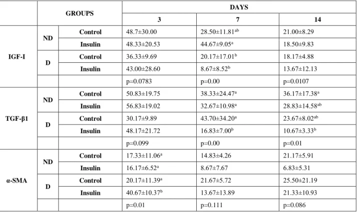
Histopathological findings: Epithelial ulceration with exudation areas in the epidermis, and granulomatous inflammation or active chronic inflammation areas containing histiocytes and polymorphonuclear leukocytes in the deep dermis, and areas of fat necrosis in the
subcutaneous fat tissue were observed in varying degrees in all groups (Figure 1). Granulomatous and active chronic inflammations were very severe, and epithelial ulceration, exudative areas and fat necrosis were relatively high particularly in the NDI group compared to the other
groups on day 3. On the 3rd day of treatment, angiogenesis
starting from the periphery of the wound was seen in all groups. On day 7, angiogenesis disappeared in most places and replaced with to a significant increase in fibroblasts and collagen fibers in DI and NDI groups.
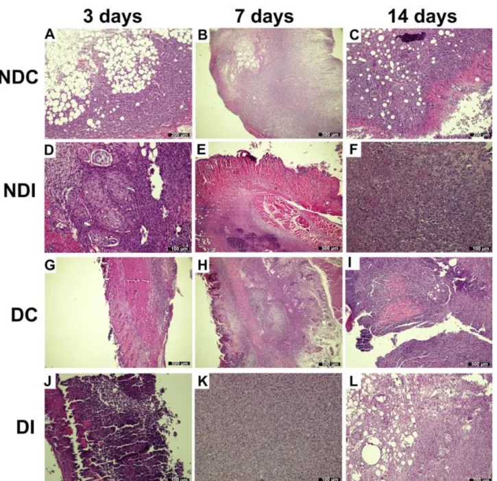
Immunohistochemical findings: Intense and diffuse
immunoreactivity for IGF-I and TGF-β1 was observed in all groups. A significant difference was only found on day
7 in tissue cross-sections marked with IGF-I primary antibody. There was a significant decrease in IGF-I-positive cell counts in the Group DC and DI (Figure 2).
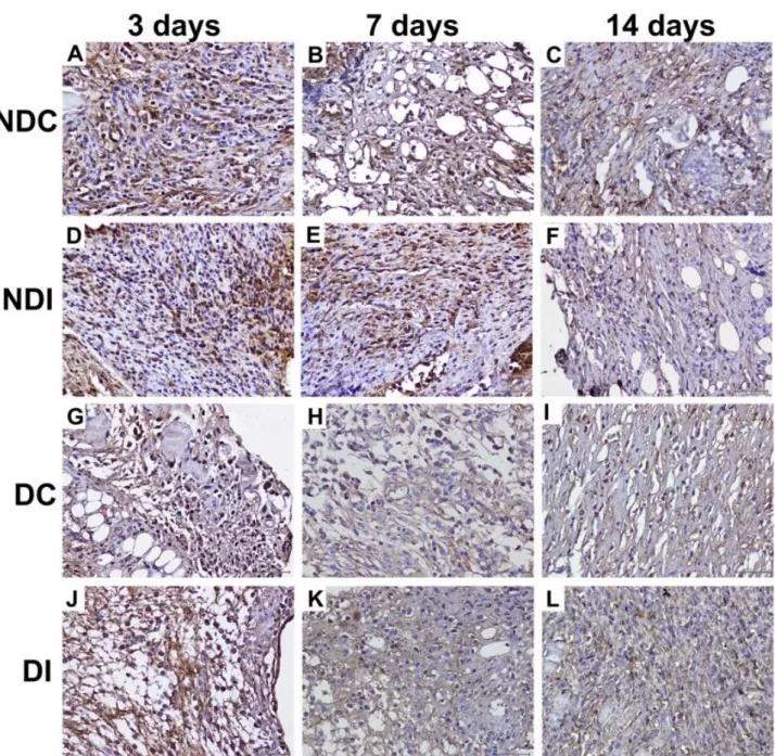
On day 14, a significant decrease was found in cells
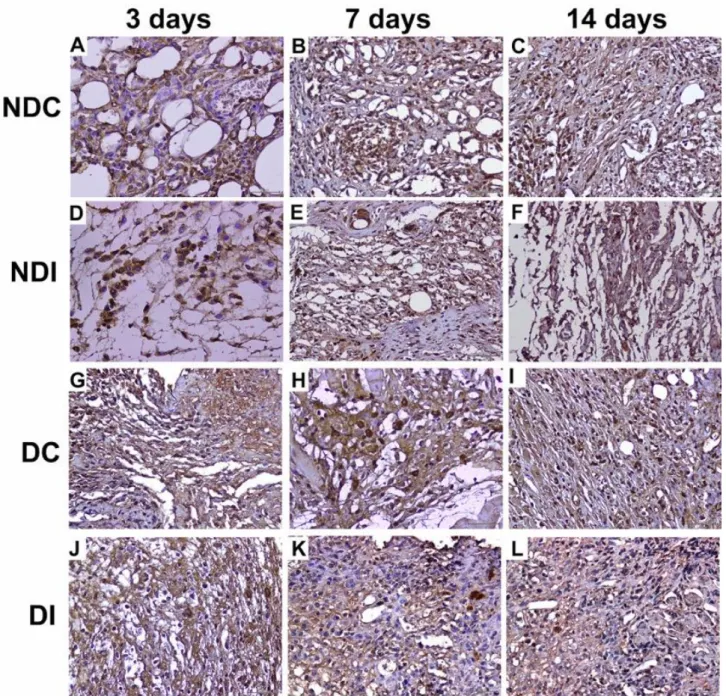
expressing TGF-β1 protein in the DI group, while there was no significant difference in other groups (Figure 3). An evaluation of anti-αSMA, which exhibits intense cytoplasmic immunoreactivity, revealed a significant increase 3rd day of treatment in the DI group, but no
significant difference in other groups. Intense and diffuse activity was observed particularly in the immunohistochemical examination showing the presence of anti-αSMA (Table 2, Figure 4).
Table 1. Digital data for wound area measurements and statistical differences. Tablo 1. Yara alanı ölçümlerine ilişkin sayısal veri ve istatistiksel farklılıklar.
DAYS
GROUPS
Nondiabetic (mm2) Diabetic (mm2) p value
Control Insulin Control Insulin
3 75.46±3.43 a 67.71±9.59 a 71.92±3.37 a 55.17±5.86 b 0.000
7 70.13±3.56 a 53.25±2.04 b 63.08±6.59 ab 30.75±8.57 c 0.001
14 39.91±1.53 a 28.33±5.83 b 45.92±4.47 a 20.29±3.39 c 0.01
a-c: Different values in each row indicate statistical significance (p<0.05). n=6
Table 2. Count of cells exhibiting IGF-I, anti-TGF-β1, and anti-αSMA immunopositive (+) activity. Tablo 2. IGF-I, anti-TGF-β1ve anti-αSMA immunopozitif (+) aktivite gösteren hücre sayısı.
GROUPS DAYS 3 7 14 IGF-I ND Control 48.7±30.00 28.50±11.81 ab 21.00±8.29 Insulin 48.33±20.53 44.67±9.05a 18.50±9.83 D Control 36.33±9.69 20.17±17.01b 18.17±4.88 Insulin 43.00±28.60 8.67±8.52b 13.67±12.13 p=0.0783 p=0.00 p=0.0107 TGF-β1 ND Control 50.83±19.75 38.33±24.47 a 36.17±17.38a Insulin 56.83±19.02 32.67±10.98a 28.83±14.58ab D Control 30.17±9.89 43.70±34.20a 23.67±8.02ab Insulin 48.17±21.72 16.83±7.00b 10.67±3.33b p=0.099 p=0.00 p=0.01 α-SMA ND Control 17.33±11.06 a 14.83±4.26 21.17±5.91 Insulin 16.17±6.52a 8.67±7.67 6.83±5.31 D Control 20.17±11.39 a 21.67±5.72 25.50±21.19 Insulin 40.67±10.37b 13.67±13.89 21.33±10.93 p=0.01 p=0.111 p=0.086
a, b: Different characters in each column indicate statistical significance (p<0.05). ND: Nondiabetic, D: Diabetic
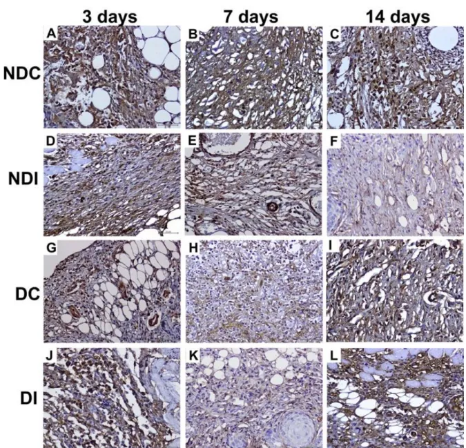
Figure 1. Histopathological changes occurring in wound healing process. A. Fat necrosis at 3rd days in Non-Diabetic Control (NDC),
B. Active chronic inflammation at 7th days in NDC, C. Fat necrosis and epithelial ulceration at 14th days in NDC, D. Granulomatous
inflammation at 3rd days in Non-Diabetic Insulin (NDI), E. Exudative areas at 7th days in NDI, F. Active chronic inflammation at 14th
days in NDI , G. Epithelial ulceration at 3rd days in Diabetic Control (DC), H. Epithelial ulceration and granulomatous inflammation
at 7th days in DC , I. Epithelial ulceration at 14th days in DC, J. Exudative areas 3rd days in Diabetic Insulin (DI), K. Active chronic
inflammation at 7th days in DI, L. Granulomatous inflammation and fat necrosis at 14th days in DI, (White Arrow: Granulomatous
inflammation, Dye: H-E).
Şekil 1. Yara iyileşme sürecinde gözlenen histopatolojik değişiklikler. A. NDK 3. günde yağ nekrozu, B. NDK 7. günde aktif kronik yangı, C. NDK 14. günde yağ nekrozu ve epitelde ülserasyon, D. NDİ 3. günde granulamatöz yangı, E. NDİ 7. günde eksudat, F. NDİ 14. günde aktif kronik yangı, G. DK 3. günde epitelde ülserasyon, H. DK 7. günde epitelde ülserasyon ve granulom yapıları, I. DK 14. günde epitelde ülserasyon, J. Dİ 3. günde eksuda alanları, K. Dİ 7. günde aktif kronik yangı, L. Dİ 14. günde granulamatöz yangı ve yağ nekrozu, (Non-diyabetik + Kontrol (NDK); Non-diyabetik + İnsulin (NDİ); Diyabetik + Kontrol (DC); Diyabetik + İnsulin (Dİ), Beyaz ok: Granulom yapıları, Dye: H-E).
Figure 2. Cells exhibiting IGF-I immunoreactivity in the wound site. (Non-Diabetic Control (NDC); Non-Diabetic Insulin (NDI); Diabetic Control (DC); Diabetic Insulin (DI), White Arrow: IGF-I (+) cells, Dye: IGF-I primary antibody, Bars=50 μm).
Şekil 2. Yara bölgesinde IGF-I immun aktivite gösteren hücreler. (Non-diyabetik Kontrol (NDK); Non-diyabetik İnsülin (NDİ); Diyabetik Kontrol (DK); Diyabetik İnsülin (Dİ), Beyaz ok: IGF-I (+) hücreler, Boya: IGF-I primer antikor, Bar=50 μm).
Figure 3. Cells exhibiting TGF-β1 immunoreactivity in the wound site. (Non-Diabetic Control (NDC); Non-Diabetic Insulin (NDI); Diabetic Control (DC); Diabetic Insulin (DI), White Arrow: TGF-β1 (+) cells, Dye: IGF-I primary antibody, Bars=50 μm).
Şekil 3. Yara bölgesinde TGF-β1 immun aktivite gösteren hücreler. (Non-diyabetik Kontrol (NDK); Non-diyabetik İnsulin (NDİ); Diabetik Kontrol (DK); Diabetik İnsulin (Dİ), Beyaz ok: TGF-β1 (+) hücreler, Boya: IGF-I primer antikor, Bar=50 μm).
Figure 4. Cells with α-SMA immunoreactivity in the wound site. (Non-Diabetic Control (NDC); Non-Diabetic Insulin (NDI); Diabetic Control (DC); Diabetic Insulin (DI), White Arrow: α-SMA (+) cells, Dye: IGF-I primary antibody, Bars=50 μm).
Şekil 4. Yara bölgesinde α-SMA immunreaktiviteye sahip hücreler. (Non-diyabetik Kontrol (NDK); Non-Diabetik İnsülin (NDİ); Diyabetik Kontrol (DK); Diyabetik İnsülin (Dİ), Beyaz ok: α-SMA (+) hücreler, Boya: IGF-I primer antikor, Bar=50 μm).
Discussion and Conclusion
Diabetic wounds are still one of the most significant problems both in human and veterinary medicine (22, 31). There is limited data about topical administration of NPH insulin both in studies with animal models and in diabetic wound cases encountered in human medicine (3, 7, 23, 25, 32). Madibally et al. (25) administered insulin through subcutaneous injection in burn injuries in rats and reported beneficial effects on burn wound healing. Kargın et al. (21) administered insulin using liquid carriers directly to the surroundings of experimentally created open wounds in rats and reported faster wound healing. Lima et al. (23) administered regular insulin in ointment form to the excisional wound created in diabetic rats and demonstrated that insulin helped healing in diabetic patients. All these data support the positive effect of
insulin on wound healing, but in different ways of administration and different forms or doses of insulin. In our study, we investigated the effectiveness of the topical application of NPH insulin prepared in ointment form (5 IU/g) on wound healing in Group D and ND in experimentally opened wounds.
Our clinical findings indicate that wound healing occurred faster in the insulin groups than the control groups. It was also remarkable that scar tissue formation was almost entirely eliminated. This is a clinical evidence of the contribution of topical NPH insulin to heal wounds through an easy way of preparation and administration.
Sources addressing the topical effect of insulin (7, 10, 14, 22, 25) indicate that insulin initially stimulates local angiogenesis, and thus provides a favorable environment for wound healing. In our study,
angiogenesis occurred quicker in the insulin groups than in the control groups. Madibally et al. (25) subcutaneously administered insulin in NPH form to cases with burn injury for 7 days and found that infiltration of inflammatory cells at the wound site decreased, while vasodilatation increased on the 4th day of treatment. This study showed
statistically significant improvement in both area and in depth of ulcer with the help of insulin therapy and thus established the role of insulin in wound healing with increased collagen content. In our study, histopathological findings were consistent with the literature.
IGF-I, which is one of the growth factors and was investigated in our study, is produced by numerous tissues in the body and is found abundantly in circulation (27).In this study, although there was no significant difference between the groups in terms of IGF-I immunoreactivity 3rd
and 14th days of treatment, there was a significant increase
in IGF-I immunoreactivity in the NDI group compared to the NDC group on day 7. Growth factors are reported to be present around the wound during healing at levels that may vary throughout the healing process (8). The increase in the NDI group showed that insulin supports mitosis around the wound. No statistically significant difference was found between the DI and DC groups on this day. As Ömeroğlu et al. (30) reported, this can be explained by the fact that IGF-I involvement is weaker in diabetic wounds than in normal wounds.
TGF-β1 activates fibroblasts to ensure differentiation and proliferation (16, 18, 19). Increased myofibroblastic activity has a closing effect on wound margins from periphery to the center on all sides, allowing wounds with tissue loss to close not only due to cell proliferation, but also due to a contractile effect (10, 18, 19, 22, 26). In this study, insulin-applied sub-groups (NDI and DI groups) had higher values compared to their own control groups on the 3rd day of treatment (p<0.05). TGF-β1 immunoreactivity was lower in the insulin groups
compared to their own control groups on day 14. The lowest value found on the day in question was in group DI (p<0.05). It can be suggested that wound healing reached the desired level since TGF-β1 immunoreactivity was no longer needed after healing took place.
In our study, there was a statistically significant increase in α-SMA immunoreactivity in the DI group compared to other groups on day 3 (p<0.05), while there was a decrease in all groups on day 7 compared to day 3 (p<0.05). There was an increase in α-SMA immunoreactivity in all groups except the NDI group on day 14. Studies showed that wound contraction continues until day 39 at a steady rate and that healing may be associated not only with myofibroblast cell counts, but also with contraction transmission between the cells (33). α-SMA immunoreactivity, which occurred remarkably in the DI group in our study, showed that insulin supports healing in this way. The stagnation in α-SMA
immunoreactivity in the NDI group at 14th days was
associated with the fact that the wounds had already closed in this group. In addition, a study reporting that bFGF experimentally co-administered with α-SMA positively affected healing shows that a number of growth factors work together until the process of wound healing is completed (1). For this reason, the activity in growth factor levels should not be evaluated unilaterally.
In the biology of wound healing, activity and re-epithelization of inflammatory components in the area is reported to last 1 to 2 days, fibroblast and myofibroblast activity 4 to 14 days, and the maturation phase continues for 2 weeks to months, depending on the size of the wound (9, 10, 11, 20, 31). In our study, wounds were clinically healed by day 14 in the NDC and NDI groups, and although wound healing had not yet been completed in the diabetic group, the wounds healed faster in the DI group compared to the DC group. We thereby established that insulin accelerated healing in terms of granulation tissue. In this experimental model, healing occurred earlier particularly in the DI group; further investigation of wound healing effects of insulin in diabetic humans is indicated.
In conclusion, we facilitated wound healing with topical administration of NPH insulin as an ointment form, which has a positive effect on the healing in complicated wounds with tissue loss as well as formation of granulation tissue and epithelization, due to faster completion of all phases of the healing process in open wounds. As a result, the created wounds closed faster, and scar tissue formation after healing was almost entirely eliminated. Therefore, aesthetic features were also restored at the wound site. In addition to the aforementioned benefits, due to the shortened healing process, medication expenses would be reduced, duration of treatment would be shorter and hospitalization time would be reduced, which provides an economic advantage.
Acknowledgements
This study received support from the Kafkas University Office of the Coordinator for Scientific Research Projects. (Project No: KAÜ- BAP-2010-VF-66).
References
1. Akasaka Y, Ono I, Tominaga A, et al. (2007): Basic
fibroblast growth factor in an artificial dermis promotes apoptosis and inhibits expression of alpha-smooth muscle actin, leading to reduction of wound contraction. Wound
Repair Regen, 15, 378-389.
2. Akbarzadeh A, Norouzian D, Mehrabi MR, et al. (2007):
Induction of diabetes by streptozotocin in rats. Indian J Clin
Biochem, 22, 60-64.
3. Apikoglu-Rabus S, Izzettin FV, Turan P, et al. (2010):
Effect of topical insulin on cutaneous wound healing in rats with or without acute diabetes. Clin Exp Dermatol, 35,
4. Armstrong DG, Lavery LA (2005): Negative pressure
wound theraphy after partial diabetic foot amputation: a multicentre, randomised controlled trial. Lancet, 366,
1704-10.
5. Aydin F, Kaya A, Karapinar L, et al. (2013): IGF-1
increases with hyperbaric oxygen theraphy and promotes wound healing in diabetic foot ulcers. J Diabetes Res, doi:
10.1155/2013/567834.
6. Bairy KL, Abhinav R, Satyam SM (2014): Evaluation of
burn wound healing activity of topical regular insulin in non-diabetic and streptozocin-induced diabetic rats. Int J
Pharm Sci, 6, 127-130.
7. Belfield WO, Golinsky S, Compton MD (1970): The use
of insulin in open-wound healing. Vet Med Small Anim
Clin, 65, 455-460.
8. Bennett NT, Schultz GS (1993): Growth factors and
wound healing: Part II. Role in normal and chronic wound healing. Am J Surg, 166, 74-81.
9. Carrico TJ, Mehrhof AI Jr, Cohen IK (1984): Biology of
wound healing. Surg Clin North Am, 64, 721-733.
10. Darby IA, Bisucci T, Hewitson TD, et al. (1997):
Apoptosis is increased in a model of diabetes-impaired wound healing in genetically diabetic mice. Int J Biochem
Cell Biol, 29, 191-200.
11. Gabbiani G (2004): The evolution of the myofibroblast
concept: A key cell for wound healing and fibrotic diseases.
J Gerontol, 52, 280-282.
12. Gal P, Kilik R, Mokry B, et al. (2008): Simple method of
open skin wound healing model in corticosteroid-treated and diabetic rats: Standardization of semi-quantitative and quantitative histological assessments. Vet Med Praha, 53,
652-659.
13. Galiano RD, Tepper OM, Pelo CR, et al. (2004): Topical
vascular endothelial growth factor accelerates diabetic wound healing through increased angiogenesis and by mobilizing and recruiting bone marrow-derived cells. Am J
Pathol, 164, 1935-1947.
14. Goren I, Müller E, Schiefelbein D, et al. (2009): Akt1
controls insulin-driven VEGF biosynthesis from keratinocytes: implications for normal and diabetes-impaired skin repair in mice. J Invest Dermatol, 129, 752-764.
15. Greenway SE, Filler LE, Greenway FL (1999): Topical
insulin in wound healing: a randomised, double-blind placebo-controlled trial. J Wound Care, 8, 526-528.
16. Grotendorst GR, Rahmanie H, Duncan MR (2004):
Combinatorial signaling pathways determine fibroblast proliferation and myofibroblast differentiation. FASEB J,
18, 469-479.
17. Gül Satar NY, Cangül İT, Topal A, et al. (2014): Effects
of Ankaferd Blood Stopper (ABS) and topical tripeptide copper complex (TCC) on wound healing in rats: An experimental study. Kafkas Univ Vet Fak Derg, 20, 545-551.
18. Hinz B (2007): Formation and function of the myofibroblast
during tissue repair. J Invest Dermatol, 127, 526-537.
19. Hinz B, Celetta G, Tomasek JJ, et al. (2001):
Alpha-smooth muscle actin expression upregulates fibroblast contractile activity. Mol Biol Cell, 12, 2730-2741.
20. Hunt TK (1990): Basic principles of wound healing. J Trauma, 30, 122-128.
21. Kargın S, Taştekin D, Kılıç K, et al. (2015): The effects of
topical insulin application on wound healing. Eur J Gen
Med, 12, 302-306.
22. Koç B, Özaydın İ, Özba B, et al. (1990): Clinical
observations on the application of local insulin and pinch
graft in gangrenous-open wound. 2nd National Veterinary
Surgery Congress, 60-65, October 1-2, Alata, Mersin, Turkey.
23. Lima MHM, Caricilli AM, Abreu LL, et al. (2012):
Topical insulin accelerates wound healing in diabetes by enhancing the AKT and ERK pathways: A double-blind placebo-controlled clinical trial. PLoS One, 7, doi:
10.1371/journal.pone.0036974.
24. Liu Y, Petreaca M, Yao M, et al. (2009): Cell and
molecular mechanisms of keratinocyte function stimulated by insulin during wound healing. BMC Cell Biol, 10, doi:
10.1186/1471-2121-10-1.
25. Madibally SV, Solomon V, Mitchell RN, et al. (2003):
Influence of insulin therapy on burn wound healing in rats.
J Surg Res, 109, 92-100.
26. Moulin V, Auger FA, Garrel D, et al. (2000): Role of
wound healing myofibroblasts on re-epithelization of human skin. Burns, 26, 3-12.
27. Mutsaers SE, Bishop JE, McGrouther G, et al. (1997):
Mechanisms of tissue repair: From wound healing to fibrosis. Int J Biochem Cell Biol, 29, 5-17.
28. Ng PC, Hinz B, Swartz MA (2005): Interstitial fluid flow
induces myofibroblast differentiation and collagen alignment in vitro. J Cell Sci, 118, 4731-39.
29. Oruç E, Kara A, Can İ, et al. (2012): Caspase-3 and CD68
Immunoreactivity in lymphoid tissues and haematology of rats exposed to cisplatin and L-carnitine. Kafkas Univ Vet
Fak Derg, 18, 871-878.
30. Ömeroğlu S, Çam M, Erdoğan D, et al. (2003):
Immunohistochemical demonstration of insulin like growth factor-I localisation in the skin wounds of streptozotocin administered experimental diabetic rats. Duzce Med J, 5, 5-8.
31. Özaydın İ (2004): Traumatic Wounds. 128-137. In: Özaydın İ (Ed), Veterinary Emergency Clinic: First Aid, Transporting, Medical Intervention. Eser Ofset, Erzurum. 32. Rezvani O, Shabbak E, Aslani A, et al. (2009): A
randomized, double-blind, placebo controlled trial to determine the effects of topical insulin on wound healing.
Ostomy Wound Manage, 55, 22-28.
33. Stadelmann WK, Digenis AG, Tobin GR (1998):
Physiology and healing dynamics of chronic cutaneous wounds. Am J Surg, 176, 26-38.
34. Taçyıldız HI, Mızraklı S, Aban N, et al. (1998): The effect
of collagenase in wound healing. Turkish J Trauma Emerg
Surg, 4, 7-11.
35. Thorey IS, Hinz B, Hoeflich A, et al. (2004): Transgenic
mice reveal novel activities of growth hormone in wound repair, angiogenesis and myofibroblast differentiation. J
Biol Chem, 279, 26674-26684.
36. Wu Y, Chen L, Scott PG, et al. (2007): Mesenchymal stem
cells enhance wound healing through differentiation and angiogenesis. Stem Cells, 25, 2648-2659.
37. Yoruk O, Dane S, Ucuncu H, et al. (2009): Stereological
evaluation of laryngeal cancers using computed tomography via the Cavalieri method: Correlation between tumor volume and number of neck lymph node metastases.
J Craniofac Surg, 20, 1504-07.
Geliş tarihi: 29.01.2017 / Kabul tarihi: 04.06.2017
Address for correspondence:
Başak KURT
Kafkas University, Faculty of Veterinary Medicine, Department of Surgery, TR 36100, Kars, Turkey. Phone: 904742426836