Evaiuation of subclinical liver lesions in goats by ultrasonographic
and biochemical analyses*
Ramazan GÖNENCIL, Ramazan DURGUT
2,Suat ERDOGAN
3iMustafa Kemal University, Veterinary Faculty, Department of Surgery, Hatay; 2 Mustafa Kemal University, Veterinary Facu!ty. Department of Internal Mcdicinc, Hatay; 'Mustafa Kemal University, Vcterinary Faculty, Department of Biochemisıry. Hatay
Summary: This study included 75 Damascus and its crossbreed goats from different age and sexes in the Hatay province. Following the history, all the animals were subjected to clinical, ultrasonographic and biochemical examinations. Thc clinical signs were generally norınaL. Ultrasonographically, parenchymal lesions in 17, biliary system abnorınalities in 8 and both biliary and pa-renchymal abnormalities in 23 animals were detected. In these 48 goats with hepatic lesions; 26 parenchyma! hyperechogenicity,
ı7
cysts and iO masses of parenchyma, and 25 wall thickenings, 3 foldings and 3 sediments of gallbladder were observed alone or to-gether. LDH levels in goats with and without hepatic lesions were higher than norınal refercnce range, whereas ALT. ALP. AST, 001'. B UN, TP, CB. CHO, albumine and glucose concentrations were in normal referencc ranges. In this study. it is concluded that Iesions could be observed ultrasonographically before clinical signs and biochemical abnormalities manifes\.Key words: Biochemical analyses, goat, liver, ultrasonography
Keçilerde subklinik karaciğer lezyonlarının uıtrasonografik ve biyokimyasalolarak
değerlendirilmesi
Özet: Bu çalı~ımı Hatay'ın çe~itli yerlerinden temin edilen deği~ik ya~ ve cinsiyenen 75 Şam keçisi ve melezIeri üzerinde ya-pıldı. Oerekli anamnez alındıktan sonra, bütün hayvanlar klinik, ultrasonografik ve biyokimyasal muayenelere tabi tutuldu. Klinik bulgular genellikle normal idi. Ultrasonografik olarak, hayvanların ITsinde parenşimal, 8'inde bilier sistem ve 23'iinde de hem pa-renşimal hem de bilier sistem lezyonlarına birlikte rastlandı. Bu lezyonlu 48 keçide 26 papa-renşimal hiperekojenite. 17 kisı, iO kitle, 25 safnı kesesi duvarında kalınla~ma, 3 katlanma ve 3 sediment oluşuımı tek başına ya da diğer lezyonlarla birlikte gözlendi. ALT. ALP.
AST, 001', BUN, TP, CB, CHO, albumin ve glukoz konsantrasyonları lezyonlu ve Iezyonsuz keçilerde normal iken. sadece LDH
her ikisinde de yüksek bulundu. Bu çalı~ma ile karaciğer lezyonlarının klinik ve biyokimyasal anormallikler ortaya çıkmadan önce ultrasonografik olarak gözlenebileceği sonucuna varıldı.
Anahtar kelimeler: Biyokimyasal analizler, karaciğer, keçi, ultrasonografi
Introduction
Liver is highly suseeptible for parenehymal,
vas-cular and biliary system lesions. Baeterial, ehemieal,
vi-ral, toxic or immune-mediated insults may eause foeal or
eliffuse hepatic abnormalities or lesions (8-10,12).
Diagnostic and prognostie use of ultrasonography is
one of the most important and prominent tools for the de-tection and reeognition of many types of foeal parenehymal lesions such as eysts, hemorrhages, haematomas, abscesses,
neerosis, nodular hyperplasia, granulomas and neoplasia
(3,5), The liver is weıı suited for ultrasonographie evalua-tion beeause of its large size and a uniform parenehymal appearanee on ultrasound seans. On the other hand, due
to the faet that ultrasonography is least valuable for
re-eognition of diffuse forms of the disease, other diagnostie
The main inelieations for seanning the biliaı'y system
are to rule out extrahepatie obstruction, to eleteet biliary
calculi, anel to visualize thiekening of the gaııblaeleler
waıı, masses or inflammatory diseases assoeiateel with
gaııbladder and biliary traet. Enlargement of the hepatic
veins, obstruetion of the eaudal vena eava by clots or
masses, eongenital and acquired vaseular abnormalities
can also be imaged (6,7,13).
Hepatoeyte damage may result in the release of
enzymes that may indicate the hepatoeyte integrity or bile
exeretions into the eireulation or failure to produee and
exerete them. The bioehemieal tests are useful in the
assessment of hepatic funetions, diagnosis and the severity
of liver disease. Theyare also valuable diagnostie
in-Ramazan Gönenci - in-Ramazan Durgut - Suat Erdoğan
Results
i i
i
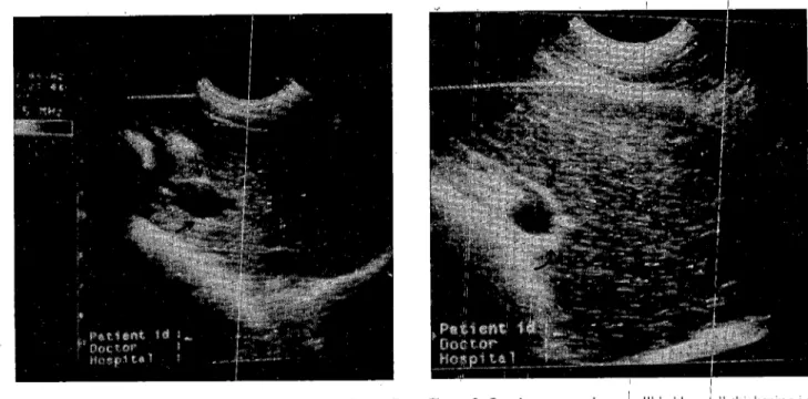
Figure 2. On ultrasonography. a Igaııbladder~all ıhickening is seen, which is shown by the cur~ed arrows. T1heregion shown by the arrow heads is diffuse hyp6rechoic liverıparenchyına.
i
i
i i i i i i iThere was no signs showing theıliver disorders such
i i
as, ieterus, weight lose, aseites, diarrpoea and abÇlominal
pain over the livers on clinical
i
examinatio,b. Fewabscesses were identified in 20 at~imals, whiJ:h were
thought to be eaused by easeous IYI~lPhadenitis tlhaı they
. i
experienced previously, and the majqrity of thesq animals
had pneumonia.
i
iOn ultrasonographie exanıination, most of thıbanimals, i
(n=48) had hepatie lesions includingi parenehyım\l, biliaı'y or both, and the rest of the animaı1 (27) appea:l'ed to be normal. Only par~nchymal lesions ;.yere seen in! 17 go ats
including parenehymal hyperech6genieity, e!ysts and
masses, the frequency of whieh: were 10,
?,
and 4,respeetively. Parenchymal tissue ~ith hyperecl;~ogenieity
had an increased eehogenieity, *hieh eitheı; foeal or
diffuse. Only biliary system abnor~ıalities werJ seen in 8
goats, inc1uding thiekening, foıding and se,bimenı of
i i
gallbladder wa11,the frequency of ıwhieh were
;6.
iand 1,respeetively. The appearanee ofı the gallbla'dders was
aneehoie, having thiek wa11s With hypere(:lhogenieity
(Figure 1). On the other hand, i[he 23 goat~ had both parenehymal and biliary lesions tqgether (Figu!re 2).
The liver of 17 goats hati cysts with anechoic
architecture and a thin wall (Fi!Aıre 3). The ı.longiıudinal~i ~
,
,
seans of goats showeel 10 mUltiliocal hypnecrOiC masses
i i
i
i i i i i i i , i 'Anova test in SPSS programme (version 9.05) w<ls used
i i
for statistieal analysis. i .
Figure
ı.
Ultrasonographic appearance of gallbladder waııthickening. The arrows show the gallbladder wall thickening. Note that hepatic parenchyma appears almost normaL.
Materİals and Methods
This study was carried out in 75 Damascus and its crossbreed goats from different age and sexes in Hatay
province. After taking the anamnesis, they were all
subjectcd to clinical, laboratory and ultrasound exan1İnations. The animals were kept in stand position for ultrasound seans after general examination. A smaIl area of their hair
between last two intercostal space in the right side was
elipped. In the imaging proeedure, the liver was
evaluated for typical signs of hepatic degeneration in size
and echogenicity in order to deteet focal or multifocal
liver lesions. The hepatic ultrasound parameters were
evaluated routinely including size, margin, echogenieity, vascularity, gallbladder and perihepatic abnormalities.
For biochemical evaluations, 10 ml of blood samples were taken from the jugular vein, and sera were separated
by eentrifugation. Samples were analysed for serum
alkaline phosphatase (ALP), alanine aminotransferase
(ALT), aspartate aminotransferase (AST), gamma-glutanıyl
transferase (OOT), lactate dehydrogenase (LDH), blood
urea nitrogen (BUN), albumin (Alb), total protein (TP), conjugated bilirubin (CB), triglyceride (TO), glueose and
cholesterol (CHO) in AMS Autolab analyser, using
Biomedical System and Audit Diagnostie kits. One-W ay
The aim of this study was to investigate and
evaluate the subelinieal foeal liver lesions using
ultra-sonographie examination and bioehemieal analyses in
clinically normal goats in Hatay provinee. 48
Fıglll'L' 3. Appearances of a cyst ıC) and a mass (M) with a diffııse and increased hepatic echogenicity.
Figııre 4. An egg-shaped mass shown by the arrows with increased hepatic echogenicity.
in parenchyma, which are rounded and with thin and
welI-defined or irregular walls. Hypoechoic masses were differentiated from the gallbladder by their location, the
absence of contraction and gallbladder neck (Figure 4).
Folding of the gallbladder on itself was also detected in 3
goats which was differentiated from a mirror-image
artifact because there is no diaphragm between two
galIbladders (Figure 5). Gallbladder lumen filled with
sediment was apparent and agitated with back-and forth
Figııre 5. Appcanıııce of a folciing in the galibideleler "ith \\dll thiekening shown by the arrows. Smail part of the foleling is possible dilated bile dllCL Note also that there is a eyst shown by "C" elose to the diaphragm.
Table I. Nıımbers of animals with biliary, parenehyma! or both lesions, and other ultrasonographic finelings, whieh were observed alone or together.
Lesions Biliary Parenchymal Biliary+ Total
system tissue ParenchYll1al
(n=8) (n=17) (n=23) (n=48) Hypereehogenieity LO In 2n Wall thiekening n ~L) 25 eyst 7 LO 17 Mass 4 (i 10 Folding ı 2 3 Sedimentation i 2 3 Total 8 21 55 X4
The biochemical analyses of the blood senını
resulted in that the activities of AST, ALT, ALP and
GGT, and the !evel of glucose, CB, TP, Alb, BUN anel CHO were found to be in normal ranges whereas the activity of LDH was higher than the normal reference
range although there was no significant difference
(p>O.05) between goats with and without hepatic lesions (Tabı e 2).
Dİscussİon and Condusion
Many clinical signs, such as ictenıs, weight lose,
ascites, diarrhoea and abdominal pain, which are regarded
not to be pathognomonic, may be present with liver
disease (4,10). None of these symptoms were eletecteel in
the study presented. Pathologic changes in the liver may
Ramazan Gönenci - Ramazan Durgut - Suat Erdoğan
Blood serum parameters
SO
i
i i i ii
i
i
Table 2. Blood senım bioehemİeal analyses of goats with and wİthout subeliniea] hepatİe 1esions dİagnosed by ul\rasound.Hepatİe lesions (n=48) Non hepatie lesion!s (n=27) i
, . i
X :t Sx Min-Max X :t Sx Mın-Max;
Ultrasound is an important tool in detecting focal
liver abnormaIities such as cyst, hemalThage, granuloma
and neoplasia because of the uniform background
provided by the normal parenchyma. These focal lesions
can not be differentiated from each other, because they
may Jook similar in echogenicity, size and appearance on
scans (8,9,12). However, cysts might be distinguished
from the others. There are two reports supporting this
notion that the sensitivity and specificity of uItrasound
examination to detect hydatid cysts in goats was
82.1-97.65% and hepatic cysts are usually detected
incielentally without clinical signs (5,11). They showed no clinical signs in animals with 17 hepatic cysts in the
study, anel they were differentiated from gallbladder by
its lack of contraction on ultrasound as researchers
reported (2,3,5,11). A mass may appear moderately
circumscribed, exceeding the normalliver margin with an
echogenicity, which had slightly more-mixed appearance
than normal liver. The increases in hepatic echogenicity
may be due to fibrosis, dystrophic caIcification or
cirrhosis 0,6,8,9). No clinical and biochemical
abnor-malities or ascites were observed in these animals,
implying that the echogenic architectme of the liver
parenchyma might be due to fibrosis or dystrophic
calcification.
The normal gallbladder wall is visualized, the neck of the gallbladder is usually not visible in normal animals
though. Thickening of the wall is a nonspecific finding
seen sametimes with acute and chronic hepatitis,
cholecystitis, cholangiohepatitis or hypoalbuminemia,
less frequently right heart failme, septic conditions, and
neoplasia (1,7-9). Thickening of the gallbladder imaged
1/6-484 : - i 12-66 ; 351-726 : 14-5i : 62-358 ; 37-97 ; 40-101 ! i 0.01 -03~ 18-59
!
6.00-8.6(r 1.IO-2.9(r) 12-49 ! i 244.65:t 12.31 31.34:t 1.65 498.38:t 13.66 32.95:t1.17 i86.78:t20.29 63.22:t2.17 62.56:t2.19 0.14:tO.09 30.86:t1,47 6.79:t093 2.12:t0.76 29.97:t1.54was lost about 75% (4,10).
i i
i
!
i
i i i ii
i iin this study can be possibly resuIt,ed from chıl'onic or
mild hepatitis or cholecystitis. It sho~ıld be notedi that the liver has a large reserve capacity, al?d clinical s{gns and biochemical parameters can occm w~en the liver Ifunction
i
:
i i
In this study, the amount of gallbladder ı~ediment
was not found to be pathognomonid:. The sedinrent may
be seen in healthy non-fasting anin~als as wellj as those
i
with illness such as icterus, chronic hepatitis, fi~rosis and
caIculi (8,10)
:!
It is reported that folding of gallbladder ob'ıserved in
~~,
i
this study may be that gallbladddr dilated Oli partially
folded on itself (8,9). i :
In chronic hepatitis, GGT, AUP and LDH linereases, and biliary damage and hyperplasih were seenl(4,10). In
i i
this study, GGT and ALP wereı found nor~nal levels
indicatina that liver functional capacitiy was niot entİrely
b i i
lost, whereas LDH levels wer9 higher than normal
reference range. This increase in ILDH may ılesult from
i
an other organ damage or disease! In conclusi;on, results presented here suggest that subcliı~ical Iiver lei~ions could be detected commonly with ultra'sonographici evaIuatian
i
even if they may be clinically liJormal, and: [heir
bioc-d~ il .
hemjcal parameters are alsa foun ito be nOrı11<:1ın goats.
i i
i
i i
References
:
I. Biller DS, Kantrowitz B, ;MiyabayaShi! T ( 1992)
Ultra.l'onography ofdittiıse liver1diseuse. J Vcit Inlem Med.
6,71-76.
i;
2. Blanton RE, Wachira TM, Zeyhlc EE, ~.iorogc EM, Magambo ,IK, Schantz ı~M (1998):; Oxjı'ndazo!e
treatment for eystie hydatid dikease iıı Ilan(mllv iıı/ected
animals. Antimicrob Ab"ents Chbmother. 42. GOi-605.
i
:
ii
i i 137-415 14-55 376-692 18-57 127-209 41-87 42-84 0.07-0,40 17-50 5.8-9.3 1.3-2.9 11-43 280.04:t15.98 30.07:t1.75 504.19:t17.1 i 33.00:t 1.85 176.00:t14.75 65.30:t2.12 63.30:t2.62 0.16:tO.01 29,45:t1.64 6.77:t0.78 2.29:t0.89 25.73:!: 1.94 Max: Maximum AST (U/L) ALT (U/L) LDH (U/L) GGT (U/L) ALP (U/L) CHO (mg/dı) Glueose (ıng/dı) CB (mg/dı) BUN (mg/dı) TP (g/dl) Alb (g/dl) TG (mg/dı) Min: MinimumAntimicrob Agents Chemother, 43,2263-2267.
4. Kaneko
n,
Harvey .JW, Hruss ML (1997): ClinicalBiochemistry oj"Domestic Aııimals. 5ıh ed. Academic Press,
Philadelphia.
5. Maxson AD, Waehira TM, Zeyhle EE, Fine A, Mwangi TW, Smith G (ı996): The use oj"u!trasound to study the
prevaleııce oL"hydaıid cysts iııthe right lung and liver of
sheep aııd goats iıı Turkana, Kenya. Int J Parasitol, 26.
i335.i338.
6. Nyland TG, Hager DA (I 985): Sonography (ii"the li ver,
galtbladder aııd splem. Vet Clin North Am Smail Anim
Pract, 15, 1123.1148.
7. Nyland TG, Hager DA, Herring DS (1989): Soııography
ol"the li ver. galtbladder aııd spleeıı. Semin Vet Med Surg
(Smail Aninı), 4, 13.3i.
8. Nyland TG, Mattoon .lS, Wisner ER(ı995): Ultrasonography
oL the Li ver. 52.73. In: TG Ny1and, JS Mattoon (Eds),
Veterinary Diagnostic Ultrasound. WB Saunders Co, Philadelphia.
9. Partington BP, BilIer DS (1996): Liver. ıo5.130. In: RW Green (Ed), Sımılı Animal Ultrasound. Lippincott-Raven, Philadelphia.
913-920. In: BP Smith (Ed), Large Aninıa! Internal Medicine. 2nd edition. Mosby, Philadelphia.
1L Sage AM, Waehira TM, Zeyhle EE, Weber EP, Njoroge E, Smith G (1998): Evcduation ol"diagııostic ıdtrasouııd as
a mass screening technique fCi/' the detectioıı ol" hydatid
cysts in the liver and lung ol sheep aııd goats. Int J
Parasitol, 28, 349-353.
12. Singh AK, Behari .J (ı994): Ultmsowıd ııoıılillellrih'
parameter (B/A) iııbiological tissues. Indian J Exp BioL
32, 28i-283.
13. West H.J (1991): Evaluatioıı ol total senıııı bile acid
cOlıcentmtioJlS for the diagnosis ol hepMobiliary diseuse iıı
caltle. Res Vet Sci, 51, 133.140.
Received 16 April 2002 / Accepted 13Ma)' 2002
Correspondenee address:
Dr. Ramazaıı Göııenci,
Mustqfa Kemal Üııiversitesi, Veteriııer Fakültesi Cerrahi Anabilim Dalı,