IMAGE SECTION Section Editor: Brian D. Hoit, M.D.
Giant Caseous Calci
fication on Tricuspid Annulus
Mimicking Cardiac Metastasis in a Patient with Colon
Cancer
Mahmut Yesin, M.D.,* C€uneyt Toprak, M.D.,* Macit Kalcßık, M.D.,† Emrah Bayam, M.D.,* Mehmet _Inanır, M.D.,* and Mehmet €Ozkan, M.D.*‡
*Department of Cardiology, Kosuyolu Kartal Heart Training and Research Hospital, Istanbul, Turkey;
†Department of Cardiology, _Iskilip Atıf Hoca State Hospital, Cßorum, Turkey; and ‡Division of Health Sciences, Ardahan University, Ardahan, Turkey
(Echocardiography 2015;32:1885–1886)
Key words: caseous calcification, tricuspit valve annulus, colon cancer Case Presentation:
A 65-year-old female patient, with a history of hypertension, anemia, diabetes mellitus, and colon cancer, was referred to our hospital for car-diology consultation due to her complaints of
shortness of breath at exercise. She had been treated for pneumonia three months earlier; however, her symptoms had not been resolved. Her blood pressure was 118/57 mmHg, heart rate was 82 beats/min, body temperature was
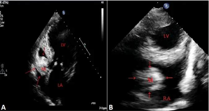
Figure 1. A. Transthoracic echocardiography apical four-chamber view demonstrated large mass on the tricuspid valve and B. modified short-axis view with the help of x-plane utility revealed the mass in a different plane. LA = left atrium; LV = left ventricle; RA= right atrium; M = mass; the arrows show the calcified mass.
Data sharing: No additional data. Funding Sources: No funding.
Address for correspondence and reprint requests: Mahmut Yesin, M.D., Denizer Cad. Cevizli Kvs. No:2 Cevizli, Kartal/Istanbul, Turkey. Fax: +(90)216-4596321;
E-mail: [email protected]
1885
© 2015, Wiley Periodicals, Inc.
36.5°C, and respiratory rate was 15 per minute. On physical examination, there was a grade 3/6 systolic murmur on cardiac apex. Atrialfibrillation was diagnosed in the 12-lead surface electrocar-diography. Chest x-ray revealed a cardiothoracic index of 0.5, with obliteration of the costodi-aphragmatic sinuses and right pleural effusion. Transthoracic echocardiography showed a mod-erate-to-severe mitral insufficiency with preserved left ventricular systolic function and a large ovoid mass, located at the posterior side of the tricus-pid valve annulus (Fig. 1A,B). The tricustricus-pid mass was thought to be an intracardiac metastasis of
the colon cancer. However, there was no meta-bolic activity on the mass with positron emission tomography (PET). Subsequently, the diagnosis of caseous calcification of the tricuspid annulus was suspected which was supported by the car-diac computed tomography (CT) findings (Fig. 2). The lesion was described by CT as calci-fied, connected with posterior part of the tricus-pid annulus and not reducing the leaflet mobility. As caseous calcifications are usually benign1,2and the patient has an end-stage colon cancer, surgery was not planned and the patient was followed up conservatively. Although mitral annular calcifications are common, tricuspid annular caseous calcification is first to be reported in the current literature. This interesting case illustrates the differential diagnosis between caseous calcification and a metastatic mass with the help of cardiac CT, PET, and transthoracic echocardiography. Transesophageal echocardio-graphy may not provide additional information in such cases with tricuspid masses.3
References
1. Martinez-de-Alegria A, Rubio-Alvarez J, Baleato-Gonzalez S: Caseous calcification of the mitral annulus: a rare cause of intracardiac mass. Case Rep Radiol 2012;2012:596962. 2. Harpaz D, Auerbach I, Vered Z, et al: Caseous calcification
of the mitral annulus: aneglected, unrecognized diagnosis. J Am Soc Echocardiogr 2001;14:825–831.
3. Kalcßik M, Yesin M, G€ursoy MO, et al: An unusual mass of tricuspid valve in an adult patient: Blood-filled cyst. Echocardiography 2015;32:1199–1202.
Figure 2. The MDCT views: a tumor-like mass with a hypo-dense central area and peripheral hypo-dense calcification in the tri-cuspid valve annulus (arrows).
Yesin, et al.