Address for Correspondence/ Yazışma Adresi: Ebru Evren, MD Baskent University Faculty of Medicine, Department of Medical Microbiology, Ankara, Turkey E-mail: [email protected]
©Telif Hakkı 2015 Gazi Üniversitesi Tıp Fakültesi - Makale metnine http://medicaljournal.gazi.edu.tr/ web adresinden ulaşılabilir. ©Copyright 2015 by Gazi University Medical Faculty - Available on-line at web site http://medicaljournal.gazi.edu.tr/
doi:http://dx.doi.org/10.12996/gmj.2015.54
Canaliculitis due to Gemella Haemolysans and Porphyromonas Asaccarolytica
Gemella Haemolysans ve Porphyromonas Asaccarolytic’ın Neden Olduğu Kanalikülit
Emine Sen
1, Ebru Evren
2, Ufuk Elgin
11 Ulucanlar Eye Education and Research Hospital, Ankara, Turkey
2 Baskent University Faculty of Medicine, Department of Medical Microbiology, Ankara, Turkey
ABSTRACT
Objective: We reported a canaliculitis case due to Gemella haemolysans and Porphyromonas asaccarolytica.
Methods: After the diagnosis of canaliculitis, the unusual finding of yellowish-greenish material supplied from canalicular curettage and canaliculotomy was sent to microbiology laboratory for the presence of aerobic, anaerobic bacteria and fungi. Systemic and topical antimicrobial treatment was administered.
Results: Bacteriological stains revealed polymorphonuclear leukocytes and gram-positive cocci. Facultative anaerobe Gemella haemolysans and anaerobe Porphyromonas asaccarolytica were identified according to bacteriological cultures. Routine cultures were negative for fungi. Recurrence was not observed at two years follow-up.
Conclusion: To our knowledge, this is the first reported case of canaliculitis due to both Gemella haemolysans and Porphyromonas asaccarolytica and is also remarkable ocular infection case occurred after any surgery and/or trauma.
Key Words: Canaliculitis, Gemella haemolysans, Porphyromonas asaccarolytica
Received: 03.10.2015 Accepted: 04.17.2015
ÖZET
Amaç: Gemella haemolysans ve Porphyromonas asaccarolytica’nın neden olduğu bir kanalikülit olgusunu sunmaktır.
Yöntemler: Kanalikülit tanısı konulduktan sonra yapılan kanalikülotomi ve kanalikül küretajından elde edilen sarımsı- yeşilimsi materyal aerobik, anaerobik bakteri ve mantar incelemesi için mikrobiyoloji labaratuarına gönderildi. Sistemik ve topikal antimikrobiyal tedavi başlandı.
Bulgular: Bakteriyolojik boyamada polimorfonükleer lökositler ve gram pozitif kok izlendi. Fakultatif anaerop Bakteriyolojik kültürlere göre Gemella haemolysans ve anaerop Porphyromonas asaccarolytica olarak tiplendirildi. Mantarlar için rutin kültürler negatifti. 2 yıllık takipte tekrarlama izlenmedi. Sonuç: Bildiğimiz kadarıyla, bu çalışma Gemella haemolysans ve Porphyromonas asaccarolytica ‘nın neden olduğu bildirilmiş ilk vakadır, aynı zamanda herhangi bir cerrahi ve/veya travma geçirmeden ortaya çıkmış bir oküler enfeksiyon olması da dikkat çekicidir.
Anahtar Sözcükler: Gemella haemolysans, Kanalikülit, Porphyromonas asaccarolytica
Geliş Tarihi: 10.03.2015 Kabul Tarihi: 17.04.2015
INTRODUCTION
Primary canaliculitis is a rare chronic disease usually caused by various aerobe and anaerobe microorganisms such as Staphylococcus and Actinomyces species. Primary canaliculitis accounts for 2% of all lacrimal diseases (1,2).Clinical findings are swelling and redness of punctum or canaliculus and also mucopurulent punctal regurgitation on palpation of the canaliculus. Although clinical findings are well defined, many cases can be misdiagnosed and easily omitted (3).
We reported a case of canaliculitis caused by Gemella haemolysans and Porphyromonas asaccarolytica.
CASE REPORT
A 84-year-old woman presented to our department with complaints of epiphora and mucopurulent discharge of the left lower eyelid. The clinical findings were redness and swelling of medial canthus and the lower lid, and also conjunctival hyperemia. Physical examination revealed mucopurulent punctal regurgitation with an unusual yellowish-greenish material on palpation of the canaliculus. The patient was referred to the department of oculoplasty as chalazion. According to existing findings a diagnosis of canaliculitis was made. There was no history of scratching caused by a dog, previous surgery, trauma, and punctum plug application.
Written informed consent was obtained from the patient. Canalicular curettage and canaliculotomy were performed, yellowish-greenish material was sent to microbiology laboratory for the presence of aerobic, anaerobic bacteria and fungi (Figure 1). At the end of the surgery cefuroxime axetil was used for canalicular irrigation.
Gram and Giemsa stains revealed polymorphonuclear leukocytes and gram-positive cocci. The culture revealed facultative anaerobe Gemella haemolysans and anaerobe Porphyromonas asaccarolytica. Routine cultures were negative for fungi.
We continued systemic ampicillin+sulbactam 2×1 and topical fortified penicillin 100.000 U/ml 8×1 antibiotic treatments after the surgery for ten days. No recurrence was observed during the postoperative 2-year follow-up period.
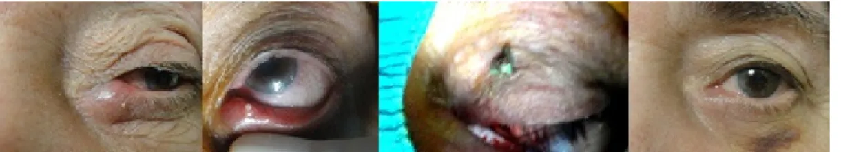
Figure 1 (a) Preoperative appearance of the patient (b) Secretion of the mucopurulent material upon pressure application © Yellowish-greenish material secretion from the canaliculit (d) Postoperative appearance of the patient
DISCUSSION
Lacrimal canaliculitis is an inflammation of the canalicular system of the eye. Punctal pouting, eyelid swelling and redness, mucopurulent discharge, as well as in some cases sulfur granules, are well-defined clinical findings of primary canaliculitis. Hence, it is a rare disease; it may be misdiagnosed as conjunctivitis, mucocele, dacryocistitis, blepharitis, and chalazion (1,4). Our patient was also referred to oculoplasty department for pre-diagnosis of chalazion.
Various bacteria, fungi, and viruses can cause canaliculitis. Most commonly isolated microorganisms are Actinomyces and Staphyloccus species (5).The bacterial etiology of the canaliculitis is still controversial. Some authors argue that the microbiology of primary canaliculitis does not appear to be evolving while others claim that the microbiological profile of canaliculitis is changing. Several different bacteria including Streptococcus spp, Propionibacterium species, Arcanobacterium haemolyticum or mixed bacteria were determined as the causal agents of canaliculitis beside the common species (3,6).Gemella haemolysans and Porphyromonas asaccarolytica were recovered from our patient.
The first case of ocular infection caused by Gemella haemolysans which is a normal commensal organism found in the oral cavity and upper respiratory tract was reported by Ritterband et al. in a patient with keratitis and endophthalmitis (7). Infectious crystalline keratopathy, postoperative endophthalmitis after trabeculectomy and cataract surgery caused by Gemella haemolysans were also reported (8,9).
The first ocular infection with Porphyromonas gingivalis that are a part of the normal oral flora of several mammalian species was reported in 2004 by Rudolph et al. in an infiltrative keratitis patient scratched by his dog (10).
In our patient’s history there was no sign of scratching caused by a dog or postoperative endophthalmitis after trabeculectomy and the cataract surgery. Suggested treatment methods-canaliculotomy and curettage- were also performed for the treatment of our patient. Despite these methods a punctal fibrosis was observed. To reduce the risk of recurrence, we used systemic and topical antibiotic treatment after the surgery. No recurrence was observed in two years of the postoperative follow-up.
To our knowledge, this is the first reported case of canaliculitis due to both Gemella haemolysans and Porphyromonas asaccarolytica. Contrary to common agents, various microbiological agents causing canaliculitis should be observed.
Studies with longer follow-ups are needed to determine whether the course of canaliculitis due to Gemella haemolysans and/or Porphyromonas asaccarolytica is different from that of other causative agents.
Conflict of Interest
No conflict of interest was declared by the authors. REFERENCES
1. Gogandy M, Al-Sheikh O, Chaudhry I. Clinical features and bacteriology of lacrimal canaliculitis in patients presenting to a tertiary eye care center in the Middle East. Saudi J Ophthalmol 2014;28:31-5.
2. Bharathi MJ, Ramakrishnan R, Shivakumar C, Meenakshi R, Lionalraj D. Etiology and antibacterial susceptibility pattern of community-acquired bacterial ocular infections in a tertiary eye care hospital in south India. Indian J Ophthalmol 2010; 58:497-507.
3. Lin SC, Kao SC, Tsai CC, Cheng CY, Kau HC, Hsu WM et al. Clinical characteristics and factors associated the outcome of lacrimal canaliculitis. Acta Ophthalmol 2011; 89: 759–63.
4. Liyanage SE, Wearne M. Lacrimal canaliculitis as a cause of recurrent conjunctivitis. Optometry 2009; 80:479-80.
5. Bharathi MJ, Ramakrishnan R, Shivakumar C, Meenakshi R, Lionalraj D. Etiology and antibacterial susceptibility pattern of community-acquired bacterial ocular infections in a tertiary eye care hospital in south India. Indian J Ophthalmol. 2010 58:497-507.
6. Kaliki S, Ali MJ, Hanoavar SG, Chandrasekhar G, Naik MN. Primary canaliculitis:clinical features, microbiological profile, and management outcome. Ophtal Plast Reconst Surg 2012; 28:335-60.
7. Ritterband D, Shah M, Kresloff M, Intal M, Shabto U, Seedor J. Gemella haemolysans keratitis and consecutive endophthalmitis. Am J Ophthalmol 2002; 133:268-9.
8. Kailasanathan A, Anderson DF. Infectious crystalline keratopathy caused by Gemella haemolysans. Cornea 2007; 26:643–4.
9. Nalamada S, Jalali S, Reddy AK.Acute postoperative endophthalmitis by Gemella haemolysans Indian J Ophthalmol: 2010;58:252-3.
10. Rudolph T, Welinder-Olsson C, Lind-Brandberg L, Stenevi U. 16S rDNA PCR analysis of infectious keratitis: a case series. Acta Ophthalmol Scand 2004; 82:463-7.