UDK 577.1 : 61 ISSN 1452-8258
J Med Biochem 36: 309 –313, 2017 Original paper
Originalni nau~ni rad
IS THERE A REAL CORRELATION BETWEEN RED CELL DISTRIBUTION
WIDTH AND PERIPHERAL ARTERIAL DISEASE?
DA LI POSTOJI STVARNA KORELACIJA IZME\U RASPODELE ERITROCITA
I PERIFERNOG ARTERIJSKOG OBOLJENJA?
Enes Duman1, Sevsen Kulaksızoglu2, Egemen Çifçi1, Mehmet Ozulku3 1Department of Radiology, Bas ¸kent University School of Medicine, Konya, Turkey 2Department of Biochemistry, Bas ¸kent University School of Medicine, Konya, Turkey 3Department of Cardiovascular Surgery, Bas ¸kent University School of Medicine, Konya, Turkey
Address for correspondence: Sevsen Kulaksızoglu
Baskent Universitesi Konya Uygulama ve Aras¸tırma Hastanesi, Biyokimya, Hocacihan Mah, Saray Cad, No:1, Selçuklu 42080, Konya, Turkey
e-mail adress: sevsenkªyahoo.com Summary
Background: Few data is available concerning the associa-tion between peripheral arterial disease (PAD) and red cell distribution width (RDW). In this study, we analyzed the relationship between RDW and atherosclerosis of the ves-sels other than coronary arteries in patients who had under-gone digital substraction angiography (DSA).
Methods: This study included 730 patients who had under-gone DSA. Patients were divided into two groups according to their angiographic images. The association between RDW and atherosclerosis of peripheral arteries was analy-zed. The relationship between atherosclerosis and smo-king, hypertension (HT), diabetes mellitus (DM), hs-CRP, hemoglobin, white blood cell (WBC), triglyceride, total cho-lesterol, HDL and LDL cholesterol levels was assessed. Results: Atherosclerosis was observed more common in male and patients with older age, HT, DM and smoking (p<0.001). hs-CRP and WBC levels were both in signifi-cantly positive association with atherosclerosis (p<0.05). However, there were no significant differences in the RDW levels, hemoglobin, triglyceride, total cholesterol, LDL and HDL cholesterol levels in the groups (p>0.05).
Conclusions: Our results seem to demonstrate that older age, male gender, HT, DM and smoking are powerful risk factors for PAD. In contrast to the previous reports, RDW levels are found not to be associated with atherosclerosis of peripheral arteries.
Keywords: red blood cell distribution width, peripheral arterial disease, digital substraction angiography, athero -sclerosis
Kratak sadr`aj
Uvod: Ima nekoliko podataka koji govore o vezi izme|u peri fernog arterijskog oboljenja i raspona raspodele eritro -cita (RDW). U ovom izu~avanju analiziran je odnos izme |u RDW i ateroskleroze krvnih sudova osim koronarnih arte rija u pacijenata koji su bili podvrgnuti digitalnoj angigrafiji (DSA).
Metode: Ovo izu~avanje obuhvatilo je 730 pacijenata pod vrgnutih DSA. Pacijenti su podeljeni u dve grupe prema angigrafskoj slici. Analizirana je veza izme|u RDW i atero -skleroze perifernih arterija. Pra}ena je povezanost izme|u ateroskleroze i pu{enja, hipertenzije (HT), diabetes mellitusa (DM), hsCRP, hemoglobina, leukocita (WBC), tri gli -ce rida, ukupnog holesterola, i nivoa HDL i LDL holeste rola. Rezultati: Ateroskleroza je bila ~e{}a kod mu{karaca i paci-jenata starije dobi, sa HT, DM i pu{a~a (p<0,001). Nivoi hs-CRP i WBC bili su u zna~ajnioj pozitivnoj korelacijhi sa aterosklerozom (p<0,05). Me|utim, nije bilo zna~ajnih razlika u nivoima RDW, hemoglobina, triglicerida, ukupnog holesterola i nivoa LDL i HDL holesterola u grupama (p>0,05).
Zaklju~ak: Rezultati ukazuju da su starije doba, mu{ki pol, HT, DM i pu{enje zna~ajni faktori rizika za PAD. Nasuprot ranijim istra`ivanjima, nije na|eno da su nivoi RDW pove -zani sa aterosklerozom perifernih arterija.
Klju~ne re~i: {irina raspodele eritrocita, periferno arte -rijsko oboljenje, digitalna angiografija, ateroskleroza
Introduction
Red blood cell distribution width (RDW) reflects the variation of red blood cell volume. It can easily be measured by routine analysis of whole blood count (1). Increased RDW results from heterogeneity of erythrocyte size and erythrocyte fragmentation in the circulation (2). Factors that cause increased eryth-rocyte size heterogeneity include iron, vitamin B12 and folate deficiency, decreased erythrocyte life span and impaired erythropoiesis. Increased RDW is also related to oxidative stress and release of cytokine in response to inflammation (3) RDW has been demonstrated to be associated also with cardiovascu-lar and pulmonary diseases (4). Several studies have shown that high RDW is associated with coronary artery atherosclerosis in hypertensive patients and acute myocardial infarction (5, 6).
Peripheral arterial disease (PAD) is associated with increased mortality and morbidity (7). It is impor-tant to identify factors that might contribute to such high morbidity and mortality in PAD. Patients with PAD show high levels of inflammation and oxidative stress, which play roles in the progression of atherosclerotic diseases (8). Whether RDW is predictive of mortality in patients with PAD is unknown. A recent study demonstrated that increased RDW is associated with greater mortality in patients with PAD identified by noninvasive lower-extremity arterial testing (9). Several studies have been done to assess the relationship bet-ween RDW and coronary artery atherosclerosis. However, few data is available concerning the associa-tion between PAD and RDW. In this study, we analyzed the relationship between RDW and atherosclerosis of the vessels other than coronary arteries in patients who had undergone digital substraction angiography (DSA). Our study is the first to use an invasive method, DSA, for peripheral arterial evaluation.
Methods
A total of 730 patients, who underwent DSA (Artis Zee, Siemens Germany, and Integris V, Philips Netherland) in our interventional radiology unit bet-ween 2013 and 2014 were included in this study. All patients provided written informed consent and the local institutional ethics commitee approved the study protocol. Patients had undergone aortography (n:92), pelvic-iliac angiography (n:142), lower exremity angi-ography (n:202), renal angiangi-ography (n:122), carotid angiography (n:134) and upper extremity angiog-raphy (n:38). We divided patients into two groups according to their angiographic images. Patients who had extensive stenosis and atherosclerosis or occlusi-on were included in group 1. Patients with no stenosis and atherosclerosis or who had normal findings were included in group 2.
The patients’ demographic characteristics inclu-ding age, sex and smoking status were acquired
thro-ugh a standardized questionnaire. The mean age of the subjects in group 1 was 64.72±10.56 (427 males and 133 females), and the mean age of the subjects in group 2 was 54.86 ± 13.96 (105 males and 65 females). Smoking was defined as at least one cigarette daily for 1 year. Hypertension (HT) was defined as systolic blood pressure (SBP) 140 mmHg and (or) diastolic (DBP) 90 mmHg or previous diag-nosis of hypertension with use of antihypertensive medication (10). Diabetes mellitus (DM) was defined as a fasting blood glucose level 6.99 mmol/L, a ran-dom glucose measurement >11.1 mmol/L or hemo-globin A1c>0.065 proportion of total hemoglobin or a previous diagnosis with any use of oral antidiabetic agent and/or insulin (11). Exclusion criteria included previous cardiovascular disease, the presence of hemolytic, hepatic and renal diseases that could affect white blood cells (WBC), red blood cells (RBC) or hemoglobin. Patients who received erythrocyte suspension for any reason within the past 6 months were not considered for the study.
RDW, hemoglobin and WBC levels were meas-ured using Abbott CELL-DYN 3700 automated CBC analyzer. The normal reference range for RDW in our laboratory is 11.6% to 15.5%. Serum total choles-terol, LDL and HDL cholescholes-terol, triglyceride and hs-CRP levels were also determined.
Statistical analyses were performed with Stati -stical Package for the Social Sciences software (Version 9.0; SPSS, Inc., Chicago, IL). For continuous variables, Kolmogorov-Smirnov test was applied to test normal distribution. Mann-Whitney U test was applied to compare ages, LDL cholesterol, triglyc-eride, RDW, hemoglobin, WBC and hs-CRP levels between two groups as the data was not distributed normally. Independent samples t test was used to compare total cholesterol and HDL cholesterol levels because of normal distribution. The chi-square test was used for comparison of categorical variables. For continuous variables; numeric values are expressed as mean±SD. A p value of <0.05 was considered statis-tically.
Results
Aortography (thoracic or abdominal) was per-formed in 92 patients. Although 26 of these patients had no stenosis or occlusion, they had extensive ath-erosclerosis. 15 patients had abdominal aortic steno-sis. 18 patients had abdominal aortic occlusion. 59 of these patients were included in group 1. 33 of these patients were included in group 2.
Pelvic and iliac angiographies were performed in 142 patients. 20 patients had no atherosclerosis. 66 patients had stenosis. 56 patients had total occlusion. We enrolled 122 of these patients in group 1 and 20 of these patients in group 2.
Lower extremity angiography was performed in 202 patients. 138 patients had undergone femoral angiography. Of the patients who had undergone femoral angiography, 16 patients had no atheroscle-rosis. 22 patients had stenosis. 100 patients had total occlusion. 64 patients had undergone knee angiography. Of the patients who had undergone knee angio -graphy, 4 patients had no atherosclerosis. 8 patienst had stenosis. 52 patients had total occlusion. We enrolled 182 of these patients in group 1 and 20 of these patients in group 2.
Renal angiography was performed in 122 patients. 49 patients had normal findings. 68 patients had stenosis. 5 patients had total occlusion. So 73 of these patients were enrolled in group 1 and 49 of these patients in group 2.
Carotid angiography was performed in 134 patients. 27 patients had normal findings. 87 patients had stenosis. 20 patients had total occlusion. 107 of these patients were included in group 1 and 21 of these patients in group 2.
Upper extremity angiography was performed in 38 patients. 21 patients had normal findings. 7 patients had stenosis. 10 patients had total occlusion. 17 of these patients were enrolled in group 1 and 21 of these patients in group 2.
The baseline characteristics are shown in Table 1. 190 patients had DM. 165 of these diabetic patients were in group 1 and 25 of these diabetic
patients were in group 2. A total of 558 patients had HT. 464 of these patients were in group 1. 94 of these hypertensive patients were in group 2. Number of the patients who had smoked was 456. And 396 of these patients were in group 1, the others in group 2.
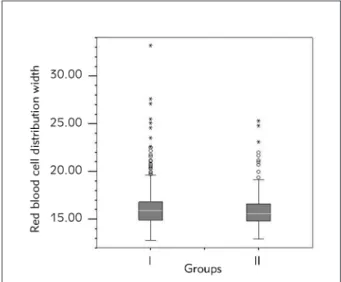
As shown in in Figure 1, the analysis of RDW val-ues revealed no significant differences in any of the groups (p=0.192). However, atherosclerosis was observed more common in male patients compared
Table I Median and demographic levels of RDW and other parameters in each group.
Figure 1 Red Blood Cell Distribution Width.
Groups Group I (n=560) Group II (n=170) P value Male/female 427/133 105/65 Chi-square=15,552;p<0.001 Age (y)* 64.72 ± 10.56 54.86 ± 13.96 Z=-8,712; p<0.001
Total cholesterol (mmol/L)* 4.83± 1.17 4.79± 1.49 t=-0.363; p=0.717
HDL cholesterol (mmol/L)* 1.0± 0.24 1.024 ±0.26 t=0.428; p=0.669 LDL cholesterol (mmol/L)* 2.99 ± 0.86 2.92 ± 0.84 Z=-0.449; p=0.652 Triglyceride (mmol/L)* 1.75 ± 0.95 1.66 ± 0.76 Z=-0,451; p=0.653 RDW* 16.15 ± 2.03 16.04 ± 2 Z=-1,306; p=0.192 HT +/- 464/96 94/76 Chi-square=58,557; p<0.001 DM +/- 165/395 25/145 Chi-square=12,332; p<0.001 Smoking +/- 396/164 60/110 Chi-square=72,546; p<0.001 Hemoglobin (g/L) 130.26 ± 20.14 130.45 ± 20.23 Z=-1,167; p=0.243 WBC count (×109/L) 9.06 ± 3.55 8.27 ± 2.77 Z=-1,167; p=0.003 hs-CRP (nmol/L) 312.76 ± 490.96 212.95 ± 277.91 Z=-1,971; p=0.049 *Values are means ± standard deviation
*DM: Diabetes Mellitus; HDL: high-density lipoprotein; HT: hypertension; LDL: low-density lipoprotein; RDW: red blood cell dis-tribution width; hs-CRP: high-sensitive C-reactive protein; WBC: white blood cell.
to female patients (p<0.001). Patients with older age had particularly extensive atherosclerosis in peripheral arteries (p<0.001). Patients who had DM, HT and smoking were more prone to atherosclerosis when compared to the control group (p<0.001). And also, hs-CRP and WBC levels were both in significantly positive association with atherosclerosis (p<0.05). But there were no significant differences in hemoglo-bin, serum total cholesterol, LDL and HDL cholesterol and triglyceride levels in these groups (p>0.05).
Discussion
The prevalence of PAD is increasing worldwide. It is well known that the morbidity and mortality of patients with PAD remain high despite current advan -ces in revascularization techniques (12). Identification of risk factors for atherosclerotic PAD will provide insights into underlying pathophysiologic mecha-nisms and facilitate the development of diagnostic and therapeutic approaches.
PAD prevalence and incidence are age related. With aging of the population, it seems likely that PAD will increase in the future. And also, PAD seems to be higher among men than men. In our study, athero-sclerosis was observed more common in male patients compared to female patients (p<0.001). Patients with older age had particularly extensive ath-erosclerosis in peripheral arteries (p<0.001). Our result is in agreement with Criqui and associates, who observed that PAD incidence rises >10% among patients in their 60s and 70s (13). Smoking, HT and DM are particularly strong risk factors for PAD. In our study, patients with HT, DM and smoking had increased atherosclerosis in peripheral arteries (p<0.001).
High levels of inflammation and oxidative stress play roles in the progression of atherosclerosis in PAD. hs-CRP is one of the most well-established bio-markers of chronic inflammation. hs-CRP produced by tissue macrophages and vascular smooth muscle cells could appear in the atherosclerotic plaques (14). Jia and associates showed that coronary artery athe-rosclerotic patients have higher hs-CRP levels than non coronary artery atherosclerotic patients (15). In our study, hs-CRP and WBC levels were both in signi-ficantly positive association with atherosclerosis in peripheral arteries (p<0.05). Although the underlying biological mechanisms remain unclear, RDW is also assessed as a marker of chronic inflammation and oxidative stress (3). Recent studies have demonstra-ted that high RDW is associademonstra-ted with poor clinical out-comes in patients with heart failure, coronary artery
disease, pulmonary hypertension and PAD (9, 16). Inflammatory cytokine release in heart failure and acute myocardial infarction might affect bone marrow function and inhibit erythrocyte maturation induced by erythropoietin. Therefore , RDW becomes elevated (17). And also increased oxidative stress directly damages erythrocytes and shortens their survival, resulting in high RDW (18). According to Almer et al. (19) coronary artery disease is significantly related with inflammatory response of the vascular endothe-lial injury, while RDW is involved in the process of inflammation. Jia et al. (15) and associates demons-trated that higher RDW levels were associated with carotid artery stenosis and intimal medial thickness. They assessed atherosclerosis and intimal medial thickness via ultrasound. Also, Wen conducted a study to demonstrate the relationship between RDW and carotid artery atherosclerosis by using carotid ult-rasonography (20).
Recently, Ye and al. (9) and associates reported a study which evaluated the utility of RDW to predict mortality in patients with PAD. They used ankle-brac-hial index which is an established noninvasive test for peripheral arterial disease. They demonstrated that higher RDW is associated with greater all-cause mor-tality in patients with PAD identified in the noninvasive vascular laboratory. And also, Zalawadiya et al. (21) demonstrated a strong and independent relationship between RDW and PAD. These findings are particu-larly noteworthy as RDW appears to be an emerging cost-effective biomarker. However, in our study, no statistically significant difference was found between two groups in terms of RDW levels. To date, RDW has been evaluated in various clinical settings both in PAD and other conditions before. However, this is the first study to use an invasive method, DSA, for peripheral arterial evaluation. DSA is stil considered the referen-ce standard in vascular imaging (22).
However, there are some limitations of our study that have to be mentioned. Lack of data on serum folate, vitamin B12and iron levels exists in our study. Therefore considering the data on the levels of serum iron, folate and vitamin B12should also be assessed. And also, the patients are not examined for any mor-bidity or mortality event. In this context, a longer peri-od of prospective observation may provide more prognostic information.
Conflict of interest statement
The authors stated that they have no conflicts of interest regarding the publication of this article.
Received: Fedruary 23, 2017
Accepted: May 6, 2017
References
1. Karnad A, Poskitt TR. The automated complete blood count. Use of the red blood cell volume distribution width and mean platelet volume in evaluating anemia and thrombocytopenia. Arch Intern Med 1985; 145: 1270–2. 2. Bessman JD, Gilmer PR Jr, Gardner FH. Improved classi-fication of anemias by MCV and RDW. Am J Clin Pathol 1983; 80: 322–6.
3. Tsuboi S, Miyauchi K, Kasai T, Ogita M, Dohi T, Miyazaki T, et al. Impact of red blood cell distribution width on long-term mortality in diabetic patients after percu -taneous coronary ıntervention. Circ J 2013; 77(2): 456– 61.
4. Hampole CV, Mehrotra AK, Thenappan T, Gomberg-Maitland M, Shah SJ. Usefulness of red cell distribution width as a prognostic marker in pulmonary hypertension. Am J Cardiol 2009; 104: 868–72.
5. Li ZZ, Chen L, Yuan H, Zhou T, Kuang ZM. Relationship between red blood cell distribution width and early-stage renal function damage in patients with essential hyper-tension. J Hypertens 2014; 32(12): 2450–5.
6. Songoi MB, Guarda NS, Rodel AP, Zorzo P. Prognostic value of red blood cell distribution width in prediction of in-hospital mortality in patients with acute myocardial infarction. Clin Lab 2014; 60(8): 1351–6.
7. Hirsch AT, Criqui MH, Treat-Jacobson D, Regensteiner JG, Creager MA, Olin JW, et al. Peripheral arterial disease detection, awareness and treatment in primary care. JAMA 2001; 286: 1317–24.
8. Steg PG, Bhatt DL, Wilson PW, D’Agostino R Sr, Ohman EM, Rother J, et al. One-year cardiovascular event rates in outpatients with atherothrombosis. JAMA 2007; 297: 1197–206.
9. Ye Z, Smith C, Kullo IJ. Usefullness of red cell distribution width to predict mortality in patients with peripheral artery disease. Am J Cardiol 2011; 107(8): 1241–5. 10. Chobanian AV, Bakris GL, Black HR, Cushman WC,
Green LA, Izzo JL Jr, et al. Joint national committe on prevention detection. Evaluation and treatment of high blood pressure. National Heart, Lung and Blood Institute: National high blood pressure education pro-gram coordinating committe. Hypertension 2003; 42(6): 1206–52.
11. Kuzuya T, Nakagawa S, Satoh J, Kanazawa Y, Iwamoto Y, Kobayashi M. Report of the committee on the classifica-tion and diagnostic criteria of diabetes mellitus. Diabetes Research and Clinical Practice 2002; 55: 65–85. 12. Vartanian SM, Conte MS. Surgical intervention for peri
-pheral arterial disease. Circ Res 2015; 116(9): 1614–28.
13. Criqui MH, Aboyans V. Epidemiology of peripheral artery disease. Circ Res 2015; 116(9): 1509–26.
14. Trifunovi} D, Stankovi} S, Marinkovi} J, Banovi} M, \u -ka novi} N, Vasovi} O, Vujisi}-Te{i} B, Petrovi} M, Ste -panovi} J, \or|evi}-Diki} A, Beleslin B, Nedeljkovi} I, Te{i} M, Ostoji} M. Oxidized low density lipoprotein and high sensitive c-reactive protein in non-diabetic, pre-dia-betic and diapre-dia-betic patients in the acute phase of the first myocardial infarction treated by primary percutaneous coronary intervention. J Med Biochem 2015; 34: 160– 9.
15. Jia H, Li H, Zhang Y, Li C, Hu Y, Xia C. Association between red blood cell distribution width (RDW) and carotid artery atherosclerosis (CAS) in patients with pri-mary ischemic stroke. Arch Gerontol Geriatr 2015; 61(1): 72–5.
16. Rhodes CJ, Wharton J, Howard LS, Gibbs JS, Wilkins MR. Red cell distribution width outperforms other potential circulating biomarkers in predicting survival in idiopathic pulmonary arterial hypertension. Heart 2011; 97: 1054– 60.
17. Malandrino N, Wu WC, Taveira TH, Whitlatch HB, Smith RJ. Association between red blood cell distribution width and macrovascular and microvascular complications in diabetes. Diabetologia 2012; 55: 226–35.
18. Grant BJ, Kudalkar DP, Muti P, McCann SE, Trevisan M, Freudenheim JL, et al. Relation between lung function and RBC distribution width in a population-based study. Chest 2003; 124: 494–500.
19. Almer G, Frascione D, Pali-Scholl I, Vonach C, Lukschal A, Stremnitzer C. Interleukin-10: An anti-inflammatory marker to target atherosclerotic lesions via PEGylated liposomes. Molecular Pharmaceutics 2013; 10(1): 175– 86.
20. Wen Y. High red blood cell distribution width is closely associated with risk of carotid artery atherosclerosis in patients with hypertension. Exp Clin Cardiol 2010; 15: 37– 40.
21. Zalawadiya SK, Veeranna V, Panaich SS, Afonso L. Red cell distribution width and risk of peripheral artery disease analysis of National Health and Nutrition Examination Survey 1999–2004. Vasc Med 2012; 17(3): 155–63. 22. Bosma J, Dijksman LM, Lam K, Wisselink W, van Swijn
-dregt AD, Vahl A. The costs and effects of contrast-enhanced magnetic resonance angiography and digital substraction angiography on quality of life in patients with peripheral arterial disease. Acta Radiol 2014; 55(3): 278–86.