Director: Ord. Prof. Ş. Akçay, the Department of Animal Pathology, Veterinary Faculty, Ankara, Turkey
Seminoma of The Testieles in An Ankara Goat
kM. PAMUKÇU
As far as I am aware there has been no record in literature of
seminoma in goats. The case of seminoma of the testicles which we
observed recently in an Ankara goat might be of interest and fili
the gap on this particular subject.
The lack of reports of seminoma is not surprising because
the-re athe-re few male and female 'of this species of animal which the-reach
cancer age. There is little record of tumors in goats. However, in
an excellent review of literature regarding the tumors of the
domes-tic animals,
Tamesehke (30)
stated that of 5245 tumor cases of
dif-ferent animals recorded, 13 cases belonged to the goat whose
tes-ticles were not involved in any case öf tumors, and he also pointed
out that the incidence of tumors in the goat was not higher than
0.02 percent. The cases of melanoma occurring endemic in the
An-kara goat raised in South Africa and K ongos was not included in
this figure.
Saminoma of the testicles in mammals, other than man, is well
known and especially in the dog (17, 7, 4, 11, 9, 21, 6, 8, 20, 34, 30
27, 35) and horse (19, 9, 1, 21, 22, 14, 30). However, there is little
record of testicular tumors in bulls, rams, mules, boars and cats
(13, 15, 30, 34).
İ
n birds they appear to be rare, although
Champy
and Lavedan
(4, 5) observed similar appearance in regenerating
testes of partially castrated common fowl.
İ
n the case they studied
the histological structure was somewhat different from those of the
mammalian seminomas. They based this difference in the structure on
the minimum capacity of the spermatogonia cells to propogate and lack
of Sertoli's cells to the testis of birds. Cappellato (2) produced similar
condition in the chicken with the injection of Zinc chlorid solution.
Rewell
(25) claimed to observe seminoma in the testes in a collared
turtle dove (Streptopelia Risoria).
İ
t resembled very closely the
se-minoma of mammals.
There is a great difference of opinion existing regarding tumors
of the testicle. Primary malignant testicular tumors are not very
common. The structure of the tumors arising from spermatic cells
of the testicle is very complex and shOws a great variety even in
the same section. Therefore, the classification of testicular tumors
of animals based on the histogenesis is somewhat difficult. However,
the testicular tumors may be divided in three main types as
semi-noma, sustentacular teli tumor and interstitial cell tumor (20)..
Teratomas in the testes of the domestic animals vary from one
species to another. Teratomas are commoner in the testis of horses
than seminoma (F2, 33, 30). Cases of seminomas and teratomas in
the bull testes were equally observed (30). Well described cases of
teratomas in the canine testis are unknown. Mulligan (20) stated
that
("(t II is is understandable since twinning is no problem in the
dog as it is in man, whose testicles exhibit a hingh incidence of
varying complexity and of chorion • epithelioma, as well as
semino-ma, but a low incidence of sustentacular teli and interstitial teli
tumors ).
Seminoma, as Chevassu first named, takes its origin from the
spermatic cells of tubules, spermatogonia, or spermatocytes (12, 2,
10), This tumor is called seminal carcinoma, spermatocytoma,
carci-noma macrocellulare solidum and embryonal carcicarci-noma (34, 31, 12,
2).
Martin et al(18) stated thad "it would be better to name the
classsical type of tumor described by
Chevassuegonioma", the
sper-matocytic type being the true seminoma". But some authors, as
Roth(26) and Ewing (cited by Willis 34) consider that seminomas
are of teratomatous origin.
Seminomas develop in advanced age in the animals (8, 7, 14).
They are very common in old dogs (34). Mulligan (20) reported the
age distribution of 36 dogs with 39 testicular neoplasms and found
that the highest incidence of the tumor was relatively late in the
cancer age (9-16 year). Seminoma usually appears in horses late in
second decade e. g. 18 (3, 14, 19) There are very few records in the
other domestic animlas regarding seminomas. Therefore, we do not
know at what age they develop the tumors. This disease is rare in
man before the age of 30 years and develops in early age in the
fifth decade (34).
The tumor is reported both in scrotal and ectopic testes of dog
and horse (20, 8, 29, 1). But displacement of testis appears to be a
predisposing factor to the development of seminomas (34, 8, 1).
Bi-lateral growths are seen in the dogs (20).
İ
nfection and injury of the testis has often been blamed as a
cause of seminomas.
İ
t seems to us these are not of primary impor-
tance in causation of seminomas, for seminomatous conditon is rare
in the bulls, whose testes have tuberculosis and brucellosis. As
long as seminomas develop in the crytorchid testis, we can not
bla-me the trauma as an unique cause of this kind of tumor.
Seminoma is malignat tumor and it spreas by metastasis early and
extensively. There is lymph spread to the abdominal lymph nodes
and blood spread to the lung, liver and other internal organs.
İ
n
the dog iliac and periaortic lymph nodes are usually involved in
in metastasis (20).
Pellat
(23) points out that testicular cancer in
horse and dogs spreads very quickly.
Courteou
(8) described a
se-minoma case in a cyrptorchid testis in a dog at the age of 15 years
which extensive metastatic tumors were found in the serous
membrane of the thoracic cavity.
Benoit
(1) reported that the
pros-tate and the regional lymph nodes in an 18 years old horse have
involved metastasis whose right cryptorchid testis was seminomatous.
Mathias
(19) described that a seminoma case of an 18 years old
horse in which similar microscopic apperance were observed
in the omentuin, the serous membrane of the gut and the spleen.
Both epididymes were also involved. The right spermatic cord was
greatly enlarged. Willis (34) observed frequently direct
Ppread
to
the epididymis and the cord.
Boyd
(2) stated that "an important though puzzling feature of
malignant tumors of the testicle is the occurrence of a positive
Asch-beim • Zondek test in the urine. Willis (34) thinks" that the problem
is a more complex one, however, as shown by the following facts:
a) Seminomas and testiclar teratomas devoid of
chorionepitheli-oma • like tissue can produce similar, though usually not so intesive,
endocrine disturbances, b) Gynecomastia occurs in many conditions
other than testicular tumours including testicular atrophy, adrenal
cortical tumours, pituitary tumours or hepatic disease''. Sofar there
is no record regarding seminoma of the testis of domestic animals
which elaborate gonodotrophins. However,
Rewell
(24) points out that
a tubular adenoma case of the testis in a dog was associated with
oestrogenic type of hypertrophy of the prostate. Mulligan (20) also
recorded that some sustentacular cell tumors elaborate estrogen.
Case Report
The subject was a tumor of the testes in an Ankara goat with
un-known age and history. The tumorous testes were sent to us by the
meat inspector, Dr. F. Borluk, of the abattoir of Ankara. Both
scro-tal testes were involved in the tumorous condition. They weighed
2525 grams all together. Their size increased greatly. The skin cove-
ring the right testis became ulcerative. The uleer localized near the
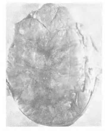
raphe scroti. The surface of the testes was smooth. The cut
sec-ton was firm and lobulated and showed necrotic and
hemorrha-gic foci (Fig. 1). Both epididymes were tumorous and enlarged in
size. The scrotum was firmly attached to the testes.
İ
t was not
possible to skin off the testicles. The spermatic cords were
cut very short when the speciemen was sent in. Therefore, we
could not notice whether they were involved in the tumorous con•
dition. The exapmles were taken from the differaent parts of both
testicles. They were fixed in neutral formaline and embedded in
paraffin. The cuts were 5 microns in thickness. They were stained
with Hematoxylin - Eosin, van Gieson and Sudan IV.
The microscopic picture was variable. The tumor was made of
well or ill defined aggregation of large rounded polyhedral cells
(Fig. 2). Each cell contained a single large spherical nucleus which
has filamented uneven chromation and often two basophilic
nucleo-li. Many mitotic figures were observed in almost every section (Fig
2) Some cells had two or more nuclei located at one pole or both.
The cytoplasma of the cell was devoid of any distinctive structure.
The cells stained with different intensities. The similarity of the
cells to sperrnatocytes was observed in the sections studied. These
cells sometimes produced compact foci simulating carcinoma (Fig.
2). These masses of anaplastic cells were sparated into the large
foci by a delicate of collagenic connective tissue. Here and there a
well developed capillary network supplying the blood was observed.
in some section the tumor cells were very anaplastic and
semini-ferous tubules disappeared entirely (Fig. 2). Every now and again
a small and dark stained cell collection like lymphosarcoma were
noticed.
The stroma of the tumor consisted of the strands of connective
tissue and blood vessels accompanied by a variable number of
lymphocytes.
The tumor cells resembling the original ones infiltrated into the
subcutis of the scrotum and the epididymes. The speciemen we
stu-died consisted of the testicles only. Therefore, we do not know
whether the regional lymph • nodes and the internal organs were
af-fected metastasis or not. Unfortunately, these points remain
undi-cided.
Discussion
A testicular tumor
in an Ankara goat with unknown age and
history was found to be a true seminoma. Both testicles were
scro-tal and affected with the tumorous condition. As far 1 kr
ı
ow there
has been no record of a true seminoma case in the goat If so, this
first case of seminoma in an Ankara goat will supplement the
Innes's list of tumors regarding the testicular ones in domestic
ani-mals (13). Tamaschke (30) reported that of 5245 tumor cases in
dif-ferent domestic animals, only 12 were in goats. This f
ı
gure shows
that reports on the tumors of the goat are very meagner. This should
not be interpreted that the goat is highly resistant to the tumorous
condition, because there are fewer male and female of this species
of animal which reach cancer age.
The microscopical strucuture of the tumor we studied was the
same as human seminomas and the cells of tumor had taken their
origin from the spermatogonia. This result agrees with the
state-ment of Willis (34), Milligan (20),
İ
nnes (13), Foot (10) and
İ
nnes
and his coworker (12) and it is further evidence against of the cla
ims of Eving (cited by Willis 34) and Roth (26) that seminomas are
of teratomatous origin. The tumor showed the noalignant features
consiting of anaplasia, numerous bizzare mitotic figures and dil'.
ference in stain characteristics. The tumor cells invaded intG+ the
subcutis of the scrotum and the epididymes.
Summary
1 — A seminoma of the testicles in an Ankara goat was obser-
ved
2 — Both scrotal testes, epididymes and scrotal skin showed a
seminomatous condition.
3 — The tumor of the goat was the same as a human seminoma.
4 — Whether the regional lymph odes and the internal organs
were affected with metastasis remains undicided.
Fig. 1 Serninonıa, testis, Ankara goat with unknown ege.
Nacrotic and Iternorrhage feci are sCeDill the eentral rwrt of the cut seetion.
Fig. 1. Senıinouni, same case as in Fig. 1.
Direktör : Ord. Prof. Ş. Akçay, Veteriner Fakültesi Patolojik Anatomi Kiirstistl
Bir tiftik Keçisinin Testis'lerinde görülen Seminoma
A. M. PAMUKÇU
Bir çok araştırmalarımıza rağmen literatürde keçilere ait tek bir seminom olayı kaydma rastlanmamıştır. Bu sebeple son zamanlarda bir tiftik keçisinde müşahede ettiğimiz bir seminom vak'asını, lite-ratürde mevcut olan bu eksikliği giderme gayesile neşretmeyi mu-yarık bulduk.
Keçilerde seminom olayına ait bir kayde rastlanmamasının se-bebi, muhtemelen bu hayvan nev'inden pek az bir kısmının kanser için elverişli yaşa kadar yaşamış olması ve ekserisinin bu yaşa ulaşamadan mezbahaya sevk edilmiş bulunmasiyle ilgilidir. Esasen keçinin diğer tümörlerine ait neşriyatta pek azdır. Evcil hayvanla-rın çeşitli tümörlerine ait neşriyatı derleyen Tamasehke (30) topla-dığı 5245 tümör olayından ancak 13 nün keçilerde görüldüğünü ve bu olayların hiç birinde testis'lerin afetzede olmad ığını kaydetmiş ve keçilerde tümor nisbetinin ancak % 0,02 olduğunu da bildirmiş -tir. Yalnız bu nisbete, cenubi Afrika ve Kongo'da yetiştirilen tiftik keçilerinde andeınik olarak görülen melanom vak'alarının dahil ol-madığını ilave etmiştir.
Insandan başka diğer memeli hayvanlarda da testislerde semi-noma rastlanmıştır. Hayvanlar arasında bilhassa köpekte (17, 7, 4, 11, 9, 21, 6, 8, 20, 34, 30, 27, 35) ve at'ta (19, 9, 1, 21, 22, 14, 30) çokça müşahede edildiği yazılmıştır. Buna mukabil boğa, koç, katır, domuz ve kedide de tek tük vak'alar halinde görüldü ğü bildirilmiş -tir (13, 15, 30, 34). Seminoml, kuşlarda nadiren müşahede edilen bir tümördür. Champy ve Lavedan (4,5) natamam kastre edilmiş bir horozun rejenere olan testislerinde benzeri görünü şte tümöre rastlandıklarını yazmışlardır. Etüd ettikleri seminom olayında histo-lojik yapılışın memeli hayvan seminomundan az çok farklı olduğ u-nu görmüşler ve bu farkı, kuşlarda spermantogonie hücrelerinin üreme kapasitesinin az olmasıyle ve kuşların testislerinde Sertoli hücrelerinin ademi mevcudiyeti ile izaha çalışmışlardır. Cappellato (3) tutya klorid solisyonu ile tavuklarda eksperimentel olarak semi-
nom tevlit edebilmi
ş
tir.
Rewell
(25) bir kumruda (Streptopelia
Hisö-ria) testislerde seminoma rastlad
ığı
n
ı
ve bu tümörün yap
ı
l
ış
ba-k
ı
m
ı
ndan memeli hayvan seminomundan farks
ı
z oldu
ğ
unu
bildir-mi
ş
tir.
Testis tümörlerinin mahiyeti hakk
ı
nda bi
ı
birinden farkl
ı
bir çok
fikirler ileri sürülmü
ş
tür. Testislerde primer ve malignan tabiatta
tümörlere pek az rastlan
ı
r. Spermatik hücrelerden men
ş
eini alan
tümörler, sturuktur bak
ı
m
ı
ndan kompleks oldu
ğ
u gibi ayni kesitte
bile bir çok varyasyonlar gösterirler. Bu sebeple hayvanlarda
tes-tislerde görülen neoplasmalar
ı
histogenesis'e dayanarak klasifiye
etmek oldukça güçtür. Bununla beraber test
ı
küler tümörleri,
semi-noma, sustentaculer ve interstitiel hücreli tümörler diye üç esasl
ı
tipe ay
ı
rmak mümkündür (20).
Hayvan türleri testis Teratomlar
ı
bak
ı
m
ı
ndan birbirinden farkl
ı
-d
ı
rlar. Atlarda testislerde teratomlar, seminomlara nazaran daha
fazla görülürler (32, 33, 30). S
ığı
rlarda ise teratom ve seminom ayni
ço
ğ
unlukta mü
ş
ahede edilir (30). Köpeklerde iyice tavsif edilmi
ş
te-ratom olay
ı
na rastlanmam
ış
t
ı
r. Mulligan (20) na göre bunun sebebi,
insanlarda oldu
ğ
u gibi köpeklerde ikizli
ğ
in bir problem te
ş
kil
etme-mesidir. Insan testisinde yüksek nisbette kompleks yap
ı
l
ış
ta
tera-tomla beraber Chorionepithelioma ve seminomaya rastlan
ı
r. Fakat
buna mukt
ı
bil insanlarda Sustentaculer hücreli tümörle, interstitiel
hücreli tümörler az görülür.
İ
lk defa Chevassu taraf
ı
ndan seminoma ad
ı
verilen testis
tümö-rü,
tubulus seminiferus hücrelerinden, Spermatogonie,
Spermatocytelerden al
ı
r (12, 2, 10). Bu tümöre, seminal carcinoma,
spermatocytoma, carcinoma macrocellular solidum ve embiryonal
carcinoma ad
ı
da verilmi
ş
tir (34, 31, 12, 2).
Martin
ve arkada
şı
(18)
Chevassu taraf
ı
ndan seminom olarak adland
ı
r
ı
lan klasik
spermatosi-tik tümör tipine gonioma ad
ı
verilmesini tavsiye etmektedir.
Roth
(26) ve
Ewing
(Willis taraf
ı
ndan site edilmi
ş
34) gibi bilginler
se-minomlar
ı
n teratom men
ş
eli tümörler oldu
ğ
unu iddia etmi
ş
lerdir.
Seminom, ya
şı
ilerlemi
ş
hayvanlarla husule gelir. Ve ya
ş
l
ı
kö-peklerde çokça görülür (34). Mulligan (20) köpekte 39 testis tümörü
olay
ı
ndan 36 s
ı
nda hayvanlar
ı
n ya
ş
nisbetlerini tesbit etmi
ş
ve
tü-mörün en fazla ileri ya
ş
larda (9-16) meydana geldi
ğ
ini görmü
ş
tür.
Atlarda seminoma ekseriya lö ya
şı
ndan sonra ve bilhassa 18 ya
şı
n-da görülür (3, 14, 19). Di
ğ
er evcil hayvanlarda seminom olaylar
ı
pek
az kaydedilmi
ş
tir. Bu sebeple biz, bu hayvanlar
ı
n hangi ya
ş
larda
seminoma yakaland
ı
klar
ı
n
ı
kat'iyetle bilmiyoruz. Bu hastal
ı
k
insan-larda 30 ya
şı
ndan evvel nadiren görülür. Ve ekseriya 50 ya
şı
ndan
sonra meydana ç
ı
kar (34).
At ve köpekte hem scrotal ve hem de ektopik testislerde bu tü-
48
möre rastlanıldiğı yazılmıştır (20, 8, 29, 1), Fakat testislerin yer de-ğiştirmesinin seminom teşekkülünde hazırlayıcı bir sebep olduğu zannedilmektedir (34, 8, 1). Köpeklerde bilateral seminomlara rast-lanmıştır (20).
Testis enfeksiyon ve traumalarının seminom husulünde mühim rol oynadığı üzerinde de israrla durulmuştur. Fakat kanaatımaca bu sebepler, seminomların teşekkülünde birinci derecede önemli rol-oynamazlar. Çünkü tüberküloz veya Brucellosis'e yakalanm ış boğa testislerinde seminom olayları oldukça nadir görülür. Bu tümör, cryptorchid hayvaniarda da görüldüğüne göre traumaları, bu tümö rün teşekkülünde esaslı bir sebep olarak sorumlu tutmak mümkün değildir.
Seminoma, malignan bir tümördür. Pek erkenden ve yaygın metastazlara sebep olur. Lenf yoluyle abdominal lenf yumrularma, kan yoluyle de akciğer, karaciğer ve diğer visceral organlara yayı -hr. Köpeklerde Lymphonodi lumbalis aortici ve Lymphonodi
larda sık sık metastaslara rastlanmıştır (20).
Pellat
(23) köpek ve at'da testis kanserinin çabuk yayıldığını bildirmiştir.Uourteau
(8) 15 yaşında cryptorchid bir köpekte seminomun göğüs boşluğunu ör-ten serozalara yaygın metastaslar yaptığını yazmıştır.henoit
(1) 18 yaşında bir beygirde sağ cryptorchid olan testiste seminom olayına rastladığını ve tilmbrün Prostat bezine ve regional lenf yum-rularma metastas yoluyle yayıldığını açıklamıştır.Mathias
(19) 18 yaşında bir beygirde seminom gördüğünü ve omentum, bağırsak se-rozasında ve dalakta ayni mikroskopik yapılışta sekonde• tümörlere rastladığını bildirmiştir. Bu hayvanda her iki Epididymis'in tihnör-lerle bezen<iiğini ve sıı . Funiculus spermaticus'un çok genişlediğini ilave etmiştir. Willis (34) ekser seminom olaylarında tümörün Epi-didymis ve Funiculus spermaticus'a direkt olarak yayıldığını bildir- miştir.Boyd'a (2)
göre, testis malignan tümörlerine mübtela olanların id-radarı= Aschheim Zondek testi yönünden müsbet oluşu bu tü-mörlerin en önemli ve fakat şaşırtıcı özelliklerinden biridir. Willis (34) seminom ve testis teratomlarının Ch•ion • epithelioma'ya ben-zer dokulardan yoksul olmalarına r4men ekseriya çok şiddetli ol mayan hormonal bozukluklara ve Gynecomastie'ye sebep olduğunu bildirmiştir. Fakat Gynecomastie'nin testis tümörlerinden başka tes-tis atrophie'lerinde, adrenal bezin cortical tümörlerinde, Ilypophyse tümörlerinde ve karaciğer hastalıklarında da görülebildiğini ilave etmiştir. Evcil hayvanlarda, seminomanın Gonadohophin ifraz etti-ğine dair elimizde bir kayıt mevcut değildir. YalnızRewell
(24) adındaki araştırıcı, bir köpekte testis adenom olayında prostat bezin-de östrogenik tipte bir hypertrophie'ye rastladığını yazmıştır.Mul-
figan (20) da köpekte baz
ı
sustentaculer hücre tümörlerinde
Estro-jen ifraz edildi
ğ
ini kaydetmi
ş
tir.
Olayımız
Etüdümüze mevzu te
ş
kil eden tümör, ya
şı
ve anamnezi bizce
malum olmayan bir tiftik keçisinin testislerinden al
ı
nm
ış
t
ı
r.
Testis-ler, Ankara mezbahas
ı
Veterineri Bay Fahri Borluk taraf
ı
ndan bize
gönderilmi
ş
tir. Scrotal olan her iki testiste tümöröz üremelere
rast-lanm
ış
t
ı
r. Ve a
ğı
rl
ı
klar
ı
2525 gram
ı
bulmu
ş
tu. Hacimleri çok
büyü-mü
ş
olup sa
ğ
testisi örten deri üzerinde ve Raphe testis'e yak
ı
n
bir k
ı
s
ı
mda geni
ş
bir ulser mevcuttu. Di
ğ
er k
ı
s
ı
mlarda testislerin
d
ış
yüzü düz görünü
ş
te idi. Kesit yüzü, sert, lobuler görünü
ş
te,
nek-rotik ve hemoraj
ı
k fuayelerle bezenmi
ş
ti (
Ş
ekil : 1). Her iki
epidid-ymis tümöröz görünü
ş
te ve çokça bilytimU
ş
til. Scrotum, testislere
s
ı
ms
ı
k
ı
yap
ış
m
ış
olup derinin yüzülmesi mümkün de
ğ
ildi, Testisler
bize gönderilirken Funiculus spermaticus'lar çok k
ı
sa kesilmi
ş
ol-du
ğ
undan bunlar
ı
n tümörlü olup olmad
ığı
tesbit olunamam
ış
t
ı
r.
Her iki testisin çe
ş
itli yerlerinden kesitler al
ı
narak neutral
for-maline'de tesbit edilerek parafin kesitleri yap
ı
lm
ış
t
ı
r. Preparatlar
Hematoxylin-Eosin, van Gieson ve Sudan 1V. ile boyanm
ış
t
ı
r.
Tümöriin mikroskopik yapılışı :
Tümörün sturukturu çok de
ğ
i-ş
iklik göstermekte idi. Tümör, büyük, yuvarlak polyhedral
hücre-Ierden yap
ı
lm
ış
t
ı
. Bu hücreler bazan s
ı
n
ı
rlar
ı
belli, bazan belirsiz
topluluklar halinde gözüküyordu (
Ş
ekil: 2). Her bir hücrede, büyük,
yuvarlak tek bir nucleus mevcuttu. Nucleus içerisinde kromatin
granüleri gayri muntazam bir yay
ı
lma göstermekte idi. Bir çekirdek
içeri
ş
inde ekseriya iki nucleolus vard
ı
. Hemen her kesitte bir çok
say
ı
da mitotik figürlere rastlamak mümkündü (
Ş
ekil: 2). Baz
ı
hac-relerde bir veya iki polda lokalize olan iki veya daha fazla say
ı
da
nticleus mü
ş
ahade edilmekte idi. Hücrelerin cytoplasmas
ı
mütebariz
bir sturukturdan mahrum olup s
ı
n
ı
rlar
ı
hemen hemen belirsiz gibi idi.
Hücrelerde boya alma kabiliyetleri de
ğ
i
ş
iklik gösteriyordu. Baz
ı
lar
ı
çok koy
ı
lya boyand
ığı
halde di
ğ
erleri aç
ı
k renkte idiler. Tümör
hüc-relerinin Spermatogonie'lere benzerlikler! kolayca fark edilmekte idi.
Bu hücreler, bazan kompakt fuayeler halinde bulunarak adeta bir
karsinomu and
ı
r
ı
yordu. Bu fuayeler, zay
ı
f bir kat
ı
lgan doku ile bir
birinden ayr
ı
lm
ış
t
ı
. Kesitlerin ötesinde berisinde iyi
ş
ekillenmi
ş
ka-pillar damarlara rastlanmakta idi. Baz
ı
kesitlerde tümör hücreleri
çok az differansiye olmu
ş
olup tubulus seminiferuslar tamamen
kaybolmu
ş
tu (
Ş
ekil: 2). Baz
ı
kesitlerde ise küçük ve koyuya
boyan-m
ış
hücre kümelerine rastlanmakta idi. Bu gibi fuayeler, adeta bir
Lymphosarcomu and
ı
rmakta idi.
Tümörün stromas
ı
kat
ı
lgan dokudan ve kan damarlar
ı
ndan ya
p
ı
lm
ış
olup yer yer lymphocyte'lerle enfiltre olmu
ş
tu.
Tümör hücreleri, Scrotum'un subcutis tabakas
ı
içerisine ve
epi-didymis'ler içine do
ğ
ru yay
ı
lm
ış
ve buralarda enfiltratif tümör odak
lar
ı
n
ı
n te
ş
ekkülüne sebep olmu
ş
tu. Etüdümüzü ancak testisler
Üze-rinde yapabildi
ğ
imizden dolay
ı
rejiyonal lenf yumrularin
ı
n ve iç
or-ganlar
ı
n metastasa maruz kal
ı
p kalmad
ığı
n
ı
bilmiyoruz. Bu noktalar
malesef bizce
dekaranl
ı
k kalm
ış
t
ı
r.
Diseussion
Yaşı
ve anamnezi bilinmeyen bir tiftik keçisinin testislerinde
görülen tümörün hakiki bir seminom oldu
ğ
u tesbit olunmu
ş
tur.
Tü-mör scrotum içerisinde bulunan her iki testiste
ş
ekillenmi
ş
tir.
Literatürde keçilerde seminom olay
ı
na ait hiç bir kay
ı
t mevcut
de
ğ
ildir. Bu sebeple bu seminom olay
ı
m
ı
zla evcil hayvanlar
ı
n testis
tilmörlerine ait
İnnestaraf
ı
ndan yay
ı
nlanan listeye yeni bir hayvan
türünü ilave etmi
ş
bulunmaktay
ı
z (13).
Tamasehke(30) literatürde
çe
ş
itli hayvan nev'ilerinde
ş
imdiye kadar görülen 5245 tümör
ola-y
ı
ndan ancak 13 ntin keçilerde mü
ş
ahede edildi
ğ
ini yazm
ış
t
ı
r. Bu
rakam bize keçilerde tümörler hakk
ı
nda yay
ı
nlar
ı
n ne kadar az
ol-du
ğ
unu gösterir. Fakat bunun keçilerin tümörlere kar
şı
çok
muka-vim oldu
ğ
u
ş
eklinde tefsir edilmemesi de laz
ı
md
ı
r. Zira bu hayvan
nev'inden pek az di
ş
i ve erkek kanser te
ş
ekkülü için elveri
ş
li olan
ileri ya
ş
a vas
ı
l olur.
Etüd edilen tümörtla histolojik yap
ı
l
ışı
insan seminomuna
ben-zemektedir. Ve tümör hücrelerinin men
ş
elerini spermatogonie'lerden
alm
ış
olduklar
ı
görülmü
ş
tür. Elde olunan bu sonuç, Willis (34),
Mul-ligan (20),
İnnes (13), Foot (10) ve İnnes ve arkadaşı(12) in dü
ş
ün-celerini desteklemekte ve seminomlar
ı
n teratom men
ş
eli tümörler
oldu
ğ
unu bildiren
Ewing(Willis taraf
ı
ndan site edilmi
ş
34) ve
Roth(26)
ı
n iddialar
ı
na kar
şı
di
ğ
er yeni maküs bir delil te
ş
kil
etmekte-dir. Tesbit etti
ğ
imiz seminom olay
ı
malignan bir karakter
göster-mi
ş
ve tümör hücrelerinde anaplasie, müteaddit mitotik figürler ve
boya alma kabiliyetlerinde farklar mü
ş
ahede edilmi
ş
tir. Tümör hile
releri scrotum'un subcutis tabakas
ı
na ve epididymis'ler içerisine
en-filtre oldu
ğ
u ve bunlar
ı
n normal strukturlar
ı
n
ı
bozdu
ğ
u
görtil-mü
ş
tür.
Ozet
1 — Bir tiftik keçisinde testislerde bir seminom olay
ı
mü
ş
ahede
edilmi
ş
tir.
2
-Her iki scrotal testiste, epididymis'de ve scrotum'da
semi-nomatöz üremelere rastlanm
ış
t
ı
r.
3 - Tümör hücrelerinin men
ş
elerini, spermatogonie'lerden ald
ığı
görülmii
ş
tür.
4 - Regional lenf yumrular
ı
ve visceral organlar
ı
n tümör
me-tastazlar
ı
na maruz kal
ı
p kalmad
ığı
bizce tesbit olunamam
ış
t
ı
r. Zira
testisler yaln
ı
z olarak muayene için bize gönderilmi
ş
tir.
References
1- Benoit, R. Contribution â l'etude des tumeurs malignes. Schweiz Arch,
Tierhe-ilk., 71, 17-23, 1929 Jahresbericht Vet. Med., 49, 460, 1929.
2 - Boyd, W.: A Text- Book of Pathology. Lea and Febiger, Philadelphia, ed. 5, pp. 670- 673, 1947.
3 - Cappellato, M.: Produzione speritnentale di un seminoma maligno nel testicolo
del gallo. Boll. Soc. İtal. Biol. Sper. 15, 896-899, 1940. Jahresbericht Vet. Med.,
68, 419, 1941.
4 - Champy, Ch, et Lavedan, J. P, : Production de tumeurs par regeneration eut-retenue dans les testicules dee oiseaux. C. R. Acad. Sci. Paris. 206, 99-100, 1938. Jahresbericht Vet. Med., 64, 427, 1939.
5 - Champy, Ch., et Lavedan, J. P. : Seminomes par regeneration testiculaires chez les oiseux. Bull. Assoc. franc, Etudes Canc., 28, 508-526, 1939. Jahresbe-richt Vet. Med. 66, 37, 1940.
6- Coeu, M. : Deux cas exceptiounels de seminomes chez le chien. Bull. Acad. Vet. France, 11, 114-121, 1938. Jahresbericht Vet. Med. 63, 527, 1938.
7 - Colella, C. : Sul cosidetto Orchidoma in un cane. Nuovo Ercolani, 41, 496-504, 1936. Jahresbericht Vet. Med. 61, 499, 1937,
8 - Courteau, R. : Sur nne turneur testiculaire ectopique du chien avec enorme metastase dans la pleure. Rey. Path. Comp. et Hyg. Gen., 38, 1114-1118., Jah• resbericht Vet. Med. 64, 427, 1939.
9 -Dobberstein, J.: Der Krebs der Haussaeugetiere. Berl. Tieraerztl. Wchschrift. 7, 100-102, 1937.
10 - Foot, N. Ch. : İdentification of Tumore. J. B. Lippincott, Co. Philadelphia, pp.
215-229, 1948.
11 - Haas, B. K. and Milaknis, A.: Testicular Neoplasms. Vet. Med., 47 (2), 79-80, 1952.
12 - İnnes, J. R. M., Harvey, W. F. and Dawson, E. K.: Debatable tumours in
human and atılma' Pathology. III. Seminorna. Edinburgh med, J. N. S. 45,
86-42, 1938. Jahresbericht Vet. Med. 63, 272, 1938.
13- İnnes, J. R. M. : Tnmors of the testis. North Am. Veterinarian, 83 (9), 623-625,
1952.
14- Kitt, TH. : Lehrbuch der Pathologischen Anatomie der Haustiere. Verlag von
Ferdinand Enke, Stuttgart, aufl. 4, Bande 2, S. 599-601, 1911.
15 - Knabe : Hodenkrebs beim Eber. T. R. 37 Jahrg. Nr, 80; S. 636, 1931.Müuch.
Tieraerztl. Wschrift. 45, 589, 1932.
16 -Kuhlencordt, F. und Scriba, K.: Zur "Spontanheilung„ primaerer
flodenges-chwillste. Frankfurter Zeitachrift f. Path., 62 (3), 316-325, 1951.
17- Kulich. R. : Über das Vorkommen von Hodengeschuıtilaten beim Hunde und
Be-ren chirurgische Behandlung. Vet. med. Diss. Wien. Beri. und Mtinch Tieraerztl. Wchschrift. 45/46, 340, 1942.
18 — Martin, J. F. and Feroldi, J, : Le t:ı tninome spermatocytaire La Semaine des Haspitaux, Paris, 25/73, 2980-2982, 1949. Excerpta Medica, Section V. 8 (8), 895, 1950.
19— Matthias, D.: Seminom bel einem Pferde. Beri. und Münh. Wchschrift., 20, 242 —248, 1941.
20— Mulligan, R. D. : Neoplasms of the dog. Williams and Wilkins, Co., Baltimore, pp. 96-109, 1949.
21 D. : Patho-histologische Sutidien tiber Hodengeschwülste der Haustiere. J. Jap. Soc. Vet, Sci., 9, Engel. Zusammenfassung, 286-287, 1930. Jahresbericht Vet. Med. 50, 498, 19P0.
22— O' Connor J. J. : Tumours of the testicles. Dollar's Veterinary Surgery. Alex»• der Eger, Inc., Chicago, ed. 4, P. 747, 1950.
23 — Pellat, P. : Lee tumeurs malignes du testiole chez le cheval et chez le chien,
Diss. Lyon, 99, 1983. Jahresbericht Vet. Med. 54, 563, 1934.
24 — Rewel, R. E. : Tubular adenoma of the testes and oestrogenic activity. J. Path.
Bact., 59 (1-2), 321-324, 1947.
25 — Rewell, R. E. : Seminoma of the testis in a collared turtle dove (Streptopelia
risoria). J. Path. Bact., 60, 155, 1948.
26 —Roth, F. : Nar die basartigen Hadengewaechse, insbesondere dee
Chorionepithe-nom und die Möglichkeit der Spontanhellung des primaeren Hodenteratoids, mit einem Beitrag zur Frage des Diabetes insipidrs. Ztschrift f. KrebsforFchung, 57 (1), 21-69, 1950.
27 — Schlotthauer, C. R., Mc Donald, J. R. and Bollman J. L.: Testicular tumors
in dogs. J. Ural., 40, 539-550, 1938.
28 — Smorlesi, L. : Perforazione del digiuno per infiltrzione metastatica da semi ııoma
destruente i due testicoli. Archivio De Vecchi per l'Anatomia Pathologica e la
Medicina Clinica, Florence 1312, 749-756, 1949. Excerpta Medica, Section V. 3 (10) 3347, 1950.
29 Tagliavini, A. : Di alcuni tumori del tesdclo del cane. Profilassi 9, 184 bis 147
1936. Jahresbeıicht Vet. Med. 61, 187, 1937.
30 — Tamaschke, Ch. : Beitraege zıır vergleichenden Onkologie der Haussitugetiere.
Wissenschaftl. Ztschrift der Humbolt - Universitaet, Berlin. Mathematisch -natur-wissenschaftliche Reihe. Nr. 1, Jahrgarg 1. Heft 2, 37-77, 1951/1952.
31 — Walther, R. E. : Krebsmetastasen. Benno Schwabe und Co., Basel. S. 428-465,
1948.
82 — Wâllis, R. A. : A teratoma of a horse's testis. J. Path. Bact., 46 (1), 198-200,
1938.
33 —Willis, R. A. and Rudduck, H. B. : Testicular teratomes in horses. J. Path.
Bact., 55 (2), 165-171, 1948.
84— Willis R. A. : Pathology of tumours. Butterworth, Co., London, pp. 554-585, 1948,
35 — Zuçkerman, S. and McKeown, T.: The canine prostate in relation to normal and