© 2008
DEÜ TIP FAKÜLTESİ DERGİSİ
CİLT 22, SAYI 2, (MAYIS) 2008, S: 83 - 88Anastomosis Between The Temporal Branch Of
Facial Nerve And Auriculotemporal Nerve
NERVUS FACIALIS’IN TEMPORAL DALI İLE NERVUS AURICULOTEMPORALIS ARASINDAKİ
ANASTOMOZ
Funda AKSU, Nazlı GÜLRİZ ÇERİ, Candan ARMAN, Süleyman TETİK
Department of Anatomy Dokuz Eylül University, Medical School
Funda AKSU
Dokuz Eylül Üniversitesi Tıp Fakültesi Anatomi AD 35340 İnciraltı, IZMIR Tel: (232) 4124350 e-posta: [email protected] SUMMARY
We observed an anastomosis between the temporal branch of facial nerve and auriculotemporal nerve during the routine dissection in the Laboratory of the Anatomy Department.
Facial mimic muscles receive the proprioceptive nerve fibers by the skin branches of trigeminal nerve which are connecting with facial nerve branches. In our case, facial nerve and auriculotemporal nerve connection was so close to orbicularis oculi muscle and it is thought to serve for the same purpose.
The knowledge of anastomosis of facial nerve between auriculotemporal nerve has significant importance during ophtalmic and plastic surgery at temporal region. Sympathetic and parasympathetic nerve fibers of auriculotemporal nerve can be injured by surgery procedures at temporal region. In this study, we discussed anatomic significance of this anastomosis and measured the distances of anastomotic branch between anatomic landmarks.
Key words: Auriculotemporal nerve, facial nerve, anastomosis ÖZET
Anatomi bölümü laboratuarımızda yapılan rutin diseksiyon sırasında, nervus fasialis’in temporal dalı ile nervus aurikulotemporalis arasında oluşmuş bir anastomoz göz-lemledik.
Yüzdeki mimik kasları, proprioseptif duyuya ait sinir liflerini, nervus fasialis dalları ile bağlantıları olan nervus trigeminalis’in deri dalları yoluyla alırlar. Örneğimizde, nervus fasialis ile nervus aurikulotemporalis arasındaki bağlantı, muskulus orbikularis okuli’nin çok yakınındaydı ve bu da bize bu bağlantının aynı amaca hizmet ettiğini düşündürdü. Nervus fasyal ve nervus aurikulotemporal sinir arasındaki anastomozların iyi bilinmesi, temporal bölgedeki oftalmik ve plastik cerrahi girişimleri sırasında önemlidir. Nervus aurikulotemporalis’in sempatik ve parasempatik sinir dalları, temporal bölgedeki cerrahi girişimler sonucu hasar görebilir. Bu çalışmada, sözü edilen anastomozun önemini tartıştık ve anastomoz dalı ile anatomik noktalar arasındaki uzaklıkları ölçtük.
Anahtar sözcükler: Nervus aurikulotemporalis, nervus fasialis, anastomoz
Auriculotemporal nerve (ATN) is one of the posterior branches of the mandibular nerve. Generally, it arises by two roots. It turns upward with the superficial temporal artery between the auricula and the condyle of the man-dible, it is covered by parotid gland and ascendens over the zygomatic arch and divides into superficial temporal
branches. The superficial temporal branches accompany the superficial temporal artery up to the vertex of the skull, they supply the skin of the temporal and frontal region and communicate with the facial and zygomaticotemporal nerves (1).
Facial nerve (FN) supplies the motor innervation for facial mimic muscles, it arises from stylomastoid foramen and divides into two main branches in parotid gland as temporofacial and cervicofacial branches. These main branches give rise to temporal, zygomatic, buccal, man-dibular and cervical branches (1).
FN and ATN connections are rare in literature. Williams at al. reported that superficial temporal branches of ATN innervates the temporal region skin and unites with FN branches (2).
There are articles suggesting the connections in parotid gland (3,4). However, there isn’t any study showing the connection in temporal region at orbicularis oculi muscle (OOM) level and defining its relation with anatomic land-marks. In this study, we defined the anatomic relation of anastomosis between the FN’s temporal branch and ATN, with the OOM and measured its distance to various ana-tomic structures and evaluated its functional importance.
CASE REPORT
The cadaver of a 62 years old man, which was fixed with formalin, was dissected with a microscope (Zeiss. OPMI- PICO/S 100, Germany) at Dokuz Eylül University Medical School Anatomy Department Laboratory. His left facial nerve’s temporal branch and ATN’s superficial temporal branch were dissected (Figure 1). After removing the skin and subcutaneous tissue and finding the trunk of the FN in parotid gland, the temporal branches of the temporofacial part were dissected. Main ATN branch was found in front of tragus and dissected superiorly by caring (respect) the relation between superficial temporal branches and superficial temporal artery (STA). Anasto-mosis between the superficial temporal branches of ATN and temporal branches of FN was investigated at temporal region (Figure 2). Lengths of anastomotic branches and the distance from origin and entering point to OOM to tragus, nasion, top of the helix and vertical line passing from lateral canthus were measured. Anatomic relation of STA worth the ATN branches just from the superior edge of parotid gland was evaluated.
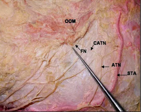
Figure 1. Photographic representation of the anastomosis of facial nerve and auriculotemporal nerve
1. OOM: Orbicularis oculi muscle, 2. FN: Facial nerve, 3. ATN: Auriculotemporal nerve, 4. CATN: Communicating auriculotemporal nerve, 5. STA: Superficial temporal artery.
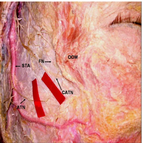
Figure 2. Anastomosis of facial nerve and auriculotemporal nerve.
1. OOM: Orbicularis oculi muscle, 2. FN: facial nerve, 3. ATN: Auriculotemporal nerve, 4. CATN: Communicating auriculotemporal
nerve, 5. STA: Superficial temporal artery.
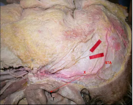
ATN was divided into two branches in front of tragus and behind the STA. Anterior branch crosses the STA superiorly, 43.3 mm. above the tragus and directs to the temporal region (Figure 3). An anastomotic branch with a 15.9 mm. lenght was seen, arising from anterior ATN branch. This branch was named as communicating ATN (CATN) referring to previous studies (2, 3). Origin of the CATN was 70 mm far from the top point of helix and 39 mm far from the vertical line passing from lateral canthus (Figure 4). CATN was connected with a thin, superficial branch arising from the temporal branch of facial nerve at a distance 3.2 mm from OOM, 74.5 mm. from tragus, 72.9 mm. from nasion, 80.5 mm. from the top of helix, 28.5 mm. from the vertical line passing from lateral canthus (Figure 4). New branch which is composed of CATN and temporal branch of FN, enter the OOM, at 75.9 mm. from tragus, 71.0 mm. from nasion, 82.6 mm. from the top of helix, 25.0
mm from the vertical line passing from lateral canthus (Figure 5).
DISCUSSION
We observed an anastomosis in our case between ATN and FN temporal branch at temporal region. This anastomosis was at superior border of OOM, different from classical literatures and previous studies. A tiny branch from FN’s temporal branch connected with a 15.9 mm. long anastomotic branch divided from ATN, at superior-exterior side of OOM.
Andersen et al. studied with 20 cadavers and found ATN - FN anastomosis at temporal region only in 3 of them. However, they didn’t give details about their ana-tomic locations (5).
Namking and Kwak reported that and FN connections frequently and these connections were realized only with
FN’s temporofacial main branch in parotid gland (3,4). These studies didn’t suggest the connections at temporal
region. Different hypothesis were reported about the aim of the ATN and FN connections.
Figure 3. X: The point of crossing the anterior branch of auriculotemporal nerve and superficial temporal artery,
T: Tragus, STA: superficial temporal artery.
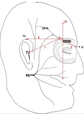
Figure 4. T: Tragus, TH: Top of helix, ATN: Auriculotemporal nerve, FN:
Facial nerve, TB: Temporal branch of facial nerve, CATN: Communicating auriculotemporal nerve, N: Nasion, OOM: Orbicularis oculi muscle, LC: Vertical line passing from lateral canthus.
1. The distance between the origin of CATN and TH 2. The distance between the origin of CATN and LC
3. The distance between the connecting point of CATN-FN and OOM
4. The distance between the connecting point of CATN-FN and T 5. The distance between the connecting point of CATN-FN and N
Trigeminal and facial nerve connections are near the motor- end plates of mimic muscles and voluntary motor impulses from cortex reach the mimic muscles by trige-minal nerve if the facial nerve is completely and persis-tently excised (4). In our case, FN and ATN connection was so close to OOM (3.2 mm) and it is thought to serve for the same purpose.
The auriculotemporal nerve communicates with the facial nerve at the posterior border of the mandibular ramus in its course. This connection is a guide for the sensory impairment of the facial nerve as well. The pain in the muscle of facial expression may occur due to the ent-rapment and compression of the auriculotemporal nerve (6).
Lei et al. defined an anatomic concept called frontal- temporal nerve triangle for avoiding the nerve injury in upper third face rhytidectomy operations. In their detailed anatomic studies, they didn’t mention any anastomosis of FN and ATN (7).
Baumel et al. observed that ATN and FN commu-nicated by two branches within the parotid gland just posterior to the masseter muscle and reported that communicating branches provide sensory fibers for the zygomatic, buccal and mandibular divisions of the facial nerve to the skin (8).
Facial mimic muscle receive the proprioceptive nerve fibers by the skin branches of the trigeminal nerve which are connecting with FN branches. It is believed that proprioception is transported with the same cutaneous nerves, innervating the skin that covers the mimic muscles (3,4,9). Also it was reported that proprioceptive sensations from OOM are important for blink reflex (3). The afferent pathway of the late response of blink reflex is mediated by FN and ATN (10). It can be suggested that, anastomosis between two nerves are working for a common function (4).
Figure 5. T: Tragus, TH: Top of helix, ATN: Auriculotemporal nerve, FN:
Facial nerve, TB: Temporal branch of facial nerve, CATN: Communicating auriculotemporal nerve, N: Nasion, OOM: Orbicularis oculi muscle, LC: Vertical line passing from lateral canthus.
1. The distance between the entrance point of new branch (which is composed of CATN and temporal branch of FN) to OOM and T
2. The distance between the entrance point of new branch (which is composed of CATN and temporal branch of FN) to OOM and N
3. The distance between the entrance point of new branch (which is composed of CATN and temporal branch of FN) to OOM and TH
4. The distance between the entrance point of new branch (which is composed of CATN and temporal branch of FN) to OOM and LC
Sympathetic and parasympathetic nerve fibers of ATN can be injured by surgery procedures at temporal region. This study can help to avoid from postoperative nerve injury, during ophtalmic and plastic surgery at temporal region.
REFERENCES
1. Standring S. Gray’s Anatomy. Thirty-ninth Edition. Else-vier Churchill Livingstone; 2005;513-514.
2. Wıllıams Pl, Warwıck R, Dyson M, Bannıster Lh Gray's Anatomy. Edinburgh: Churchill Livingstone 1989. 3. Namking M, Boonruangsri P, Woraputtaporn W, Guldner
FH. Communication between the facial and auriculo-temporal nerves. J Anat 1994;185:421-426.
4. Kwak HH, Park HD, Youn KH et al. Branching patterns of the facial nerve and its communication with the auri-culotemporal nerve. Surg Radiol Anat 2004;26:494-500. 5. Andersen NB, Bovim G, Sjaastad O. The frontotemporal
peripheral nerves. Topographic variations of the supra-orbital, supratrochlear and auriculotemporal nerves and their possible clinical significance. Surg Radiol Anat 2001;23:97-104.
6. Gulekon N, Anil A, Poyraz A, Peker T, Turgut HB, Karakose M. Variations in the anatomy of the auricu-lotemporal nerve. Clin Anat 2005;18:15-22.
7. Lei T, Gao JH, Xu DC et al. The frontal-temporal nerve triangle: a new concept of locating the motor and sensory nerves in upper third of the face rhytidectomy. Plast Reconstr Surg 2006;117:385-394.
8. Baumel JJ, Vanderheiden JP, McElenney JE. The auri-culotemporal nevre of man. Am J Anat 1971;130:431-440. 9. Last RJ. Regional and applied anatomy, 7th ed. Churcill
Livingstone, New York, 1984; 384-387.
10. Nakamura K, Sakamaki K, Sizuku H, Koike Y. Deter-mining the pathway of the blink reflex through trans-cutaneous electrical stimulation of the facial nerve over the stylomastoid foramen. ORL J Otorhinolaryngol Relat Spec 1999;61:350-354.