Vol.64: e21190530, 2021
https://doi.org/10.1590/1678-4324-2021190530 ISSN 1678-4324 Online Edition
Article - Biological and Applied Sciences
Assessment of Antioxidant and Cytotoxic Activities and
Identification of Phenolic Compounds of Centaurea
solstitialis and Urospermum picroides from Turkey
Mehlika Alper1* https://orcid.org/0000-0001-6193-346X Cennet Özay2 https://orcid.org/0000-0002-1120-6122 Hatice Güneş3 https://orcid.org/0000-0001-5191-365X Ramazan Mammadov1 https://orcid.org/0000-0003-2218-53361Muğla Sıtkı Koçman University, Faculty of Science, Department of Molecular Biology and Genetics, Muğla, Turkey; 2Izmir Katip Celebi University, Faculty of Pharmacy, Department of Basic Pharmaceutical Sciences, Izmir, Turkey; 3Muğla Sıtkı Koçman University, Faculty of Science, Department of Biology, Division of Molecular Biology and Biotechnology, Muğla, Turkey.
Editors-in-Chief: Paulo Vitor Farago Associate Editor: Jane Budel
Received: 2019.09.09; Accepted: 2020.10.07.
*Correspondence: [email protected]; Tel.: +90-252-2115588 (M.A.).
Abstract: It is known that some genera of the Asteraceae family are commonly used in Turkish folk medicine.
Several studies have investigated the biological effects of different extracts of Centaurea and Urospermum species, but studies involving the phenolic composition of C. solstitialis and U. picroides extracts are very limited. This study aimed to investigate the phenolic composition and antioxidant activity of C. solstitialis and U. picroides and evaluate their possible cytotoxic effect. RP-HPLC analysis was used to elucidate the phenolic profiles of the ethanolic extracts of flowering parts of C. solstitialis and U. picroides.The both ethanolic extracts were assessed for their antioxidant properties using DPPH, FRAP, phosphomolybdenum and metal chelating assays. Furthermore, the effect of the extracts on cell viability was evaluated against MCF-7 and PC-3 cancer cells and HEK293 cell line using the MTT assay. The most abundant phenolic
HIGHLIGHTS
The phenolic composition, antioxidant activity and cytotoxic potential of the extracts of C. solstitialis and U. picroides were investigated.
Caffeic acid was found as the most abundant phenolic compound in the extracts. Both species showed promising antioxidant activity towards different assays. The highest cytotoxic potential was observed in the extract of C. solstitialis.
and 14329.59 µg g-1 in the extracts of C. solstitialis and U. picroides, respectively. The antioxidant activity of
the extracts was found similar. Compared with U. picroides extract, C. solstitialis extract had higher potential on the inhibition of cell viability. The IC50 value of C. solstitialis on MCF cells was found as 58.53 µg mL-1.
These data suggest that the extracts of C. solstitialis and U. picroides may be considered as novel and alternative natural antioxidant and anticancer sources.
Keywords: Centaurea solstitialis; Urospermum picroides; Antioxidant activity; Cytotoxic activity; HPLC. INTRODUCTION
Turkey is one of the most floristically rich countries in the world with amazing plant diversity. Its flora consists of about 12000 vascular plants [1] and the therapeutic significance of many medicinal plants among them has not been recognized yet.
Centaurea L., which is a medicinal genus belonging to the Asteraceae family, is represented with 217 taxa 60% of which are endemic to Turkey [2]. Members of this genus were proposed to have several biological activities such as antioxidant [3,4], antimicrobial [5] and antiproliferative [6,7] effects. Centaurea solstitialis L. (yellow star thistle) which grows all over Turkey is used for herpes infections around the lips, malaria, upset stomachs, common colds, hemorrhoids, peptic ulcer and abdominal pain [8-11].
Urospermum picroides (L.) Scop. ex F. W. Schmidt (prickly golden fleece) is an annual herb of the Asteraceae family and used as a medicinal herb for the treatment of inflammatory diseases [12]. This species has been shown to have anti-inflammatory, antimicrobial, antioxidant and antiproliferative activities [12-14] and contain secondary metabolites such as sesquiterpene lactones and glycosides [15] and indole alkaloids [16].
The Centaurea and Urospermum genera represent a significant source of bioactive substances [13,17], but up to now, to the best of our knowledge, the phenolic compounds of the ethanolic extracts of C. solstitialis have not been clarified yet, and only one study has been performed with U. picroides’ methanolic extract in Spain [18]. Therefore, we were inspired to manage this study to determine the phenolic compounds of the ethanolic extracts of C. solstitialis and U. picroides using RP-HPLC. Additionally, these extracts were evaluated for their potential antioxidant activities and cytotoxic properties against MCF-7 and PC-3 cancer cells and HEK293 cell line.
MATERIAL AND METHODS Chemicals
The chemicals and reagents for the antioxidant and cell viability assays were purchased from Sigma-Aldrich (Germany), Merck (USA) and Biochrom (Germany).
Plant Materials and Extraction
C. solstitialis and U. picroides were collected in July and August, 2014 from Yerkesik, Muğla-Turkey (37°8'40'' N, 28°17'11'' E, 550 m) respectively.Taxonomic identification of the plants was confirmed by Dr. Fatma Güneş from the Department of Pharmaceutical Botany at Trakya University in Edirne, Turkey. All specimens were deposited in their herbarium (Voucher No: H.G. 902 and H.G. 901).The flowering parts of plants were air-dried (for one week)in shadow at 25 °C, and the dried flowers were powdered down to fine grains. Ethanolic extracts were produced using a Soxhlet extractor for 10 h. The extracts were filtered and vaporized by using a rotary evaporator and then lyophilized. The crude extracts were kept at -20 °C until needed. For the MTT assay, DMSO at 0.1% final concentration was used as solvent of the extracts. For the antioxidant assays, the extracts were solubilized in ethanol.
Total Phenolic and Flavonoid Contents
The phenolic and flavonoid contents of the plant extracts were determined by using the Folin-Ciocalteu protocol [19] and aluminum [20] colorimetric methods, respectively. These contents were expressed as gallic acid (mg GAEs g-1) and quercetin (mg QEs g-1) equivalents, respectively.
Phenolic Compound Analyses
Phenolic compounds of the extracts of C. solstitialis and U. picroides were analyzed by RP-HPLC (Shimadzu, Japan) as described by Caponio and coauthors [21] with some modifications. Separation was performed at 30 °C by using a reversed phase column (Agilent Eclipse XDB C-18, 250 mm × 4.6 mm, 5 μm) by a mixture of two solvents (A: the formic acid solution 3% and B: methanol) as a mobile phase. Gradient conditions were particularized at a flow rate of 0.8 mL min-1 as follows: 93% A + 7% B for 3 min, 72% A +
28% B in 28 min, 67% A + 33% B in 60 min, 58% A + 42% B in 62 min, 50% A + 50% B in 70 min, 30% A + 70% B in 75 min, 93% A + 7% B in 80 min. Phenolic compounds in the ethanolic extracts of C. solstitialis and U. picroides are expressed as μg g-1 extract, which were analyzed with a diode array detector (DAD).
Antioxidant Activity Assays
DPPH Radical Scavenging Activity
Extract solution (1 mL) was added to the 2,2-diphenyl-1-picrylhydrazyl (DPPH) radical solution (0.004% methanol, 4 mL), and after 30 min of incubation at 25 °C in the dark, the absorbances were recorded at 517 nm. DPPH radical scavenging activity is expressed as Trolox (mg TEs g-1 extract) equivalents [22].
Ferric Reducing Antioxidant Power (FRAP) Assay
Ferric reducing antioxidant power assay was applied as described by Zengin and Aktumsek [23] with some modifications. Extract solutions were added to FRAP reagent which was mixed in advance (acetate buffer– 0.3 mol L-1, TPTZ (2,4,6-tripyridyl-s-triazine– 10 mmol L-1, FeCI
3– 20 mmol L-1). After measuring the
absorbances at 593 nm, FRAP activity was expressed as Trolox (mg TEs g-1 extract) equivalents.
Total Antioxidant Capacity (Phosphomolybdenum Method)
The phosphomolybdenum method was used to evaluate the total antioxidant capacity of the extracts. To keep it short, different extract solutions were mixed with the reagent solution (0.6 M H2SO4, 28 mmol L-1
Na3PO4 and 4 mmol L-1 (NH4)2MoO4) and incubated for 90 min at 95 °C. The absorbance values were
determined at a wavelength of 695 nm [24]. Total antioxidant capacity is expressed as Trolox (mg TEs g-1
extract) equivalents. Metal Chelating Activity
Extract solutions at different concentrations were added to FeCl2(0.05 mL, 2 mmol L-1). The reaction that
started directly after adding 5 mmol L-1 of ferrozine was measured at 562 nm after being left for 10 min at
room temperature. Metal chelating activity is expressed as EDTA (mg EDTAEs g-1 extract) equivalents. [25].
Cell Cultures and MTT Assay
The cell lines were sourced from the American Type Culture Collection (ATCC, USA). MCF-7 (breast adenocarcinoma) (ATCC #HTB-22), PC-3 (prostate adenocarcinoma (ATCC #CRL-1435) and HEK293 (human embryonic kidney) (ATCC #CRL-1573) cell lines were maintained in an RPMI 1640 medium supplemented with 10% heat-inactivated fetal bovine serum, 100 units mL-1 penicillin and 100 µg mL-1
streptomycin at 37°C in 5% CO2. The cell viability was detected based on the MTT assay [26]. For this assay, cell lines were added separately at a final concentration of 4x10³ cells per well into 96-well plates as triplicates
and incubated at 37°C for 24 h. Dose course experiments for 72 h were performed at seven different concentrations (1000-15.625 µg mL-1) of each extract under the same conditions. The untreated cells were
used as a control. At the end of this 72-h incubation, the medium with the extract was removed from the wells and replaced with 100 µL of fresh growth medium. Later, 10 µL of MTT (3-(4,5-Dimethyl-2-thiazolyl)-2,5-diphenyl-2H-tetrazolium Bromide) (5 mg mL-1) solution to achieve a final concentration of 0.45 mg mL-1 was
added to every well, and the microplates were incubated for another 4 h. Then, the media containing MTT were discarded, and the formazan crystals formed by viable cells were dissolved in 100 µL of DMSO. The absorbance (Abs) for each well was recorded using a microplate reader at 540 nm. The percentage of cell viability according to the following formula:
Statistical Analysis
The results obtained in this study are expressed as mean ± Standard error (SE). Statistical analysis and data processing were performed by using GraphPad Prism 7.0 (GraphPad Software, Inc., San Diego, CA). Comparisons of the treatments among groups were analyzed by one-way ANOVA with post-hoc Tukey's test. Significance was accepted as * P 0.05, ** P 0.01, *** P 0.001, **** P 0.0001, ns-no significance (P 0.05). The IC50 values were also calculated using the GraphPad Prism software.
RESULTS
Total Bioactive Compounds and Phenolic Composition
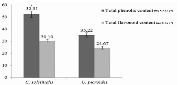
Total phenolic and flavonoid contents of C. solstitialis and U. picroides ethanolic extracts were investigated with spectrophotometric methods, and the results are presented in Figure 1. According to the data obtained, the highest total phenolic content (52.31 mg GAEs g-1) and total flavonoid content (30.10 mg
QEs g-1) were detected to be in the C. solstitialis extract. There were no significant differences between the
two plants in terms of total flavonoid contents (P> 0.05), whereas a significant difference was found in terms of their phenolic contents (P 0.05).
Figure 1. Total phenolic and flavonoid contents of C. solstitialis and U. picroides extracts. The bar represents the mean
of contents (± SE) in each species. (GAEs: Gallic acid equivalents, QEs: Quercetin equivalents). * P 0.05.
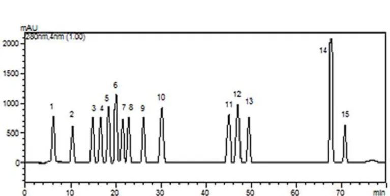
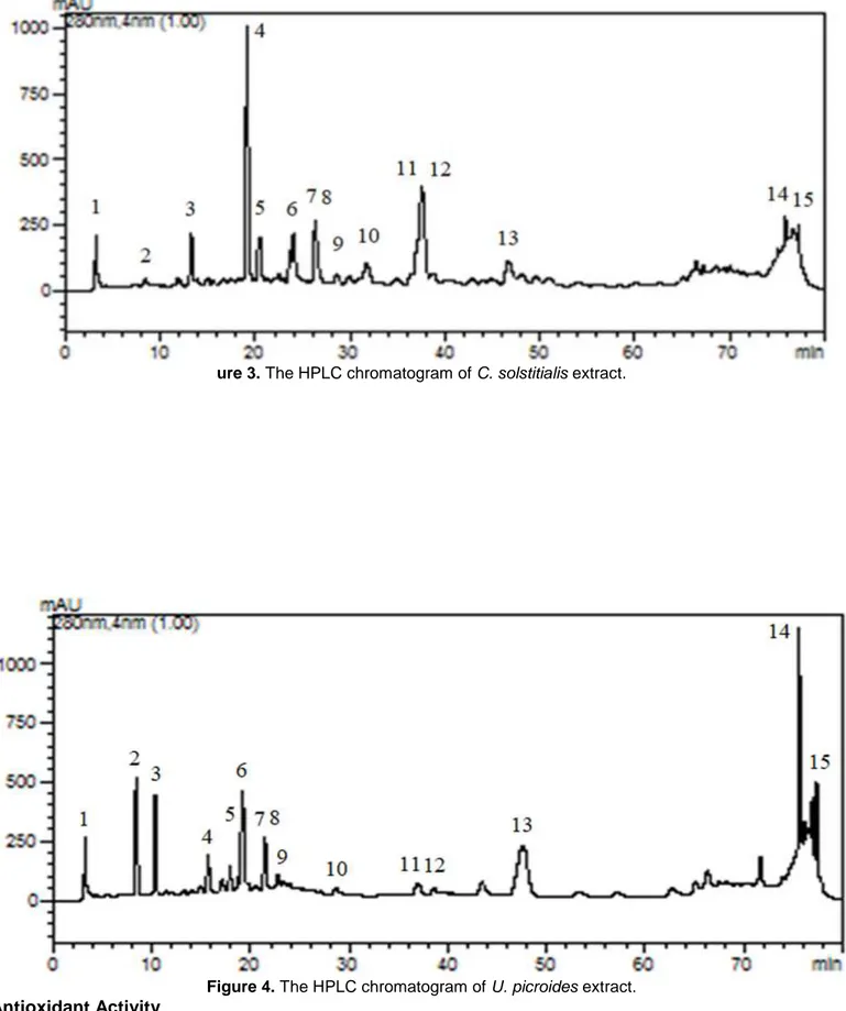
In order to identify the phenolic compounds in the ethanolic extracts of the flowering parts of both C. solstitialis and U. picroides, the RP-HPLC method was applied by using 15 phenolic compounds (gallic acid, 3,4-dihydroxybenzoic acid, 4-hydroxybenzoic acid, 2,5-dihydroxybenzoic acid, chlorogenic acid, vanillic acid, epicatechin, caffeic acid, p-coumaric acid, ferulic acid, rutin, ellagic acid, naringin, cinnamic acid, quercetin) as the standards (Figure 2). The phenolic compounds were detected in both extracts (Figures 3 and 4)with varying amounts and are given in Table 1. Caffeic acid was detected to be the most abundant phenolic compound in both extracts, and the amount of this compound was 24078.03 and 14329.59 µg g-1 extract in
the C. solstitialis extract and the U. picroides extracts, respectively. Following caffeic acid, the main phenolic compounds of the C. solstitialis extract were determined as epicatechin (3684.22 µg g-1 extract),
2,5-dihydroxybenzoic acid (3360.16 µg g-1 extract) and 4-hydroxybenzoic acid (2077.33 µg g-1 extract),
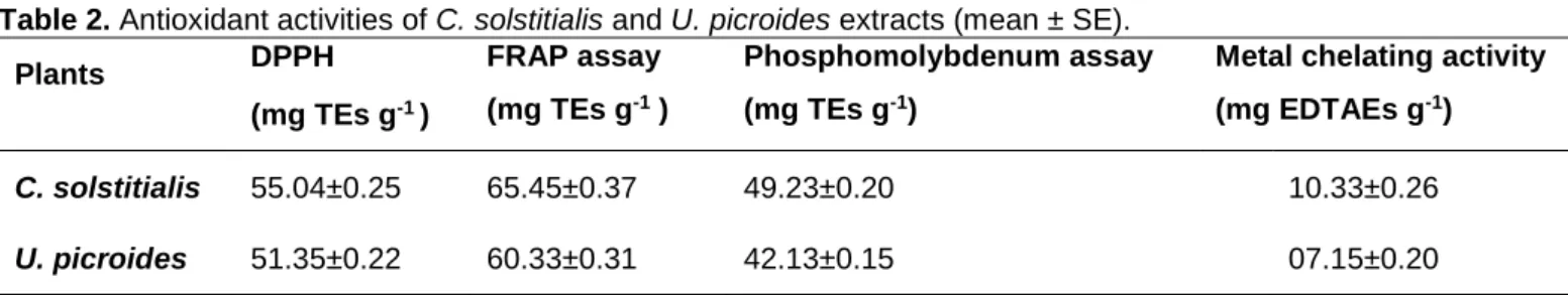
respectively. The main phenolic compound in U. picroides extract was 4-hydroxybenzoic acid (1189.72 µg g -1 extract) after caffeic acid. The amounts of epicatechin (499.33 µg g-1 extract) and 2,5-dihydroxybenzoic acid
(799.48 µg g-1 extract) in the U. picroides extract were significantly lower than that of the C. solstitialis extract.
Table 1. Phenolic compounds of C. solstitialis and U. picroides extracts (μg g-1 extract) (mean ± SE). No Phenolic Compounds RT (min) UVmax (nm) LOD (µg mL-1) C. solstitialis (μg g-1 extract) U. picroides (μg g-1 extract) 1 Gallic acid 6.8 280 0.01 92.59 ± 0.95 168.43 ± 2.06 2 3,4-dihydroxybenzoic acid 10.7 280 0.03 496.32 ± 3.15 156.27 ± 2.00 3 4-hydroxybenzoic acid 15.7 280 0.01 2077.33 ± 43.20 1189.72 ± 11.83 4 2,5-dihydroxybenzoic acid 17.2 320 0.75 3360.16 ± 51.53 799.48 ± 5.13 5 Chlorogenic acid 18.2 320 0.01 52.16 ± 0.41 285.25 ± 3.02 6 Vanillic acid 19.2 320 0.11 340.15 ± 3.84 842.25 ± 5.42 7 Epicatechin 21.3 260 0.43 3684.22 ± 77.44 499.33 ± 3.26 8 Caffeic acid 22.7 280 0.01 24078.03 ± 495.5 14329.59± 263.1 9 p-coumaric acid 26.1 320 0.01 41.00 ± 0.33 2.14 ± 0.08 10 Ferulic acid 30.1 320 0.01 127.21 ± 1.64 23.27 ± 0.52 11 Rutin 45.6 360 0.57 46.17 ± 0.36 13.40 ± 0.25 12 Ellagic acid 47.7 240 0.45 45.85 ± 0.30 344.57 ± 3.75 13 Naringin 49.7 280 0.40 120.62 ± 1.48 13.76 ± 0.36 14 Cinnamic acid 67.8 280 0.01 383.82 ± 3.06 544.20 ± 4.01 15 Quercetin 71.1 360 0.57 37.86 ± 0.27 116.29 ± 1.35
Figure 2. The HPLC chromatogram of standard phenolic compounds (1. gallic acid, 2. 3,dihydroxybenzoic acid, 3.
4-hydroxybenzoic acid, 4. 2,5-di4-hydroxybenzoic acid, 5. chlorogenic acid, 6. vanillic acid, 7. epicatechin, 8. caffeic acid, 9. p-coumaric acid, 10. ferulic acid, 11. rutin, 12. ellagic acid, 13. naringin, 14. cinnamic acid, 15. quercetin).
ure 3. The HPLC chromatogram of C. solstitialis extract.
Figure 4. The HPLC chromatogram of U. picroides extract. Antioxidant Activity
Four various antioxidant assays, DPPH, FRAP, phosphomolybdenum and metal chelating were applied to the ethanol extracts of the flowering parts of C. solstitialis and U. picroides. The outcomes of these assays are presented in Table 2. There were no significant differences between the two plants in terms of all the antioxidant assays that were applied (P >0.05). However, the C. solstitialis extract showed a slightly higher antioxidant activity than U. picroides. The results of the DPPH, FRAP, phosphomolybdenum and metal chelating assays for C. solstitialis were determined as 55.04 mg TEs g-1, 65.45 mg TEs g-1, 49.23 mg TEs
Table 2. Antioxidant activities of C. solstitialis and U. picroides extracts (mean ± SE). Plants DPPH (mg TEs g-1 ) FRAP assay (mg TEs g-1 ) Phosphomolybdenum assay (mg TEs g-1)
Metal chelating activity (mg EDTAEs g-1)
C. solstitialis 55.04±0.25 65.45±0.37 49.23±0.20 10.33±0.26
U. picroides 51.35±0.22 60.33±0.31 42.13±0.15 07.15±0.20
TEs: trolox equivalents, EDTAEs: EDTA equivalents.
Cytotoxicity of the Extracts
The effect of the extracts at different concentrations (1000-15.625 µg mL-1) on viability of the cells tested
for 72 h were determined by the MTT assay. The results of the MTT assay are graphically illustrated in Figure 5. The viability of the cancer cells (MCF-7 and PC-3) seemed to significantly decrease at higher concentrations than 125 µg mL-1, when treated with both extracts separately. The MCF-7 cell viability was
significantly decreased to about 90% when treated with the C. solstitialis extract in the concentration range between 1000 to 125 µg mL-1 or the U. picroides extract at between 1000 to 250 µg mL-1. The effects of both
extracts on PC-3 cells were generally similar to each other. No cytotoxic activity of the C. solstitialis extract was found at 31.25 and 15.625 µg mL-1 concentrations in MCF-7 cancer cells and HEK293 cells. However,
the C. solstitialis extract at 62.5 µg mL-1 had a lower cytotoxic potential against HEK293 cells in comparison
to that of MCF-7 cells. The U. picroides extract at between 125 to 15.625 µg mL-1 did not decrease the
percentage of HEK293 cell viability. According to the approximate IC50 values (50% inhibitory concentrations),
the IC50 values of both extracts were found to be lower in cancer cells than HEK293 cells. The IC50 values of
the C. solstitialis extract on MCF-7, PC-3 and HEK293 cells were 58.53, 91.47, 224.5 µg mL-1, respectively.
Moreover, the IC50 values for the U. picroides extract were 115.5, 109.8 and 464.1 µg mL-1 on MCF-7, PC-3
Figure 5. The effects of C. solstitialis (A) and U. picroides (B) extracts on cell viability. Each bar represents the percent
of cell viability after treatment with different concentrations of the extracts. Data are means (± SE) of three independent experiments. * P 0.05, ** P 0.01, *** P 0.001, **** P 0.0001, ns-no significance (P 0.05).
DISCUSSION
Although many studies have been carried out on the evaluation of the antioxidant capacity of plants, a single method which completely reflects the antioxidant capacity has not been developed. Therefore, it is necessary to fully interpret antioxidant capacity using different chemical assays. From this point of view, different antioxidant assays (DPPH, FRAP, phosphomolybdenum and metal chelating) were used in our study, and the total phenolic and flavonoid contents were calculated for each extract.
In studies evaluating the antioxidant capacity of plants, at least one free radical was used, and the studies determined the rate at which this radical is scavenged by the plant extract. The most common of these
radicals is DPPH. Ferric reducing power assay (FRAP) is often used as an indicator of electron–donating activity, which is an important mechanism of antioxidant compounds, especially phenolics [27]. The phosphomolybdenum assay is based on reduction of Mo (VI) to Mo (V) and subsequent formation of a green phosphate/Mo (V) complex. Transition metals act as a catalyst for lipid peroxidation. Therefore, chelating these metals is considered as an important antioxidant mechanism [23].
Most phenolic compounds have been reported to possess potent antioxidant activity and have anticarcinogenic, anti-inflammatory and antibacterial activities to a greater or lesser extent [28,29]. A study was conducted by Cai and coauthors [30] examined the structure-radical scavenging activity relationships of phenolic compounds from anticancer-related traditional Chinese medicinal plants. The authors reported that hydroxycinnamic acids, such as caffeic acid and chlorogenic acid were found to be rather active in scavenging radicals related to antioxidant activity. The presence of abundant amounts of caffeic acid in both plant extracts examined in this study may explain the antioxidant activity of these plants.
Several researchers showed that the major phytochemical components of Centaurea taxa were sesquiterpene lactones, terpenoids, flavonoids and acetylenes [31,32]. Previous phytochemical studies showed that Centaurea taxa contain flavonoids including quercetin, luteolin, kaempferol, salvigenin, apigenin, hispidulin, cirsimaritin, apigenin 7-O-glucoside and isokaempferide [33,34].
Formisano and coauthors [35] reviewed reports on flavonoids from the Centaureinae subtribe of the family Asteraceae and reported that only 16 of 72 recognized genera of Centaureinae have been examined for the occurrence of flavonoids. According to these data, it was seen that the majority of the genera of the Centaureinae has not been examined for their flavonoid profile yet.
The results on the chemical composition of C. solstitialis reconfirmed the value of the Centaurea genus as a source of phenolic compounds. Our results were in accordance with the phytochemical profile of the Centaurea species from the Turkish flora [36,37]. The phenolic profile of the methanolic extract of Centaurea urvillei subsp. stepposa was obtained by using RP-HPLC analysis [36], and it showed a rich phenolic content. Many earlier studies showed that the aerial parts of Centaurea species is an alternative source of phenolic compounds. Compared to other Centaurea species collected from Turkey, it was evident that the phenolic content of C. solstitialis is higher than the values found for the Centaurea species investigated by Şen and coauthors [38] (ranged from 4.82 to 12.46 mg GAEs g-1) and lower than the values found for the
Centaurea species investigated by Aktumsek and coauthors [39] (ranged from 82.27 to 175.40 mg GAEs g-1). Koc and coauthors [40] reported that the total phenolic contents in the ethanolic extracts of the flowers
and leaves of C. solstitialis were determined as 18.43 and 13.66 mg L-1, respectively. It was reported that
differences were observed in the phenolic contents of plant species belonging to the same genus depending on several factors including temperature, soil content and altitude (growing conditions) [41].
The results on total phenolic content in this study displayed a similar propensity to those of the antioxidant abilities. Accordingly, the high content of total phenolics in the extracts might explain the antioxidant properities of the extracts. These results were consistent with other results in the literature which demonstrated a strong relationship between antioxidant activities and total phenolic contents [23,42].
The DPPH radical scavenging, FRAP, metal chelating activities and the total antioxidant capacity of methanolic Centaurea lycopifolia extracts were reported previously as 52.49 mg TE g-1, 64.44 mg TE g-1,
8.63 mg EDTAE g-1 and 1.45 mmol TE g-1 extract, respectively [37]. In another study, the DPPH and FRAP
activities of the methanolic Centaurea urvillei subsp. stepposa extracts were detected as 43.35 and 52.18 TEs g-1 extract, respectively [36]. Based on these results, it may be concluded that C. solstitialis had a higher
antioxidant activity than C. lycopifolia and C. urvillei subsp. stepposa. In addition, it was reported that DPPH free radical scavenging activity, reducing power activity and also total phenolic compound of C. solstitialis (from Konya) were evaluated [43].
El-Amier and coauthors [44] detected the total phenolic and flavonoid contentsof an aqueous extract of U. picroides, which was collected from Egypt, as 9.31 and 4.24 mg g-1 dry weight, respectively. The authors
also reported that the DPPH scavenging activity of U. picroides was found as 4.14% at 500 μg mL-1. The total
phenolic and flavonoid contents of methanolic U. picroides extracts were determined by El-Amier and coauthors [14] as 19.34 and 8.28 mg g-1 dry weight, respectively. According to these results, the ethanolic
extract of U. picroides, which was collected from Turkey, had stronger antioxidant activity and more abundant total bioactive compounds than those collected from Egypt.
El-Nabawy and coauthors [13] assessed the antioxidant activity of U. picroides using the ABTS method. Inhibition of ABTS free radicals were found to be 86.1% and 82.4% for the aerial parts in the ethyl acetate and methylene chloride fraction, respectively.
Giner and coauthors [18] reported that chlorogenic acid, quercetin, luteolin, quercetin-3-galactoside, kaempferol-3-galactoside, isochlorogenic acid and luteolin-7-glucoside were isolated from the methanolic extract of U. picroides, which was collected from Spain. The outcomes of the phenolic profile of U. picroides, which was studied in this study, were in good agreement with the aforementioned results.
Plant species and their bioactive compounds have an important role in inhibiting the progression of cancer and development of clinically useful new anti-cancer agents [45,46]. In our study, the ethanolic extracts from the flowering parts of both C. solstitialis and U. picroides were assessed on MCF-7, PC-3 cancer cells and HEK293 cells for 72 h. The C. solstitialis extract caused higher inhibition in the percentage of the viabilities of MCF-7 cells (IC50 value=58.53 µg mL-1) and PC-3 cells (IC50 value=91.47 µg mL-1) compared to
HEK293 cells (IC50 value=224.5 µg mL-1). The C. solstitialis extract was found to be more effective than the
U. picroides extract (IC50 value = 115.5 µg mL-1) in reducing viability of MCF-7 cells. The potential cytotoxic
effects of both extracts on PC-3 cells were similar. Additionally, the IC50 value of the U. picroides extract
against HEK293 cells was 464.1 µg mL-1. Our results suggested that these extracts have more cytotoxic
potentials against MCF-7 and PC-3 cancer cell lines than against HEK293 cell lines, in other words the cancer cell lines used herein were more sensitive to both extracts than HEK293 cell line. In contrast, the IC50 values
of the ethanolic extract of the flowering parts of C. solstitialis on HeLa (cervix adenocarcinoma), Daudi (Burkitt’s lymphoma, CCL-213), A549 (lung adenocarcinoma) and BEAS-2B (normal bronchial epithelium) human cell lines at 72 h were reported to be 63.18, 69.27, 252.5 and 75.25 μg mL-1, respectively [7]. Erenler
and coauthors [47] stated that, at 500 μg mL-1, the methanolic extract of stem parts of C. solstitialis L. ssp.
solstitialis collected from Tokat caused higher antiproliferative activity on HeLa cells than that of the methanolic extracts of root and flower parts of the plant, whereas all extracts at 1000 μg mL-1 had about the
same antiproliferative activity on C6 (rat brain tumor) cells. Among the different fractions of aerial parts and seeds of U. picroides from Egypt, the butanol fraction from seeds and the ethyl acetate fraction from aerial parts were indicated to be very strongly cytotoxic to both MCF-7 (IC50 value=9.4±0.37 and 8.8±0.47 µg mL -1, respectively) and HePG-2 (liver carcinoma) (IC
50 value=14.7±0.85 and 10.1±0.88 µg mL-1, respectively)
cell lines [13]. Furthermore, the IC50 values of the ethanolic extract from the flowering part of U. picroides
were found to be 85.64 µg mL-1 for Daudi, 135.35 µg mL-1 for HeLa, 234.8 µg mL-1 for A549 and 109.80 μg
mL-1 for BEAS-2B cell lines [48]. These results suggest that the region where the plant is collected, part of
plant used, solvent chosen for extraction and cell lines that are tested may affect cytotoxic activity on different scales.
CONCLUSION
In conclusion, this study revealed the HPLC profile, antioxidant and cytotoxic potentials of ethanolic extracts of the flowering parts of C. solstitialis and U. picroides. Caffeic acid was found to be the highest amount in both extracts based on HPLC analysis, especially the C. solstitialis extract was a superb source of caffeic acid. Both extracts showed promising antioxidant potential towards different assays. The highest inhibitory effect on cell viability was obtained from the C. solstitialis extract in comparison to the U. picroides extract. It may be proposed that the biological activities of both species that were obtained may be ascribed to the presence of phenolic compounds. Our results showed that C. solstitialis and U. picroides may be accepted as a novel and alternative source of natural antioxidant and anticancer agents. These potential biological activities are needed to be approved via additional experiments. Future studies should focus on clarifying the substances responsible for the potential antioxidant and cytotoxic effects of the extracts. Also, detailed in vivo investigations may contribute to design of new drug or food additive formulations.
Acknowledgments: Authors would like to thank Mehmet Akif Ersoy University Scientific and Technology Application
and Research Center for HPLC analysis.
Conflicts of Interest: The authors declare no conflict of interest. REFERENCES
1. Guner A, Aslan S, Ekim T, Vural M, Babac MT. Türkiye Bitkileri Listesi Damarlı Bitkiler. Nezahat Gökyiğit Botanik Bahçesi ve Flora Araştırmaları Derneği Yayını, Istanbul; 2012.
2. Bona M. An overview to Centaurea (Asteraceae) based on herbarium specimens of ISTE. J Fac Pharm Istanbul. 2013 Mar;43(2):121-37.
3. Belkassam A, Zellagui A, Gherraf N, et al. Assessment of antioxidant effect of the essential oil and methanol extract of Centaurea dimorpha Viv. aerial parts from Algeria. Asn. 2019 Apr;6(1):54-62.
4. Yırtıcı U. The determination of antioxidant properties and enzyme inhibition effect of Centaurea fenzlii Reichardt extract. Bitlis Eren Univ J Sci. 2019 Jan;8(1):66-73.
5. Ugur A, Duru ME, Ceylan O, Sarac N, Varol O, Kivrak I. Chemical composition, antimicrobial and antioxidant activities of Centaurea ensiformis Hub.-Mor. (Asteraceae), a species endemic to Muğla (Turkey). Nat Prod Res. 2009 Jan;23(2):149-67.
6. Tugba Artun F, Karagoz A, Ozcan G, et al. In vitro anticancer and cytotoxic activities of some plant extracts on HeLa and Vero cell lines. J Buon. 2016 May-Jun;21(3):720-25.
7. Alper M, Güneş H. The anticancer and anti-inflammatory effects of Centaurea solstitialis extract on human cancer cell lines. Turk J Pharm Sci. 2019 Jun;16(3):273-81.
8. Yesilada E, Honda G, Sezik E, et al. Traditional medicine in Turkey V. Folk medicine in inner Taurus Mountains. J Ethnopharmacol. 1995 Jun;46(3):133-52.
9. Fujita T, Sezik E, Tabata M, et al. Traditional medicine in Turkey, vol. VII. Folk medicine in middle and West Black Sea Regions. Econ Bot. 1995 Oct;49(4):406-22.
10. Honda G, Yesilada E, Tabata M, et al. Traditional medicine in Turkey VI. Folk medicine in West Anatolia: Afyon, Kutahya, Denizli, Mugla, Aydin provinces. J Ethnopharmacol. 1996 Aug;53(2):75-87.
11. Yilmaz G. Some medicinal plants used as folk medicine for dermatological diseases in European Turkey. Curr Pers MAPs. 2018 Jul;1(1):48-52.
12. Strzelecka M, Bzowska M, Koziel J, et al. Anti-inflammatory effects of extracts from some traditional Mediterranean diet plants. J Physiol Pharmacol. 2005 Mar;56(Suppl 1):139-56.
13. El-Nabawy HI, Ayyad DM, Serag MS, Abdel-Mogib M. Phytochemical and biological evaluation of Urospermum
picroides. Mansoura J Chem. 2015 Aug;19-31.
14. El-Amier YA, Al-hadithy ON, Abdullah TJ. Antioxidant and antimicrobial activity of different extracts obtained from aerial parts of Urospermum picroides (L.) F.W. from Egypt. J Adv Chem Sci. 2016 Jun;2(3):299-301.
15. Balboul BA, Ahmed AA, Dsuka H. Sesquiterpene lactones and glucosides from Urospermum picroides. Phytochemistry. 1997 May;45(2):369-73.
16. Ji X, Lu G, Gai Y, Zheng C, Mu Z. Biological control against bacterial wilt and colonization of mulberry by an endophytic Bacillus subtilis strain. FEMS Microbiol Ecol. 2008 Sep;65(3):565-73.
17. Khammar A, Djeddi S. Pharmacological and biological properties of some Centaurea species. Eur J Sci Res. 2012;84(3):398-416.
18. Giner RM, Cuéllar MJ, Recio MC, Máñez S, Ríos JL. Chemical constituents of Urospermum picroides. Z Naturforsch C. 1992;47(7-8):531-34.
19. Slinkard K, Singleton VL. Total phenol analysis: automation and comparison with manual methods. Am J Enol Vitic. 1977;28:49-55.
20. Moreno MI, Isla MI, Sampietro AR, Vattuone MA. Comparison of the free radical-scavenging activity of propolis from several regions of Argentina. J Ethnopharmacol. 2000 Jul;71(1-2):109-14.
21. Caponio F, Alloggio V, Gomes T. Phenolic compounds of virgin olive oil: influence of paste preparation techniques. Food Chem. 1999 Feb;64(2):203-09.
22. Ceylan R, Katanić J, Zengin G, Matić S, et al. Chemical and biological fingerprints of two Fabaceae species (Cytisopsis dorycniifolia and Ebenus hirsuta): Are they novel sources of natural agents for pharmaceutical and food formulations? Ind Crop Prod. 2016 Jun;84:254-62.
23. Zengin G, Aktumsek A. Investigation of antioxidant potentials of solvent extracts from different anatomical parts of Asphodeline anatolica E. Tuzlaci: an endemic plant to Turkey. Afr J Tradit Complement Altern Med. 2014 Jan;11(2):481-8.
24. Berk S, Tepe B, Arslan S, Sarikurkcu C. Screening of the antioxidant, antimicrobial and DNA damage protection potentials of the aqueous extract of Asplenium ceterach DC. Afr J Biotechnol. 2011 Aug;10(44):8902-8.
25. Zengin G, Nithiyanantham S, Locatelli M, et al. Screening of in vitro antioxidant and enzyme inhibitory activities of different extracts from two uninvestigated wild plants: Centranthus longiflorus subsp. longiflorus and Cerinthe minor subsp. auriculata. Eur J Integr Med. 2015 Jun;8(3):286-92.
26. Mosmann T. Rapid colorimetric assay for cellular growth and survival: Application to proliferation and cytotoxicity assays. J Immunol Methods. 1983 Dec;65(1-2):55-63.
27. Nabavia SM, Ebrahimzadeh MA, Nabavi SF, Fazelian M, Eslami B. In vitro antioxidant and free radical scavenging activity of Diospyros lotus and Pyrus boissieriana growing in Iran. Pharmacogn Mag. 2009 Dec;5(18):122-6. 28. Baba SA, Malik SA. Determination of total phenolic and flavonoid content, antimicrobial and antioxidant activity of
a root extract of Arisaema jacquemontii Blume. J Taibah Univ Sci. 2015 Oct;9(4):449-54.
29. Huang W-Y, Cai Y-Z, Zhang Y. Natural phenolic compounds from medicinal herbs and dietary plants: potential use for cancer prevention. Nutr Cancer. 2009 Dec;62(1):1-20.
30. Cai YZ, Sun M, Xing J, Luo Q, Corke H. Structure-radical scavenging activity relationships of phenolic compounds from traditional Chinese medicinal plants. Life Sci. 2006 May;78(25):2872-88.
31. Al-easa HS, Rızk AM. Constituents of Centaurea species. Quatar Univ Sci J. 1992;12:27-57.
32. Gurbuz I, Yesilada E. Evaluation of the anti-ulcerogenic effect of sesquiterpene lactones from Centaurea solstitialis L. ssp. solstitialis by using various in vivo and biochemical techniques. J Ethnopharmacol. 2007 Jun;112(2):284-91.
33. Beltagy AM. Chemical composition and cytotoxic activity of Centaurea scoparea Sieb against four human cell lines. J Pharm Sci Res. 2015;7(3):103-7.
34. Melikoglu G, Ozsoy N, Yilmaz Ozden T, et al. Flavonoids and biological activities of Centaurea nerimaniae S. Kultur. Farmacia. 2018;66(6):1070-5.
35. Formisano C, Rigano D, Senatore F, et al. Flavonoids in subtribe Centaureinae (Cass.) Dumort. (Tribe Cardueae, Asteraceae): distribution and 13C-NMR spectral data. Chem Biodivers. 2012 Oct;9(10):2096-158.
36. Uysal S, Ceylan R, Zengin G, Aktumsek A, Zengin N, Guler GO. Phytochemical characterization of an endemic plant used as foodstuff in Turkey: Centaurea urvillei subsp. stepposa and its antioxidant properties. Int Res J Pharmacy. 2014 Aug;5(8):646-52.
37. Zengin G, Zheleva-Dimitrova D, Gevrenova R, et al. Identification of phenolic components via LC-MS analysis and biological activities of two Centaurea species: C. drabifolia subsp. drabifolia and C. lycopifolia. J Pharm Biomed Anal. 2018 Feb;149:436-41.
38. Şen A, Bitiş L, Birteksöz-Tan S, Bulut G. In vitro evaluation of antioxidant and antimicrobial activities of some
Centaurea L. species. Marmara Pharm J. 2013;17:42-5.
39. Aktumsek A, Zengin G, Guler GO, Cakmak YS, Duran A. Screening for in vitro antioxidant properties and fatty acid profiles of five Centaurea L. species from Turkey flora. Food Chem Toxicol. 2011 Nov;49(11):2914-20.
40. Koc S, Isgor BS, Isgor YG, Shomali Moghaddam, N, Yildirim O. The potential medicinal value of plants from Asteraceae family with antioxidant defense enzymes as biological targets. Pharm Biol. 2015 May;53(5):746-51. 41. Giorgi A, Madeo M, Speranza G, Cocucci M. Influence of Environmental Factors on Composition of Phenolic
Antioxidants of Achillea collina Becker ex Rchb. Nat Prod Res. 2010 Oct;24(16):1546-59.
42. Ozay C, Mammadov R. Screening of some biological activities of Alyssum fulvescens var. fulvescens known as Ege madwort. Acta Biol Hung. 2017 Sep;68(3):310-20.
43. Tekeli Y, Sezgin M, Aktümsek A. Antioxidant property of Centaurea solstitialis L. from Konya, Turkey. Asian J Chem. 2008;20(6):4831-5.
44. El-Amier YA, Abbas MA, Dawood SH. Phytotoxic effect of plant extracts from Asteraceae on germination of
Echinocloa crus-galli growth. IJDR. 2015 Jul;5(7):4926-31.
45. Iqbal J, Abbasi BA, Mahmood T, et al. Plant-derived anticancer agents: A green anticancer approach. Asian Pac J Trop. 2017 Dec;7(12):1129-50.
46. Kaur R, Kapoor K, Kaur H. Plants as a source of anticancer agents. J Nat Prod Plant Resour. 2011;1(1):119-24. 47. Erenler R, Sen O, Yaglioglu AS, Demirtas I. Bioactivity-Guided Isolation of Antiproliferative Sesquiterpene Lactones
from Centaurea solstitialis L. ssp. solstitialis. Comb Chem High Throughput Screen. 2016;19(1):66-72.
48. Alper M, Güneş H. Determination of anticancer effects of Urospermum picroides against human cancer cell lines. IJSM. 2019 Jan;6(1):28-37.
© 2021 by the authors. Submitted for possible open access publication under the terms and conditions of the Creative Commons Attribution (CC BY NC) license (https://creativecommons.org/licenses/by-nc/4.0/).