www.turkderm.org.tr
Case Report
Olgu Sunumu
©Copyright 2016 by Turkish Society of Dermatology and Venerology
Turkderm-Archives of the Turkish Dermatology and Venerology published by Galenos Yayınevi.
Abstract
Vitiligo nedeni tam olarak bilinmeyen, genetik ve genetik olmayan faktörlerin birlikte rol oynadığı edinsel bir bozukluktur. Bu hastalıkta tutulan deride melanositler ortadan kalkar, klinik olarak depigmente makül ve yamalar belirir. Becker nevüs (BN) sıklıkla unilateral dağılım gösteren, keskin ama düzensiz sınırlı hiperpigmente makül, yama veya verrüköz plakların izlendiği, üzerinde değişik derecelerde hipertrikozun bulunduğu bir hastalıktır. Patogenezi belli olmamakla birlikte hamartamatöz bir lezyon olduğu ve üzerinde androjen reseptörlerinin arttığı ileri sürülmektedir. Biz burada; önce vitiligo lezyonları, on yıl sonra ise vitiligo lezyonuna komşu, gövde arka üst sağ alanda BN lezyonu ortaya çıkan on dokuz yaşında bir erkek hastayı sunmayı amaçladık.
Anahtar Kelimeler: Becker nevüs, vitiligo, birliktelik
Introduction
Vitiligo is an acquired disorder with an unknown etiology in which genetic and non-genetic factors coexist. Melanocytes are destructed in the affected skin areas and clinically depigmented macules and patches appear on the skin1. Becker’s nevus (BN) appears as a hyperpigmented macule, patch or verrucous plaques with sharp and irregular margins and often unilateral occurrence and with associated hypertrichosis in various degrees2-6. In this report we present
a case that harbored these two lesions.
Case Report
A 19-year-old male patient presented to our outpatient clinics for the brown skin spots that appeared on the upper back portion of the trunk. The dermatological examination revealed multiple depigmented macules and patches with sharp margins scattered on the trunk and extremities. A brownish patchy and hairy lesion with sharp irregular margins was observed adjacent to the depigmented patch lesion on the upper back portion of the trunk at the midline (Figure 1). The patient reported that white lesions appeared firstly in front of his body approximately ten years ago and then disseminated
Öz
Vitiligo is an acquired disorder with an unknown etiology in which genetic and non-genetic factors coexist. Melanocytes are destructed in the affected skin areas and clinically depigmented macules and patches appear on the skin. Becker's nevus (BN) appears as hyperpigmented macule, patch or verrucous plaques with sharp and irregular margins and often unilateral occurrence and with associated hypertrichosis in various degrees. Although its pathogenesis is unknown, it is suggested to represent a hamartomatous lesion harboring androgen receptors on the lesion. In this report, we present a 19-year-old male patient who developed vitiligo lesions and then BN adjacent to the vitiligo lesion in the right upper back portion of the body ten years after the initial vitiligo lesion.
Keywords: Becker's nevus, vitiligo, co-occurrence
Address for Correspondence/Yazışma Adresi: Ayşegül Yalçınkaya İyidal MD, Ağrı State Hospital, Clinic of Dermatology, Ağrı, Turkey
Phone.: +90 472 215 10 56 E-mail: [email protected] Received/Geliş Tarihi: 07.10.2015 Accepted/Kabul Tarihi: 29.02.2016
Ağrı State Hospital, Clinic of Dermatology, *Clinic of Pathology, Ağrı, Turkey **Balıkesir University Faculty of Medicine, Department of Dermatology, Balıkesir, Turkey
Ayşegül Yalçınkaya İyidal, Özge Çokbankir
*,Arzu Kılıç
**Vitiligo ve Becker nevüs birlikteliği: Olgu sunumu
Co-occurrence of vitiligo and Becker's nevus: A case report
Turkderm - Arch Turk Dermatol Venerology 2016;50
www.turkderm.org.tr
Turkderm - Arch Turk Dermatol Venerology 2016;50
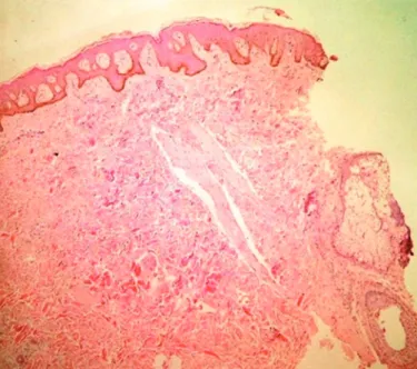
to the entire trunk, arms and legs during ten years. He used many topical therapies that were ineffective. The patient reported that a hairy brownish skin lesion also appeared on the upper back portion of his body one year prior to his admission. Biopsy specimens were obtained both from the brownish and depigmented lesions. The examination of the specimens obtained from the brownish area revealed acanthosis in the epidermis, elongation of the rete ridges, flattening at the base of the rete ridges, fusion of some of these rete ridges, melanocytic proliferation at the basal membrane, and bundles of smooth muscle unrelated to cutaneous adnexa in the dermis. Histomorphologic findings were consistent with BN (Figure 2). The histopathological examination of the specimen obtained from the depigmented area did
not show melanocytes and was consistent with vitiligo (Figure 3). The patient did not report additional complaints, and other tests showed normal findings for the nerves, muscles, and skeletal system.
Discussion
Vitiligo is an acquired disorder characterized by depigmented macules and skin patches with determined margins. Various hypotheses have been introduced although its etiology is still uncertain. Among these, autoimmune destruction of melanocytes is the most extensively studied hypothesis. Both humoral and cellular immunity have been implicated. The presence of anti-organ antibodies in vitiligo and detection of antibodies recognizing melanocytes in the sera of patients supports the role of humoral immunity; however, the presence of infiltration rich from CD8+ T cells in perilesional skin sites suggests the role of cellular immunity. Many other hypotheses have been suggested including intrinsic defects in the structure and functions of melanocytes, impairment in the free radical system, shortening in the life span of melanocytes, and changes in the membranes of the melanocytes1.
BN is characterized by hyperpigmented macules, patches, and verrucous plaques, which show unilateral occurrence and are accompanied by hypertrichosis in various degrees. The lesions often occur in the second and third decades of life. The frequency is six-fold higher in males. The disease is often acquired but can be also congenital. The lesion most commonly affects the upper quadrants of the back and front side of the body and shoulders and less commonly affects the face, neck, lower portion of the body, extremities, and gluteal area. The pathogenesis of this disorder is also not clear. However, this lesion is considered to be a hamartoma harboring ectodermal and mesodermal tissues. An increase in androgen receptors has also been suggested. This argument also explains the more common occurrence in males, onset at puberty and afterwards, hypertrichosis, dermal thickening, and presence of hypertrophic sebaceous glands2,3. The lesions sometimes
occur together with developmental anomalies of the muscles, skeleton and/or skin such as ipsilateral hypoplastic breasts, scoliosis, pectus excavatum, ipsilateral hypoplastic extremities, odontomaxillary hypoplasia; this condition is referred to as BN syndrome3,4. In addition,
Yalçınkaya İyidal et al.
Co-occurrence of Vitiligo and Becker's Nevus
Figure 3. There is complete absence melanocytes depigmented area
(hematoxylin x100)
Figure 1. A brownish patchy adjacent to the depigmented patch
lesion on the upper back portion of the trunk at the midline
Figure 2. Acanthosis in the epidermis, elongation of the rete ridges,
flattening at the base of the rete ridges, fusion of some of these rete ridges, melanocytic proliferation at the basal membrane, and bundles of smooth muscle unrelated to cutaneous adnexa in the dermis (hematoxylin x40)
www.turkderm.org.tr
Turkderm - Arch Turk Dermatol Venerology 2016;50
lichen planus, granuloma annulare, basal cell carcinoma, malignant melanoma, lymphangioma, and osteoma cutis have been reported as cutaneous lesions accompanying BN5,6. When the literature was
reviewed for the publications reporting co-occurrence of vitiligo and BN, the case reported by Gupta et al.6 showed co-occurrence of vitiligo,
BN, and lichen planus.
In the present case, vitiligo lesions first appeared when the patient was 9 years old and the BN lesion appeared ten years after the first manifestation of vitiligo lesions. A hypothesis to explain this occurrence could not be suggested due to the extremely rare co-occurrence of these two lesions. The cytokines released by CD8+ T lymphocytes in perilesional skin sites in vitiligo lesions or previously applied topical therapies may have triggered the development of BN. Herein, we report a case of patient who developed vitiligo and Becker nevus simultaneously. Additional studies are required to find an explanation for this condition.
Ethics
Informed Consent: Informed consent form was obtained from all patients.
Peer-review: Externally peer-reviewed.
Authorship Contributions
Surgical and Medical Practices: Ayşegül Yalçınkaya İyidal, Özge Çokbankir, Concept: Ayşegül Yalçınkaya İyidal, Arzu Kılıç, Design:
Ayşegül Yalçınkaya İyidal, Data Collection or Processing: Ayşegül Yalçınkaya İyidal, Analysis or Interpretation: Ayşegül Yalçınkaya İyidal, Arzu Kılıç, Literature Search: Ayşegül Yalçınkaya İyidal, Writing: Ayşegül Yalçınkaya İyidal.
Conflict of Interest: No conflict of interest was declared by the authors. Financial Disclosure: The authors declared that this study received no financial support.
References
1. Jean Ortonne JP: “Vitiligo and Other Disorders of Hypopigmentation.” In: Bolognia JL, Jorizzo JL, Rapini RP, et al: editors. 2nd ed. Spain. Mosby Elsevier, 2008;913-20.
2. Karabacak E, Aydın E, Doğan B, Göker K: Genç Erkeklerde Becker Nevüs Prevalansı. Türkderm 2013;47:80-3.
3. Danarti R, König A, Salhi A, Bittar M, Happle R: Becker’s nevus syndrome revisited. J Am Acad Dermatol 2004;51:965-9.
4. Steiner D, Silva FA, Pessanha AC, et al: Do you know this syndrome? Becker nevus syndrome. An Bras Dermatol 2011;86:165-6.
5. Dasegowda SB, Basavaraj G, Nischal K, et al: Becker’s Nevus Syndrome. Indian J Dermatol 2014;59:421.
6. Gupta S, Gupta S, Aggarwal K, Jain VK: Becker nevus with vitiligo and lichen planus: Cocktail of dermatoses. N Am J Med Sci 2010;2:333-5.
Yalçınkaya İyidal et al. Co-occurrence of Vitiligo and Becker's Nevus