IN-VITRO GENERATED MEMORY-LIKE CD8
+T
CELLS ELICITED PRONOUNCED CYTOTOXIC
EFFECT UPON ANTIGEN SPECIFIC TCR GENE
TRANSDUCTION
A THESIS SUBMITTED TO
THE GRADUATE SCHOOL OF ENGINEERING AND SCIENCE OF BILKENT UNIVERSITY
IN PARTIAL FULFILLMENT OF THE REQUIREMENTS FOR THE DEGREE OF
MASTER OF SCIENCE IN
MOLECULAR BIOLOGY AND GENETICS
By
Hakan Köksal
ii
IN-VITRO GENERATED MEMORY-LIKE CD8+ T CELLS ELICITED PRONOUNCED CYTOTOXIC EFFECT UPON ANTIGEN SPECIFIC TCR GENE TRANSDUCTION
By Hakan Köksal September 2016
We certify that we have read this thesis and that in our opinion it is fully adequate, in scope and in quality, as a thesis for the degree of Master of Science.
__________________________________
İhsan Gürsel (Advisor)
__________________________________
Güneş Esendağlı
__________________________________
Özgür Şahin
Approved for the Graduate School of Engineering and Science:
____________________________________ Levent Onural
iii
Abstract
IN-VITRO GENERATED MEMORY-LIKE CD8
+T
CELLS ELICITED PRONOUNCED CYTOTOXIC
EFFECT UPON ANTIGEN SPECIFIC TCR GENE
TRANSDUCTION
Hakan Köksal
M.S. in Molecular Biology and Genetics Advisor: İhsan Gürsel
September, 2016
Cytotoxic T cells are the important arm of immune system against malignant and virus infected cells. APC activated cytotoxic T cells survey body for threats, recognize peptide-MHC class I molecules by TCR and eliminate targets by inducing apoptosis. Following a successful clearance of danger, these cells undergo a contraction phase which eliminates 90-95% of antigen specific CD8+ T cells. At this stage, surviving cells are named as memory T cells. Maintenance of these cells are primarily depends on homeostatic cytokines, IL-7 and IL-15. Surviving memory CD8+ T cells are especially important against recurrence of malignancies and reinfections due to their quick and potent immune response. Similar memory functions and characteristic can be achieved under lymphopenic
iv
environment where abnormally low levels of lymphocytes present. This phenomenon is called homeostatic proliferation and has been shown to promote tumor clearance, reverse anergy and improve immune response against pathogens. Similar to natural generation of memory T cells, homeostatic proliferation fundamentally depends on homeostatic cytokines. Recent studies demonstrated memory-like phenotype (CD44hiCD62Lhi) can be acquired in vitro by culturing naive CD8+ T cell in the presence of anti-CD3/28 and recombinant IL-15. These in vitro generated phenotype when transferred to a mice proved to have an augmented antitumor response in vivo. In the light of these findings, we aimed to improve in vitro generation of memory-like phenotype and make it more practical and use identified improvements in tumor therapy. Our first approach is the utilization of DCs, known to be one of the most potent IL-15 secreting cells, in a co-culture setup with naive T cells. Identification of correct PRRs to trigger innate immunity and hence providing an environment suitable for transferred CD8+ T cells, should improve overall immune response and antitumor potential of CD8+ T cell. To test this idea, we made an in vitro co-culture setup. Stimulating DCs with potent IL-15 inducing ligands, c-GAMP and LPS, we achieved to improve expression of IL-15 but failed to induce memory-like phenotype in co-culture. Still we observed CD8+ T cells acquire a memory-like phenotype with an increased cytotoxic potential in the presence of recombinant IL-15. As an alternative, CD8+ T cells retrovirally transduced with an IL-15 vector to enable them to produce their own IL-15. Transduction managed to induce a memory-like progression without any additional supplementation. Similar to supplementation, IL-15 transduction improved cytotoxic potential of CD8+ T cell. Our approach can be used against any antigen with an improved immune response, eliminate the necessity of cytokine supplementation and enhance in vivo persistence of the transferred cells.
v
Özet
İN-VİTRO ORTAMDA OLUŞTURULAN CD8
+T
HÜCRELERİNE ANTİJENE ÖZGÜ TCR GENİ
TRANSFER EDİLDİĞİNDE GÖZLEMLENEN
BELİRGİN SİTOTOKSİK ETKİLER
Hakan Köksal
Moleküler Biyoloji ve Genetik, Yüksek Lisans Tez Yöneticisi: İhsan Gürsel
Eylül, 2016
Sitotoksik T hücreleri immün sistemin özellikle virüs enfekte olmuş ve malignan hücrelere karşı en önemli elemanlarındandır. Antijen sunucu hücrelerin aktive ettiği sitotoksik T hücreleri vücudu tehditlere karşı tarar ve TCR reseptörü yardımı ile tanıyıp hedef hücreyi hücrede apoptoz indükleyerek ortadan kaldırır. Tanınan tehditler ortadan kalktığında, antijene özgü olan T hücreleri kontraksiyon adı verilen bir faza girerler. Bu faz esnasında antijene özgü olan CD8+ T hücrelerinde yaklaşık 90-95% oranında ölüm gözlenir. Hayatta kalan hücreler hafıza hücreleri olarak tanımlanır ve uzun vadeli immün sistemin en önemli uzantılarındandır. Hafıza hücrelerinin muhafazaları için homeostatik sitokinlerden olan IL-7 ve IL-15 en önemli unsurlardandır. Hafıza CD8+ T hücreleri daha hızlı ve etkili yanıt
vi
verebildikleri için tekrarlayabilecek rahatsızlıklara karşı önemli bir savunma potansiyeli sunmaktadır. Sitotoksik T hücreleri lenfosit miktarı düşük olan ortamlarda homeostazi sağlayabilmek amacı ile homeostatik proliferasyon adı verilen bir proliferatif tepki gösterirler. Bu tepki sonucu hafıza hücreleri fonksiyonu ve karakteristiği (CD44+CD62L+) gösterebilen hücrelere donüşebilirler. Geçtiğimiz yıllarda yapılan çalışmalar sonucu bu ortamın in vitro ortamda da taklit edilebileceği ve hafıza benzeri T hücrelerinin oluşturulabileceği gösterilmiştir. Doğal olarak üretilen hafıza hücrelerinde olduğu gibi hafiza benzeri hücrelerin üretiminde de en önemli unsur IL-15 olduğu gözlemlenmiş ve bu sayede üretilen hücrelerin kanser tedavisinde daha etkili olabileceği gösterilmiştir. Bu bulgular doğrultusunda yaptığımız araştırmalarda bu fenotipi oluşturma yöntemini daha pratik ve/veya daha etkili yapmayı hedefledik. Bulunacak potansiyel gelişmeler kanser tedavisinde test edilecek. İlk denenen yöntem, dendritik hücreleri çeşitli ligandlar ile stimüle ederek en efektif IL-15 salgılanmasına yol açan ligand tanımlanarak bunun in vivo ortamında verilen CD8+ T hücrelerinin hafıza fenotipine dönüşmesine etkisi olup olamayacağını incelemekti. Fakat gözlemlerimiz IL-15 miktarı ile bağlantısız bir şekilde aktif edilmiş dendritik hücrelerinin gerek total salgıları ile gerek ise kendisi ile inkübe edilmiş T hücrelerinin hafıza benzeri fenotiplerinde düşüş gözlemlenmiştir. İkinci yöntemimizde ise IL-15 ifade geni retroviral olarak T hücrelerine transfer edilmiştir. Bu hücreler anti-CD3/28 ile aktive edildiğinde IL-15 takviyesi yapılan hücrelere yakın anti tümör sitotoksik etki gözlemlenmiştir. Bizim yöntemimiz ile üretilecek hafıza hücreleri herhangi bir ekstra takviyeye ihtiyaç olmadan istenilen antijene karşı dizayn edilebilir. Bunun yanı sıra burada belirtilerek üretilen hücreler transfer edilen canlıda çok daha uzun süreler dayanabilir ve uzun vadede daha etkili anti tümör fonksiyonu sergileyebilirler.
vii
viii
Acknowledgement
First, I would like to express my sincere gratitude to my advisor Prof. İhsan Gürsel for the opportunity to work in his lab and for his continuous support, patience, motivation, guidance and encouragement.
I would like to express my wholehearted thanks to Asst. Prof. Dr. Özgür Şahin and Assoc. Prof. Dr. Güneş Esendağlı for accepting to become members of my thesis jury and sparing time to evaluate and improve my thesis.
I am lucky to be a part of the Gürsel group. I thank my fellow labmates: Fuat Cem Yağcı, Tamer Kahraman, Gizem Tinçer-König, Kübra Almacıoğlu, Begüm Han Horuluoğlu, Gözde Güçlüler, Defne Bayık, Muzaffer Yıldırım, Fehime Kara Eroğlu, Bamu Bayyurt and particularly to Begüm Yıldız and Alican Savaş for their support and friendship.
It was fun to be a part of the MBG family. I would like to express my gratitude to all my instructors and friends in graduate school for their companionship and assistance.
This journey would not have been possible without the support of my family. I would like to express my deepest love and thankfulness to especially my mother Ayten, my father Aziz, and my sister Dilvin. Additionally, I’d like to express my gratitude to my other mothers Nurten and Gülten for their invaluable and everlasting support. Their love is the most significant motivation for me, which makes my accomplishments meaningful.
Finally, and most importantly, I would like to thank my dearest wife Elif Senem. Her unwavering love, support, encouragement, patience were the foundation which the past five years of my life have been built on. Her tolerance to my everyday childish pranks is a
ix
testament in itself of her unyielding devotion. Thank you for transcending me into a new being with your love.
x
Table of contents
Abstract ... iii Özet ... v Acknowledgement ... viii Table of contents ... xList of figures ... xvi
List of tables ... xviii
Abbreviations ... xix
Chapter 1 ... 1
Introduction ... 1
1.1 The immune system ... 1
1.1.1 Innate immune system ... 1
1.1.2 Adaptive immune system ... 3
1.2 T cell development ... 5
1.3 CD8+ T cell mediated immunity ... 7
xi
1.3.2 T cell mediated cytotoxicity... 9
1.4 Long term immunity ... 12
1.4.1 Memory CD8+ T cells ... 13
1.4.2 Memory T cell generation under inflammation ... 13
1.4.3 Homeostatic proliferation induced memory CD8+ T cells ... 14
1.5 Cancer immunotherapy ... 15
1.6 Aim of the study ... 16
Chapter 2 ... 18
Materials and Methods ... 18
2.1 Materials ... 18
2.1.1. General Laboratory & Cell Culture Reagents and Materials ... 18
2.1.2 Recombinants and Other Agents ... 19
2.1.3 CpG ODNs ... 19
2.1.4 PRR Ligands ... 19
2.1.5 Flow Cytometry ... 20
2.1.6 Determination of Gene Expression ... 20
2.1.6.1 Primers ... 21
2.1.7 Reagents for ELISA ... 22
2.2 Solutions, Buffers and Culture Media ... 23
xii
2.2.2 Flow Cytometry Buffers ... 23
2.2.3 Agarose Gel Electrophoresis ... 23
2.2.4 ELISA Buffers ... 24 2.2.5 MACS Buffer ... 25 2.2.6 Bacteria Solutions ... 25 2.2.6.1 Liquid Broth ... 25 2.2.6.2 Agar Plate ... 25 2.3 Methods ... 26
2.3.1 Maintenance of cell lines ... 26
2.3.1.1 PHOENIX-ECO ... 26
2.3.1.2 B16-F10 ... 26
2.3.1.3 B16-OVA ... 26
2.3.1.4 B16-Blue IFNα/β ... 26
2.3.2 Thawing of cells and cryopreservation ... 27
2.3.3 Cell counting ... 27
2.3.3.1 Haemocytometer ... 27
2.3.3.2 Flow cytometer ... 28
2.3.4 Primary murine cell suspension preparation ... 28
2.3.4.1 Mouse spleen cell suspension preparation ... 28
xiii
2.3.5 Bone marrow derived dendritic cell generation ... 29
2.3.6 CD8+ T cell separation from mouse spleen ... 29
2.3.7 CD8+ T cell activation ... 30
2.3.8 Retrovirus collection from Phoenix-Eco cells ... 30
2.3.9 Generation of memory-like CD8+ T cells ... 31
2.3.10 Retroviral transduction of CD8+ T cells ... 31
2.3.11 CD8+ T cell:B16 co-culture and antigen specific lysis ... 32
2.3.12 Flow cytometry ... 32
2.3.12.1 Fixation of cells ... 32
2.3.12.2 Surface marker staining of cells ... 33
2.3.13 Gene expression determination ... 33
2.3.13.1 Total RNA isolation ... 33
2.3.13.2 cDNA synthesis ... 34
2.3.13.3 PCR ... 34
2.3.14 Gene cloning ... 35
2.3.14.1 2-step PCR... 35
2.3.14.2 Agarose Gel Electrophoresis ... 37
2.3.14.3 Restriction enzyme digestion ... 37
2.3.14.4 Gel extraction ... 37
xiv
2.3.14.6 Transformation ... 38
2.3.14.7 Plasmid DNA purification ... 38
2.3.15 Cytokine ELISA... 39
2.3.16 B16 IFN-α/β reporter assay... 40
Chapter 3 ... 41
Results ... 41
3.1 Efforts to induce memory-like phenotype by DC co-culture ... 41
3.1.1 Identification of potential ligands to induce IL-15 secretion from DCs ... 41
3.1.2 Regardless of IL-15 expression, DCs doesn’t support memory-like progression in co-culture setup ... 45
3.2 Efforts to induce memory-like phenotype by DC conditioned medium ... 47
3.2.1 Regardless of IL-15 amount, DC conditioned medium supports effector phenotype progression rather than memory-like phenotype ... 47
3.2.2 Antibody activated CD8+ T cells elicit pronounced antigen specific cytotoxic activity upon TCR gene transduction ... 48
3.3 Efforts to induce memory-like phenotype by transducing CD8+ T cells with IL-15 expression vector ... 52
3.3.1. Antibody activated CD8+ T cells without additional IL-15 supplementation elicit pronounced antigen specific cytotoxic activity upon dual transduction of OT-I TCR gene and OT-IL-15 expression vector ... 52
Chapter 4 ... 63
xv
Chapter 5 ... 66
Discussion ... 66
Bibliography ... 67
xvi
List of figures
Figure 1.1: Nucleic acid recognizing receptors ... 2
Figure 1.2: Naïve CD4+ T cells differentiate into variety of subsets. ... 4
Figure 1.3: Diagram of T cell development ... 6
Figure 1.4: Under different conditions TCR engagement leads to separate T cell fate. ... 8
Figure 1.5: FAS ligand expressed on CD8 T cells induce apoptotic cell death upon interacting with FAS receptor (CD95) on target cell ... 10
Figure 1.6: Granzyme B induced cell death pathways. ... 11
Figure 1.7: CD8+ T cell fate upon infection or vaccination ... 12
Figure 1.8: An approach to adoptive transfer of engineered T cells ... 16
Figure 3.1: BMDC generation efficiency.. ... 41
Figure 3.2: CD8+ T cells isolation efficiency ... 42
Figure 3.3: Comparison of IL-15 mRNA transcripts in total RNA of CD11c+ BMDC. .. 43
Figure 3.4: IL-6, IL-12 and IFN-α/β production from stimulated DCs.. ... 43
Figure 3.5: DC activation is confirmed by FACS analysis. ... 44
Figure 3.6: Strongest IL-15 inducer decreases memory-like T cell phenotype regardless of peptide pulse or IL-15 supplementation. ... 46
xvii
Figure 3.7: DC conditioned mediums favored effector phenotype regardless of IL-15 supplementation. ... 48
Figure 3.8: OT-I TCR retroviral vector can efficiently be transduced to CD3/CD28
antibody activated CD8+ T cell. ... 50
Figure 3.9: rIL15 supplementation improves IFN-γ secretion in CD8+ T cell : B16 co-culture. ... 50
Figure 3.10: rIL15 enhences overall cytotoxicity potential of CD8+ T cells. ... 51
Figure 3.11: Retroviral vector transduction efficiency can be improved by concentration of virus through ultra-centrifugation. ... 53
Figure 3.12: Induction of CD8+ T cells proliferation by rIL-15. ... 54
Figure 3.13: Dual transduction allows CD8+ T cells to produce rIL-15. ... 55
Figure 3.14: Transduction of IL-15 expression retroviral vector improves memory-like phenotype. ... 57
Figure 3.15: Transduction of IL15 retroviral vector improves IFN-γ secretion in CD8+ T cell : B16 co-culture indicated ratios. ... 59
Figure 3.16: Transduction of IL15 retroviral vector improves antigen specific lysis. ... 61
xviii
List of tables
Table 2.1: Commercial name of the CpG ODN names and their properties. ... 19
Table 2.2: Commercial name of the ligands and their sources. ... 20
Table 2.3: Commercial name of the antibodies and their properties. ... 20
Table 2.4: Primers used in confirming gene expression. ... 21
Table 2.5: Primers used in cloning procedure. ... 22
Table 2.6: List of mouse ELISA antibodies and recombinants. ... 23
Table 2.7: PCR protocol for gene expression determination. ... 35
xix
Abbreviations
OAS1 2’-5’-oligoadenylate synthetase 1
AIM2 Absent in melanoma 2
ADAR1 Adenosine deaminase acting on RNA 1
ALR AIM2-like receptors
ACK Ammonium-Chloride-Potassium
Ab Antibody
APC Antigen presenting cell
AIF Apoptosis-inducing factor
APAF1 Apoptotic protease-activating factor 1
BM Bone marrow
BSA Bovine serum albumin
CAR Chimeric antigen receptor
JNK c-jun N-terminal kinase
CD Cluster of differentiation
xx
CLR C-type lectin receptor
cGAS cyclic GMP-AMP synthetase
cGAMP Cyclic guanosine monophosphate–adenosine monophosphate
CTL Cytotoxic T lymphocyte
CTLA4 Cytotoxic T lymphocyte antigen 4
DC Dendritic cell
DNA Deoxyribonucleic acid
DMSO Dimethyl sulfoxide
DN Double negative
DP Double positive
ddH2O Double-distilled water
DMEM Dulbecco's Modified Eagle's Medium
EndoG Endonuclease G
E.coli Escherichia coli
EtOH Ethanol
EDTA Ethylenediaminetetraacetic acid
ERK Extracellular signal-regulated kinase
FASL FAS ligand
xxi
FBS Fetal bovine serum
GMC-SF Granulocyte macrophage colony-stimulating factor
RAC Guanine triphosphatase
HSC Hematopoietic stem cell
ISG IFN stimulated gene
IRF3 IFN-regulatory factor 3
ITAM Immunoglobulin family tyrosine-based activation motif
IFN Interferon
IL Interleukine
KIR Killer inhibitory receptor
LPS Lipopolysaccharide
LB Lysogeny broth
MHC Major histocompatibility complex
MDA5 Melanoma differentiation associated gene 5
mRNA messenger ribonucleic acid
MAP Mitogen-activated protein
NK Natural killer
NLR NOD-leucine rich repeat receptors
xxii
NOD Nucleotide-binding oligomerization domain
ODN Oligonucleotide
PRR Pattern recognition receptor
PBS Phosphate-buffered saline
PI3K Phosphatidylinositol 3-kinase
PLCγ1 Phospholipase Cγ1
PCR Polymerase chain reaction
PD1 Programmed death 1
PI Propidium Iodide
PKB Protein kinase B
PKR Protein kinase R
RAG Recombination activating gene
RNA Ribonucleic acid
RLR RIG-I like receptor
RT Room Temperature
RPMI Roswell Park Memorial Institute
STING Stimulator of interferon genes
Th T helper
xxiii
TLR Toll-like receptor
TAE Tris-Acetate-EDTA
1
Chapter 1
Introduction
1.1 The immune system
The immune system is a vast network consisting of physical barriers, soluble factors and leukocytes which contribute to host resistance to both altered-self and non-self-threats [1]. According to specificity, there are two main branches of immunity named as innate and adaptive immunity. Innate immunity recognizes molecular patterns of pathogens (and in some cases even self danger signals) and initiates an orchestrated signaling cascades leading to primarily eradication of the insult. Another important feature of the innate immune system is to instruct effective adaptive immunity. Adaptive immunity, unlike innate doesn’t rely on already established molecular patterns rather learns to recognize unique antigen(s) or antigenic epitopes and launches an immune response accordingly. One of the distinct differences between innate adaptive immunity is that the cells of the adaptive immunity are professional cells that can memorize the antigenic epitopes and remember same antigen upon second exposure [2].
1.1.1 Innate immune system
Innate immune system consists of variety of cell types such as neutrophils, macrophages, dendritic cells (DC), basophils, eosinophils, mast cells and natural killer (NK) cells. These cells provide the first line of defense against pathogens. These cells
2
express pattern recognition receptors (PRR) which enable them to recognize pathogen and danger-associated molecules. Therefore, innate immune system can be able to differentiate from self to non-self or altered-self [3]. PRR consist of several receptor types; Toll-like receptors (TLR), C-type lectin receptors (CLR), nucleotide-binding oligomerization domain-leucine rich repeat containing receptors (NLR), AIM2-like receptors (ALR), RIG-I like receptors and cytosolic DNA sensors [4-5]. Through their PRRs, innate cells are able to recognize microbial patterns, including lipids, glycoproteins, nucleic acids or polysaccharides that exist in fungi, parasites, bacteria and virus [4-7].
Figure 1.1: Nucleic acid recognizing receptors [12].
Nucleic acid sensors can be categorized as immune sensing receptors and nucleic acid receptors. The first category consists of TLR family, RIG-I like receptor (RLR) family, melanoma differentiation associated gene 5 (MDA5), absent in melanoma 2 (AIM2) and cyclic GMP-AMP synthetase (cGAS). Among TLR family, TLR3 recognizes dsRNA, TLR7-8 recognizes ssRNA and CpG-DNA recognized by TLR9. However, TLR family doesn’t only recognize nucleic acids; TLR1-2, TLR2-6 responds to lipopeptides, TLR4 responds to lipopolysaccharides (LPS) and TLR5 recognizes flagellin [8]. In RLR family, RIG-I responds to 5’-triphosphate dsRNA, MDA5 also responds to dsRNA but if the length is greater than 2000 nucleotides [9]. Both AIM2 and cGAS recognizes dsDNA; AIM2 initiates inflammasome formation whereas cGAS responds through stimulator of interferon genes (STING) receptor [10-11]. Described
3
receptors either directly or indirectly promotes transcription factors such as nuclear factor- κB (NF- κB) and IFN-regulatory factor 3 (IRF3) which induces antiviral immune response by type I IFN and IFN stimulated genes (ISGs) [12]. Second category receptors, nucleic acid receptors, unlike PRRs they primarily depend on type I IFN and/or PRR signaling. Nucleic acid receptors have a more direct antiviral immunity and consists of protein kinase R (PKR), 2’-5’-oligoadenylate synthetase 1 (OAS1) and adenosine deaminase acting on RNA 1 (ADAR1) [12]. Similar to immune sensing receptors, nucleic acid receptors contribute to the antiviral immune response with a more direct approach such as translation inhibition or chemical modification and degradation of RNA threats [13].
Recognition of these patterns leads to an inflammatory response which aims to contain and eradicate the source of danger utilizing phagocytosis, pro-inflammatory mediators, recruitment and activation of innate and possibly the adaptive immune system [14].
1.1.2 Adaptive immune system
An adaptive immune response is induced if innate immune system is overwhelmed by an infection. Even though, this happens very rarely innate defense mechanism cannot stop the replication of pathogen and antigen accumulates. The environmental changes by innate immunity combined with antigen accumulation induces adaptive immune system. Two major sets of cells, B and T lymphocytes, can induce antigen specific response in adaptive immune system.
B lymphocytes are the precursors of antibody-secreting plasma cells. Generally, naïve B lymphocytes are activated by antigen and T helper (Th) cells which then differentiated into plasma cells. Secreted antibodies have different ways to contribute adaptive immunity. Certain pathogens requires specific molecules on target cells to enter the cells. Antibodies can bind to pathogen and neutralize them by blocking the binding sites. Moreover, antibodies can coat the surface of a pathogen to make them a target for phagocytosis. Finally, antibodies can initiate complement system after coating the surface of a pathogen. Complement proteins opsonize the pathogen by interacting the complement receptors on phagocytic cells [15].
T lymphocytes completes their development in thymus and enters into circulation. Mature T cells that have not confront to an antigen are called naïve T cells. They
4
circulate back and forth between peripheral lymphoid tissues and blood. Upon antigen encounter, naïve T cells differentiate into several different functional subsets. Naïve CD8+ T cells recognize antigen through pathogen peptides presented by MHC class I molecules. On recognition, naïve cells differentiate into effector CD8+ T cells which are capable of killing the infected cells. Therefore, CD8+ T cells play roles in limiting tumor development and growth and controlling intracellular pathogen infections [16].
Figure 1.2: Naïve CD4+ T cells differentiate into variety of subsets [17].
Upon activation, naïve CD4+ T cells differentiate into several distinct subsets of Th
cells; Th1, Th2, Th17 and iTreg. These subsets can be categorized according to their cytokine secretion patterns and associated transcription factors. (Figure 2) Th1 cells mainly contributes to macrophage and CD8+ T cell mediated immunity whereas Th2 cells are related to humoral immune system where they stimulate B cells. On the other side, Th17 contributes to protection at mucosal surfaces and its dysregulation might lead to several autoimmune disorders. On the contrary, Treg cells demonstrate immosuppressive properties in order to prevent autoimmune diseases and keep the immune response in check. In general, CD4+ T cells play roles in helping B cells in
5
antibody production, improve response of CD8 T cells and keep the autoimmunity in check [18].
1.2 T cell development
Multipotent hematopoietic stem cells are the precursors of T lymphocytes located in bone marrow. Progenitor cells migrate from bone marrow to thymus for maturation. That is why name of these cells are T lymphocytes. Thymus supplies necessary survival environment to support the differentiation of T cells from Hematopoietic stem cell (HSC)-derived lymphocyte progenitors [19]. These cells don’t express any of T cell surface markers when they enter into the thymus. Because they don’t express CD8 and CD4, they are called double negative (DN). At this point DNs can give rise to two distinct lineage of T cells. They either don’t express CD4 and CD8 at all and choose to become γ:δ T cells or express these markers and differentiate to α:β T cells [20]. DN stages takes place in outer cortex of the thymus and can be divided into four stages. (Figure 3) At the DN1 stage these cells express CD44 and Kit. When they acquire CD25 expression, they are called DN2 cells. Subsequently, expression of CD44 and Kit decreases and cells are named as DN3. At this stage if cells fail to rearrange T cell receptor β-chain locus, they can’t move to DN4 stage and die. Otherwise, they lose CD25 surface expression and proceed to DN4 stage [21]. Upon expression of pre-TCR, additional rearrangement stops and expression of CD8 and CD4 surface markers are induced. CD8 and CD4 commitment takes place in inner cortex of the thymus.
6 Figure 1.3: Diagram of T cell development [22].
At this step, cells are called double positive (DP) thymocytes. Initially, DP cells are large and have a huge proliferation capacity. Subsequently, proliferation rate decreases and cells become smaller. Consequently, α chain rearrangement begins which increases potential diversity. At this step small DP cells are tested against self-peptide: self-MHC complexes. If they don’t recognize, cells die. If self-peptide complex was successfully recognized, small DP cells mature and lose one of the surface markers becoming either CD4 or CD8 single positive thymocytes [23]. The lineage commitment, whether these cells become helper CD4+ or cytotoxic CD8+, is regulated by series of mechanisms
which include TCR and MHC interactions, transcriptional regulations and cytokine signaling [24].
7
1.3 CD8
+T cell mediated immunity
1.3.1 Priming of naïve T cells by antigen presenting cells
Immune response of T lymphocytes is one of the most important arm of whole immune system. Antigen presenting cells (APCs) display antigen peptides of infected or transformed cells via MHC class I on their surface to specifically target and activate cytotoxic T cells. Antigen recognition is a must for T cells to undergo proliferation and differentiation. Naïve T cells can transiently bind mature APCs while migrating through cortical region of the lymph node. One of the most important APC in T cell priming is dendritic cells (DCs) these cells can attach to LFA-1 and CD2 on T cells via their ICAM-1, ICAM-2 and CD58 receptors [25]. Naïve T cells mainly receive two types of signal which are depicted in Figure 1.4. These signals are categorized as activation and survival.
First signal is the recognition of specific peptide: MHC class I complex by T-cell receptors (TCR) and co-receptor CD8. Upon this recognition, SRC kinase Lck is activated and immunoglobulin family tyrosine (Y)-based activation motifs (ITAMs) are phosphorylated. Then, ZAP-70 is recruited and phosphorylated which in turn phosphorylates adaptor proteins. Following activation of phospholipase Cγ1 (PLCγ1) and the guanine triphosphatase (RAC), several mitogen-activated protein (MAP) kinases; extracellular signal-regulated kinase (ERK), c-jun N-terminal kinase (JNK) and p38 together with phosphatidylinositol 3-kinase (PI3K) and protein kinase B (PKB/Akt) are activated. All these pathways promote transcription factors related to T cell activation [26].
8
Figure 1.4: Under different conditions TCR engagement leads to separate T cell fate [26].
Second signal is co-stimulation, which is required for a pronounced immune response. One of the most well-known and effective co-stimulatory molecule is 44-kDa sized glycoprotein CD28. It is expressed by naïve and primed T cells and interacts with B7-1(CD80) and B7-2(CD86) on APCs. Co-stimulation is an indispensible since TCR recognition without a co-stimulator molecule leads to anergy (Figure 1). In general, anergic T cells aren’t able to proliferate, secrete survival factors like IL-2 and are unresponsive to immune stimuli [27]. CD28 signal improves T cell response in many ways. Production of a panel of cytokines is improved in T cells receiving CD28 stimulation both by mRNA stabilization and transcriptional activity [28]. In addition to that CD28 promotes T cell survival by upregulating BCL-XL, an anti-apoptotic BCL-2
family member [29]. Moreover, there are inhibitory signals that inactivate T cells such as cytotoxic T lymphocyte antigen 4 (CTLA-4), programmed death 1 (PD-1) and killer inhibitory receptors (KIRs). Of all, CTLA-4 is the most studied receptor expressed on activated T cells. CTLA-4 competes with CD28 as it binds to same ligand and prevents
9
T cells from receiving co-stimulation. (Figure 1.4) T cell activation is inhibited by both blocking CD28 from promoting positive signals and arresting T cells at the G1 phase of the cell cycle [30].
1.3.2 T cell mediated cytotoxicity
Activation of CD8+ T cells by APCs takes place in lymph nodes. Then, T cells enter into a rapid proliferation state and changes are observed in their surface markers. Initially, homing markers such as CD62L, a marker which allows T cells to recirculate through lymph nodes, is downregulated. Hence, T cells are allowed to leave lymph node and search for inflammation throughout the body. Moreover, integrin VLA-4 is expressed from activated T cells which allows T cells to recognize VCAM-1 expressed on inflamed tissues [31]. Therefore, activated T cells can identify and enter into the sites of inflammation.
CD8+ cytotoxic T cells also known as cytotoxic T lymphocytes (CTLs) are especially important against malignant cells and intracellular pathogens such as viruses. When cells are infected or transformed, they display antigen fragments with MHC class I complex which are recognized specifically by CD8+ cytotoxic T cells. APC activated T cells seek these cells and eliminate by inducing apoptosis. At first, discoveries led scientist to believe that changes in the cytoplasm of a CTL lead to granule exocytosis of a pore-forming protein perforin [32]. This idea is still valid today but more proteins are identified helping this phenomenon, such as granzymes.
Further studies revealed that CTLs can induce apoptosis not by just granule exocytosis of toxic materials. CTLs are also capable of inducing apoptosis through FAS, a tumor-necrosis factor receptor family member, pathway that doesn’t require pore forming material secretion onto the target cell surface [33]. Upon activation, CTLs express a ligand named FAS ligand (FASL) which can interact with FAS receptor on target cell. Stimulation of FAS receptor leads to recruitment of caspase-8 with the help of an adaptor protein called FAS-associated death domain protein (FADD). (Figure 1.5) Caspase-8 either directly activates other caspase members such as caspase-3 or cleaves BID, a pro-apoptotic BCL-2 family member to induce apoptosis. After cleavage, BID and BAX are translocated to mitochondria and induce release of cytochrome c. Finally,
10
through apoptotic protease-activating factor 1 (APAF1) caspase-9 is activated which in turn activates caspase-3 to induce apoptosis [34].
Figure 1.5: FAS ligand expressed on CD8 T cells induce apoptotic cell death upon interacting with FAS receptor (CD95) on target cell [33].
Perforin is exclusively expressed by CTLs and can be found in the cytoplasmic granules. Initially, perforin by itself is thought to be sufficient in killing cells due to its membrane damaging properties. However, it has been confirmed that even though perforin is capable of killing in vitro, they are not enough in vivo and requires other granule proteins including granzymes [35]. Granzymes are serine proteases released by cytoplasmic granules from CTLs. There are different types of granzymes that has been characterized, grazyme A,B, H, K and M. Granzyme B is the most studied and
well-11
characterized of all granzymes. It has been shown that granzyme B can be internalized by target cells via mannose-6-phosphate receptor with the help of perforin [36]. Once internalized, granzyme B can initiate cell death by variety of ways. (Figure 1.6) Similar to FAS-mediated cell death pathway, granzyme B can initiate apoptosis through caspase-3 directly or via caspase-8. Unlike FAS pathway, granzyme B promotes mitochondrial dysfunction which can potentially lead to necrosis, a more aggressive type of cell death which is not programmed unlike apoptosis [37-38]. Additionally, granzyme B can initiate a caspase-independent cell death through apoptosis-inducing factor (AIF) and endonuclease G (EndoG) [39].
Figure 1.6: Granzyme B induced cell death pathways [33].
In addition to killing function, cytotoxic T cells by secreting specific cytokines can induce immune response. Even though main way is to induce apoptosis to obliterate target cells, cytotoxic T cells can secrete IFN-γ and TNF-α in order to contribute host
12
defense. IFN-γ can inhibit viral replication and increase the expression of MHC class I molecules on adjacent cells which improves the chance of recognition by CTLs. Together with TNF-α, IFN-γ promotes macrophage recruitment and activation [40].
1.4 Long term immunity
Upon activation antigen specific CD8+ T cells initiate a proliferation cascade, only a single precursor is able to give rise to about 10,000 daughter cells in 5-8 days [41-42]. As explained in the previous section these cells leave the secondary lymphoid organs and seek for the signs of infection. Regardless of success in antigen specific CD8+ T cell immune response, number of antigen specific CD8+ T cells undergo a significant decline in all organs which is called contraction phase [41-42]. Mainly in contraction phase about 90-95% of antigen specific CD8+ T cells are eliminated to prevent a potential immunopathology. Even though, most of the antigen specific portion is eliminated, a small percentage always survives from the contraction in all tissues [43-44]. At this stage, these surviving stable antigen specific cells are called memory CD8+ T cells and they depend on homeostatic cytokines such as IL-7 and IL-15. (Figure 1.7) Memory T cells are able to induce a much more significant immune response to their specific antigen when it comes to recurrence of malignancies and reinfections [45].
13 1.4.1 Memory CD8+ T cells
Although contraction phase clears most of the antigen specific CD8+ T cells, number of surviving memory CD8+ T cells are stable. To keep them in a stable state, memory CD8+ T cells are maintained by a dynamic process depending on cytokine induced proliferation and a balanced death (memory turnover) [46-49]. Based on time and tissue which memory CD8+ T cell resides, functional and phenotypic properties of these cells may vary [50-51]. Most of the time homing molecules are analyzed to differentiate memory CD8+ T cell subsets. For effector memory T cells CD62LlowCCR7low phenotype is expected whereas in central memory phenotype expected phenotype is CD62LhiCCR7hi [52-53].
Following a successful immune response from CD8+ T cells may clear all pathogens and threats. Surviving contraction, cells acquire an early memory phenotype with low CD62L expression and IFN-γ secretion. (Figure 4) Eventually early memory cells are maintained by homeostatic proliferation related cytokines, IL-7 and IL-15 which are responsible from survival and proliferative signals respectively [49]. Depending on homeostatic proliferation related cytokines, cells acquire late memory phenotype with a higher CD62L expression and IFN-γ secretion. To keep the number of memory CD8+ T cells at a stable level, homeostatic cytokines driven basal proliferation is accompanied by a similar rate of cell death. (Figure 4) In opposed to a successful immune response, if T cells had to undergo contraction without clearing the threats, this leads to significant alterations in both functionality and phenotype of memory CD8+ T cells [54-55]. Chronic infections and inflammations might lead to deletion of CD8+ T cells of certain specificities and/or exhaustion of these cells’ effector functions [56].
1.4.2 Memory T cell generation under inflammation
Innate and adaptive system can’t be separated from each other. In general, innate immune system recognizes danger signals through their PRRs and initiates APCs to promote adaptive immune system [57]. In the meantime, innate immune cells produce variety of proinflammatory cytokines like IL-12 by macrophages and DCs, type I IFN by plasmacytoid DCs and IFN-γ by NK cells and CD8+ T cells [58]. It has been known that these proinflammatory signals are able to promote and sustain innate immune response but recently studies demonstrated that these cytokines can act directly on
14
CD8+ T cells [59-60]. Reduced expansion of antigen specific CD8+ T cells are observed when type I IFNs, IL-12 or IFN-γ receptors are deleted compared to wild-type CD8+ T cells [61-63]. Most importantly, studies demonstrated that proinflammatory cytokines especially IL-12 and IFN-γ influence the contraction phase and memory T cell generation. In BALB/c IFN-γ deficient mice reduced contraction is observed after LCMV infection [64-65]. Upon IL-12 receptor deletion contraction phase is limited in antigen-specific CD8+ T cells after infection [66]. Overall, these observations indicate
that proinflammatory cytokine composition would affect contraction phase which would directly impact memory generation.
1.4.3 Homeostatic proliferation induced memory CD8+ T cells
Under lymphopenic environment meaning abnormally low level of lymphocytes, T cells initiates a proliferative response to regain homeostasis. This phenomenon is called homeostatic proliferation and has been shown to promote tumor clearance, reverse anergy and improve immune response against pathogens [67-70]. Studies proved that when naïve CD8+ T cells transferred to a lymphopenic host, recombination activating gene (RAG) deficient mice, these cells undergo homeostatic proliferation and acquire a central memory-like phenotype indicated as CD44hiCD62Lhi. These memory-like CD8+ T cells demonstrate functional characteristics of antigen specific memory CD8+ T cells [71-72]. Conversion mechanism of naïve T cells into memory-like T cells are partially illuminated and categorized as two hits. First hit is the self-peptide-MHC recognition by TCR and second is the homeostatic proliferation related cytokines, IL-7 and IL-15 [73]. Adopting a memory-like phenotype following TCR recognition leads to improved immune response of CD8+ T cells against foreign peptide-MHC molecules [74-75].
Generation of memory-like CD8+ T cells would be helpful to induce a pronounced tumor rejection in adoptive cell therapies due to their augmented responsiveness [76-77]. Studies demonstrated that improving the degree of lymphopenia previous to adoptive transfer, allowing to T cells to acquire memory-like phenotype due to homeostatic proliferation, augments the clinical responses [78]. Even though these approaches are effective, they are fastidious especially if the person would like to collect these memory-like cells to use them directly in adoptive transfer. In order to improve the generation, Kaiser and his colleagues utilized many different approaches
15
to mimic this conversion in vitro [79]. Naturally, homeostatic proliferation is observable in secondary lymphoid tissues. For that reason, they incubated T cells on three-dimensional collagen coated ceramic replica of lymph node in the presence of IL-15 and IL-7. Hence, they discovered that IL-7 and lymph node are dispensable. Therefore, they claimed that culturing CD8+ T cells in the presence of IL-15 would be suffice for them to acquire memory-like phenotype which improves their antitumor potential [79].
1.5 Cancer immunotherapy
In the industrialized countries cancer malignancies are one the most life-threatening diseases. Even though ingrained cancer therapies such as resection of primary tumor, chemotherapy and radiotherapy, still 1 out of 4 patients dies because of the cancer [80]. In addition to already established treatment methods, new methodologies arise such as cancer immunotherapy. In opposed to other therapeutic methods, immunotherapy aims to prevent the occurrence and the recurrence of the cancer. There are two major arms of immunotherapy, active and passive. Main aim of the active immunotherapy is the induction of long-lasting antigen specific immune response [81]. Mostly this approach involves a type of vaccination which induces host’s immune response against a specific tumor antigen either an already established one or identified from patient’s biopsy [82-84]. Additionally, active immunotherapy might be a more general approach involving unspecific stimulations induced by adjuvants and cytokines. On the other hand passive immunotherapy involves supplementation of one’s own tumor specific effector cells. Unlike active immunization, passive immunotherapy is more potent but short lived and fastidious [80]. One of the most famous passive immunotherapy branch is the adoptive T cell therapy involves collecting patients’ T cells, teaching them to recognize a specific antigen and transferring back to the patient. (Figure 1.8)
16
Figure 1.8: An approach to adoptive transfer of engineered T cells [85].
T cells can be engineered in many ways. An approach is the patient material, T cells collected from patient’s cancer tissue are tested against that particular tumor. The ones with the best immune response are separated and their TCRs are cloned into a retroviral or lentiviral vector. In this approach, patient’s T cells are transduced with this vector to enable autologous T cells to recognize patient’s tumor. In another approach chimeric antigen receptor (CAR) vectors are utilized. These vectors consists of monoclonal antibody which enables T cells to recognize antigens without MHC restriction, TCR intracellular regions to induce T cell activation and activating motifs to eliminate the necessity of co-stimulatory molecules [85-86]. Considering immunotherapies can be used together with already established treatments such as chemotherapy and radiotherapy, novel immunotherapeutic approaches offer flexibility and a better treatment chances to the field.
1.6 Aim of the study
Considering the observation of Kaiser and his colleagues, we embarked on a project to improve the approach of memory-like T cells generation, make it more practical and then exploit the improvements in tumor therapy. Our first approach is to utilize bone marrow derived DCs, known to be one of the most potent IL-15 expressing cells, in a co-culture setup with T cells. Stimulating DCs with proper ligand or ligand combinations could induce pronounced IL-15 secretion that should then help to elicit memory-like T cells conversion. Therefore, we may use most potent ligands alongside of activated T cells in vivo. By this approach, we could both stimulate innate immune system to raise a response against cancer and create and environment which would help memory-like conversion and improve adoptively transferred T cells’ antitumor
17
response. Next as an alternative approach, we utilized retroviral transduction to enable T cells to express their own IL-15 to induce memory-like conversion. Consequently, memory-like phenotype generation could be improved by transducing CD8+ T cells with IL-15 expression vector without any extraneously secreted IL-15 or recombinant IL-15 usage. In future, this phenotype might be exploited in tumor therapy and might provide a viable alternative eliminating some of the limitations of classical adoptive T-cell therapy.
18
Chapter 2
Materials and Methods
2.1 Materials
2.1.1. General Laboratory & Cell Culture Reagents and Materials
Cell culture mediums DMEM and RPMI-1640, alongside of PBS, FBS, L-glutamine and 2-mercaptoethanol were purchased from Gibco, USA. For cell counting Zap-OGLOBIN® II Lytic reagent and Z-PAK Isoton II diluent used and purchased from Beckman Coulter, USA. Mouse Pan T, CD8 T cell magnetic isolation kits and all MACS equipments were purchased from Miltenyi Biotech, Germany. Other cell culture reagents Pen-Strep, Na-Pyruvate, HEPES, NEAA and Trypsin were purchased from Lonza, Switzerland. Another cell counting reagent was Trypan Blue and together with Hypure Molecular Biology Grade Water were purchased from Hyclone. Plastic materials utilized mainly in cell culture such as plates, flasks, scrapers, serological pipettes and filters purchased from Corning Life Sciences Inc., USA. In order to freeze and store the cells, Mr. Frosty container was used and purchased from Thermo Scientific, USA and 2 ml cryovials were purchased from Greiner bio-one, Austria.
19 2.1.2 Recombinants and Other Agents
Recombinants used in bone marrow derived dendritic cell generation, mouse IL-4 and GM-CSF were purchased from Tonbo, USA. Recombinant used in T cell culture, mouse IL-15 was purchased from Biolegend, USA. T cell activation materials such as anti-CD3 and anti-CD28 were purchased from Tonbo, USA.
2.1.3 CpG ODNs
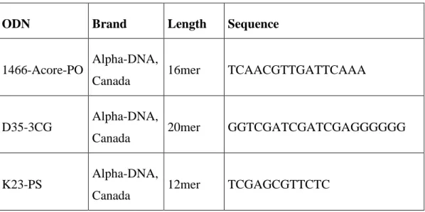
Sequences of CpG ODNs supplied from Alpha-DNA. Their length and sequences were listed in Table 2.1.
Table 2.1: Commercial name of the CpG ODN names and their properties.
2.1.4 PRR Ligands
Ligands used for DC stimulations were listed in Table 2.2.
Ligand Source Brand
c-di-GMP Synthetic Biolog, Germany
3’3’ cGAMP Synthetic Invivogen, USA
LPS E. coli Sigma, USA
3’3’ cGAMP Synthetic Invivogen, USA
ODN Brand Length Sequence
1466-Acore-PO Alpha-DNA,
Canada 16mer TCAACGTTGATTCAAA
D35-3CG Alpha-DNA,
Canada 20mer GGTCGATCGATCGAGGGGGG
K23-PS Alpha-DNA,
20
Poly(da:dt) Synthetic Invivogen, USA
R848 Synthetic Invivogen, USA
Table 2.2: Commercial name of the ligands and their sources.
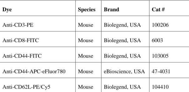
2.1.5 Flow Cytometry
Staining antibodies used in flow cytometry analysis were listed in Table 2.3. In order to perform cell fixation Medium A was used and purchased from Invitrogen, USA.
Dye Species Brand Cat #
Anti-CD3-PE Mouse Biolegend, USA 100206
Anti-CD8-FITC Mouse Biolegend, USA 6003
Anti-CD44-FITC Mouse Biolegend, USA 103005
Anti-CD44-APC-eFluor780 Mouse eBioscience, USA 47-4031
Anti-CD62L-PE/Cy5 Mouse Biolegend, USA 104410
Table 2.3: Commercial name of the antibodies and their properties.
2.1.6 Determination of Gene Expression
In order to perform RNA isolation, Trizol reagent were used and purchased from Life Technologies, USA. For cDNA synthesis, ProtoScript M-MulV cDNA Synthesis Kit and OneTaq Quick-Load 2x Mastermix were used and purchased from NEB, USA. High fidelity polymerases used in cloning procedure such as Phusion HF DNA polymerase and purchased from NEB, USA. DNA laders and 6X loading dye were used in PCR analysis and purchased from Fermentas, USA.
21 2.1.6.1 Primers
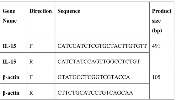
Primers used to confirm gene expression were listed in Table 2.4 and designed as follows. Mouse cDNA sequences at EnsemblTM database were used and primers were
designed by using Primer3 Input v.0.4.0 program
(http://frodo.wi.mit.edu/primer3/input.htm). Primer sequences designed to perform cloning were listed in Table 2.5. Original murine OT-I TCR retroviral vector (Plasmid #52111) was purchased from Addgene, USA.
Gene Name
Direction Sequence Product
size (bp) IL-15 F CATCCATCTCGTGCTACTTGTGTT 491 IL-15 R CATCTATCCAGTTGGCCTCTGT β-actin F GTATGCCTCGGTCGTACCA 105 β-actin R CTTCTGCATCCTGTCAGCAA
Table 2.4: Primers used in confirming gene expression.
Sequence Name Direction Sequence
EcoR-I/IL-15 F GCGCCAGAATTCACCATGAAAATTTTGAAAC CATATATG Xho-I/IL-15 R GCGTCGCTCGAGTCAGGACGTGTTGATGAAC EcoR-I/TCR-α F CAGCCCTCACTCCTTCTCTAGGCGCCGGAATT CACCATGGACAAGATCCTGACAGCATC TCR-β/T2A R CCAGCCTGCTTCAGCAGGCTGAAGTTAGTAG CTCCGCTTCCGGAATTTTTTTTCTTGACC
22
T2A/IL-15 F CTGCTGAAGCAGGCTGGAGACGTGGAGGAG
AACCCTGGACCTACCATGAAAATTTTGAAAC CATATATGAGGAATACATC
IL-15/Xho-I R GCGTCGCTCGAGTCAGGACGTGTTGATGAAC
Table 2.5: Primers used in cloning procedure.
2.1.7 Reagents for ELISA
To perform ELISA, 2HB plates were used and purchased from SPL Life Sciences, Korea. ELISA antibodies were purchased either from Biolegend, USA or Mabtech, Sweden. SA-ALP was purchased from Mabtech, Sweden as well. To develop PNPP Tablets and 5X Diethanolamine Buffer were used and purchased from Thermo Scientific, USA. Detailed information of antibodies and recombinant proteins were listed on Table 2.6.
Antibody Name Brand Cat # Working concentration
Anti-IFN-γ Ab Mabtech,
Sweden
3321-1a-20 1 μg/ml in PBS
IFN-γ Biotinylated Ab Mabtech, Sweden
3321-1a-20 1:1000 diluted in T cell buffer
Recombinant IFN-γ Biolegend, USA
575309 80 ng/ml in Blocker
Anti-IL-15 Ab LSBio,
USA
LS-C104510 1 μg/ml in PBS
IL-15 Biotinylated Ab LSBio, USA
23 Recombinant IL-15 Biolegend,
USA
566302 200 ng/ml in Blocker
Table 2.6: List of mouse ELISA antibodies and recombinants.
2.2 Solutions, Buffers and Culture Media
2.2.1 Cell Culture Media
High Glucose DMEM and RPMI-1640 (Lonza)
2, 5 or 10% FBS inactivated at 55 °C
50 g/ml Penicillin/Streptomycin
10 mM HEPES
0,11 mg/ml Na Pyruvate
1% Non-Essential Amino Acids Solution
2 mM L-Glutamine
Ingredients dissolved in 500 ml medium
Storage temperature: +4 °C 2.2.2 Flow Cytometry Buffers PBS-BSA-Na azide Buffer
500 ml 1x PBS
5g BSA (1%)
125 mg Sodium Azide (0.25%)
Storage temperature: +4 °C 2.2.3 Agarose Gel Electrophoresis 50X TAE (Tris-Acetate-EDTA)
242 g Tris (C4H11NO3)
24
57.1 ml Glacial acetic acid
Ingredients dissolved in 1 lt ddH2O and autoclaved
Storage temperature: +4 °C Working dilution: 1X 2.2.4 ELISA Buffers Blocking Buffer 500 ml 1x PBS 25 g BSA (5%) 250 μl Tween20 (0,025%) Storage temperature: -20 °C T-cell Buffer 500 ml 1x PBS 25 ml FBS (5%) 250 μl Tween20 (0,025%) Storage temperature: -20 °C Wash Buffer 500 ml 10x PBS 2.5 ml Tween20 4.5 lt ddH2O Storage temperature: +4 °C 10X PBS (Phosphate Buffered Saline)
80 g NaCl 2 g KCl 8,01 g Na2HPO4 . 2H2O 2 g KH2PO4 1 lt ddH2O pH adjustment: 6.8
25
Storage temperature: Room temperature 2.2.5 MACS Buffer 0.5% BSA 2mM EDTA Ingredients dissolved in 1X PBS Storage temperature: +4 °C 2.2.6 Bacteria Solutions 2.2.6.1 Liquid Broth 10 grams Tryptone
5 grams Yeast Extract
5 grams NaCl
Ingredients dissolved in 1lt ddH2O and autoclaved.
Storage temperature: +4 °C 2.2.6.2 Agar Plate
10 grams Tryptone
5 grams Yeast Extract
5 grams NaCl
10 grams of Agarose
Ingredients dissolved in 1lt ddH2O and autoclaved.
Chilled to ~50 °C and distributed to 20 ml per petri dish
26
2.3 Methods
2.3.1 Maintenance of cell lines 2.3.1.1 PHOENIX-ECO
PHOENIX-ECO (ATCC®, CRL-3214) is a human kidney epithelial derived cell line. Cells were sustained in a DMEM media with 10% regular FBS. Cells cultured for retrovirus packaging and production are seeded as 3x10⁶ cells on 10cm plates. These cells are stated as adherent but they are very fragile and can detach from the plate very easily. Sub-culturing performed at 90% confluence with 1X Trypsin. Cells collected at 300 g with 5 min centrifugation were seeded on appropriate density back to new plate.
2.3.1.2 B16-F10
B16-F10 (ATCC®, CRL-6475) is a murine skin melanoma cell line. These cells are strongly adherent. Sub-culturing of these adherent cells performed at %90 confluence with 1X Trypsin following fresh RPMI1640 supplemented with %10 FBS. Cells are centrifuged at 300g for 5 min and seeded on appropriate concentration back to new plate. If necessary, cells are counted by haemocytometer.
2.3.1.3 B16-OVA
B16-OVA is a murine skin melanoma cell line. This cell line is a variant of B16-F10 which stably expresses ovalbumin protein. Similar to B16-F10 these cells are strongly adherent. Sub-culturing of these adherent cells performed at %90 confluence with 1X Trypsin following fresh RPMI1640 supplemented with %10 FBS. Cells are centrifuged at 300g for 5 min and seeded on appropriate concentration back to new plate. If necessary, cells are counted by haemocytometer.
2.3.1.4 B16-Blue IFNα/β
B16-Blue IFNα/β is a murine type I IFN sensor cell line. This line has the origin of C57BL/6 murine b16 melanoma cell line. Cells can respond to type-I IFN because this line is stably transfected by a SEAP reporter gene under the control of the IFNα/β-inducible ISG54 promoter. B16-Blue IFNα/β cell line can monitor the activation of JAK/STAT/ISGF3 and IRF3 pathway. Culturing and sub-culturing are performed in
27
RPMI1640 media supplemented with 10% FBS. Sub-culturing of these adherent cells performed at %90 confluence with 1X Trypsin following fresh RPMI1640 supplemented with %10 FBS. Cells are centrifuged at 300g for 5 min and seeded on appropriate concentration back to new plate. If necessary, cells are counted by haemocytometer.
2.3.2 Thawing of cells and cryopreservation
Cryovials are taken from liquid nitrogen and immediately put in 37 °C pre-heated water bath. Vials are gently swirled in a circular motion until they are melted. After the ice in the vials melted, cells are immediately transferred into a 15 ml falcon tube and supplemented with appropriate medium until the total volume is 15 ml. Afterwards, the falcon is centrifuged at 300 g for 5 min. Supernatant aspirated and the pellet is re-suspended with appropriate fresh complete medium. Then, cells are seeded on preferred plate or flask and placed into the cell culture incubator, which is set for 37 °C with 5% CO2.
Cryopreservation can be performed when cells reach around 60-80% confluency and 80-90% viability. Cells are collected with an appropriate method and centrifuged to remove the supernatant. Then the cell pellet is re-suspended in FBS which has a 10% DMSO content. Cell mixture is immediately transferred into labeled cryovials. Cryovials are placed in Ms. Frosty Freezing Container and placed to -80 °C fridges. At minimum 24 hours later cells are transferred into liquid nitrogen tanks to be able to store them for longer periods of time.
2.3.3 Cell counting 2.3.3.1 Haemocytometer
After the primary cells and adherent cell lines are washed, centrifuged and re-suspended in 1-10 ml of fresh media. 10 μl from cell mixture are mixed with 10 μl Trypan Blue to separate live cells from dead ones. 10 μl from this Trypan Blue-cell mixture are loaded on haemocytometer and cells are counted under light microscope. Live cells can be observed as bright dots and only these bright dots are counted to calculate the total number of living cells by considering the dilution factor.
28 2.3.3.2 Flow cytometer
After the primary cells and suspension cell lines are washed, centrifuged and re-suspended in 1-10 ml of fresh media. Generally, 20 μl of cell suspension are mixed 10 ml Isoton II Diluent buffer and 2-3 drops of ZAP-OGLOBIN II Lytic Reagent was added to lyse erythrocytes if necessary. This mixture was analyzed under Flow Cytometry. Live cells were gated, apoptotic cells or cell clusters were ignored. Total number of cells are calculated considering live cell gate and dilution factor.
2.3.4 Primary murine cell suspension preparation 2.3.4.1 Mouse spleen cell suspension preparation
Cervical dislocation used to sacrifice mice (C57/BL6). The left side of the sacrificed mouse wet by using %70 ethanol. Cut open the left side of the mouse, nearly half-way between front and back legs. Spleen removed by using forceps and scissors and transferred into %2 FBS supplemented complete RPMI-1640 media. The spleen was mashed using the syringe plunger into a petri dish and washed twice with 10ml fresh RPMI-1640 medium followed by a centrifugation step at 300 g for 5 minutes. At the final step, cells were resuspended in 1ml of %5 FBS supplemented RPMI-1640 to count by Flow Cytometry.
2.3.4.2 Bone marrow suspension preparation
Cervical dislocation used to sacrifice mice (C57/BL6). Sacrificed mice pinned down to dissection board and sterilized with ethanol. Incisions made on thigh and ankle, the skin was peeled down to ankle and removed from the leg. Muscle tissue was removed from each leg by using a surgical blade. Each leg completely separated and placed into ethanol for sterilization. Bones are further cleaned from remaining muscle tissues and separated from knee joints as femur and tibia. By holding each bone fragment with a tweezer bone marrow containing red part was identified and introduced a cut immediately before the identified part. Bone marrow was flushed out with 26g insulin syringe into the 15ml Falcon tube. %2 FBS supplemented RPMI-1640 media passed through the bone until the color turns from red to white. Collected marrow cells were centrifuged at 300g for 5 minutes. Red blood cells were lysed by resuspending marrow in 1ml of ACK lysis buffer and incubated for 2 minutes. Cells were washed with %2
29
FBS supplemented RPMI-1640 media and centrifuged at 300g for 5 minutes. Cells were resuspended in %10 FBS supplemented RPMI-1640 media to count by Flow Cytometry.
2.3.5 Bone marrow derived dendritic cell generation
Bone marrow cells are seeded as 1x10⁶ cells/ml into 10cm2 plate in RPMI-1640 supplemented with 10% FBS, 20ng/ml mGM-CSF and 10ng/ml mIL-4 recombinant proteins. Three days after initial seeding half of the medium aspirated from plate and replenished with fresh medium and supplements. Six days after initial seeding cells are collected. At this time point cells can be a bit adherent in order to detach them cold 1X PBS supplemented with 2mM EDTA can be used. Then, cells are stained with CD11b/CD11c and analyzed under Flow Cytometry to confirm their dendritic cell phenotype.
2.3.6 CD8+ T cell separation from mouse spleen
After mouse spleen suspension prepared and counted, ACK lysis performed. Generally, single spleen re-suspended and incubated for 2 minutes in 2 ml of ACK lysis buffer. In order to dilute lysis buffer 15 ml falcon filled with fresh RPMI-1640 and centrifuged at 300 g for 5 minutes. Most of the pellet should be observed as white colored. There might be a thin red layer above the white pellet that wouldn’t create a problem since all other cells are seperated. Supernatant was aspirated, re-suspended in fresh RPMI-1640 and cells are counted again with the help of Flow Cytometry. After calculating total number of splenocytes, pellet can be re-suspended in MACS buffer as 40μl buffer per 107 splenocytes. Then, 10μl of Biotin-Antibody Cocktail was added to per 107 total cells and incubated for 5 minutes in the refrigerator. After incubation, 30μl MACS buffer per 107 splenocytes was added to mixture. On top of that 20μl Anti-Biotin Microbeads are added to per 107 total cells and incubated for 10 minutes again in the refrigerator. Afterwards, cells are washed with 2 ml of MACS buffer and centrifuged and supernatant removed to rid of excess magnetic beads. Pellet was re-suspended in 500μl MACS buffer. In the meantime an MS column was prepared by washing with 500μl buffer. After washing was finished, splenocyte suspension were loaded on the MS column attached to a magnet. At this step cells are separated by magnetic force and drop down with the help of gravity. After the last drop of cell suspension, 500μl MACS
30
buffer was loaded on MS column in order to wash the column. This step performed twice. Then, flow through was centrifuged and cells are re-suspended in 1ml of RPMI-1640 supplemented with 50μM 2-mercaptoethanol in order to count them via Flow Cytometry. A percentage of separated cells are used in FACS staining, specifically for CD3 and CD8 markers, to calculate the separation efficiency.
2.3.7 CD8+ T cell activation
CD8+ T cells are activated through anti-CD3 and anti-CD28 murine antibodies. This is an artificial activation mimicking the APC cell contact. Activation of CD8+ T cells are a crucial part of retroviral transduction. It has been known that anti-CD3 is better when coated whereas anti-CD28 is better in soluble form. Therefore, anti-CD3 prepare in 1X PBS at 2μg/ml and discard on the wells in appropriate amounts. Plate can be coated overnight at 4°C or 1-2 hours at 37°C. Later on wells were flooded with 1X PBS two times to remove excess antibodies. Finally, PBS were removed and separated CD8+ T cells were placed on coated wells. As indicated above, anti-CD28 was added in soluble form at 1μg/ml concentration.
2.3.8 Retrovirus collection from Phoenix-Eco cells
At Day 0 cells are collected from the plate and counted via haemocytometer. For 10cm2 plates cells are seeded as 3x10⁶ cells/plate in 9 ml DMEM supplemented with 10% FBS. On the next day, transfection mixture is prepared. 12μg retroviral vector, 3μg pCL-Eco helper plasmid and 50μl 2.5M CaCl2 are mixed. Total volume is adjusted to 500μl ddH2O. Then, while vortexing the CaCl2 mixture, 500μl 2X HEPES are added drop wise. Total mixture incubated for 20 minutes at room temperature. Afterwards, mixture transferred on seeded Phoenix-Eco cells drop wise. After about 2 hours, small crystal like structures can be observed via 20X light microscope. If there are no small dots but rather big clumps, solutions must be checked and most probably the pH of the solutions are not proper. At Day 2, transfection mixture is aspirated, plate is washed with warm 1X PBS and 10 ml of fresh DMEM with 10% FBS is added on cells for virus production. At Day 3, exactly 24 hours later medium is collected, filtered via 0.45μm and stored at -80°C. Another set of virus production can be made with a lower titer. To do that cells are feeded with another set of fresh DMEM with 10% FBS and on the next day can be collected, filtered and stored. Collected Phoenix-Eco supernatant
31
can be subjected to ultracentrifugation to condense virus particles. Exactly at 1x10⁵ g supernatant can be centrifuged for 2 hours at 4°C. Pellet is visible if supernatant is more than 30ml. Supernatant is aspirated and pellet can be re-suspended in either 1X PBS or DMEM with %10 FBS according to the volume of choice. After this step dense virus aliquots can be stored at -80°C.
2.3.9 Generation of memory-like CD8+ T cells
CD8+ T cells are separated from spleen and seeded on anti-CD3 coated wells. Soluble anti-CD28 is added to each well. IL-15 mouse recombinant protein is added in designated wells as 200ng/ml concentration. Cells are incubated for 7 days in total when medium turns to yellow cells are diluted in 1:3 ratio and supplemented with previously indicated conditions. On day 7, cells are collected from wells. In general, T cells are suspension cells meaning their detachment can be simply performed by a 1ml pipet. If cells don’t detach from the well properly, cold 1X PBS or cold 1X PBS supplemented with 2mM EDTA can be used. After collection, cells are either fixed and stained or directly stained with CD3, CD44 and CD62L FACS antibodies to determine their phenotype. In general, articles suggest many other markers alongside of CD44 and CD62L such as PD-1, CD127, Ly6c and CD25. One of the most used combination determined for memory-like phenotype is CD44hiCD62Lhi whereas effector phenotype accepted as CD44hiCD62Llo and naïve phenotype as CD44loCD62Lhi.
2.3.10 Retroviral transduction of CD8+ T cells
CD8+ T cells separated from mouse spleen is seeded on coated 2μg/ml anti-CD3 wells and supplemented with 1μg/ml anti-CD28 in RPMI-1640 with 10% FBS and 50μM 2-mercaptoethanol. For 24-well plate, cells are seeded in 500μl medium. Cells are activated for 2 days with antibodies. On day 2, cells are collected and counted with Flow Cytometry to calculate total cell number. After calculation, cells are separated 5x10⁵ cells/well in 500μl retrovirus containing medium supplemented with 10μg/ml Polybrene. For optimum efficiency each vector should be subjected to titration. After seeding the cells, plate is placed in the incubator to allow cells to regain optimal temperature and CO2 percentage. In the meantime, Nüve NF 1200R centrifuge is set to 32°C. After about an hour, plate is removed from the incubator and immediately wrapped with parafilm to keep the pH stable. Plate can be centrifuged at 1500 g for 1.5
![Figure 1.1: Nucleic acid recognizing receptors [12].](https://thumb-eu.123doks.com/thumbv2/9libnet/5798723.118108/25.892.171.798.428.669/figure-nucleic-acid-recognizing-receptors.webp)
![Figure 1.2: Naïve CD4 + T cells differentiate into variety of subsets [17].](https://thumb-eu.123doks.com/thumbv2/9libnet/5798723.118108/27.892.180.793.316.729/figure-naïve-cd-t-cells-differentiate-variety-subsets.webp)
![Figure 1.4: Under different conditions TCR engagement leads to separate T cell fate [26]](https://thumb-eu.123doks.com/thumbv2/9libnet/5798723.118108/31.892.232.711.113.512/figure-under-different-conditions-tcr-engagement-leads-separate.webp)
![Figure 1.5: FAS ligand expressed on CD8 T cells induce apoptotic cell death upon interacting with FAS receptor (CD95) on target cell [33]](https://thumb-eu.123doks.com/thumbv2/9libnet/5798723.118108/33.892.222.645.202.722/figure-ligand-expressed-induce-apoptotic-interacting-receptor-target.webp)
![Figure 1.6: Granzyme B induced cell death pathways [33].](https://thumb-eu.123doks.com/thumbv2/9libnet/5798723.118108/34.892.213.600.410.883/figure-granzyme-b-induced-cell-death-pathways.webp)
![Figure 1.7: CD8+ T cell fate upon infection or vaccination [45].](https://thumb-eu.123doks.com/thumbv2/9libnet/5798723.118108/35.892.167.805.701.949/figure-cd-t-cell-fate-infection-vaccination.webp)