Received: 28.03.2019 Accepted: 23.09.2019
Published: 21.10.2019 http://www.polradiol.com
Original paper
The accuracy of 3T magnetic resonance cholangiopancreatography
in suspected choledocholithiasis
Önder Yeniçeri
A,B,D,E, Neşat Çullu
C,D,E, Burak Özşeker
A,B,D, Emine Neşe Yeniçeri
D,E,FFaculty of Medicine, Mugla Sitki Kocman University, Turkey
Abstract
Purpose: The purpose of this study was to investigate interobserver agreement during magnetic resonance cholangio pancreatography (MRCP) evaluation and the sensitivity and specificity of MRCP obtained with 3T scanners in cases of bile duct obstruction.
Material and methods: A total of 37 patients who had MRCP and endoscopic retrograde cholangiography (ERCP) were included. Choledochal pathology was divided into two groups regarding the presence of stones as “there is stone or not”. MRCPs were performed with a 3Tesla system using respiratory triggered HASTE technique in axial and coronal planes and with T2 SPACE sequence in the coronal plane. Sensitivity, specificity, negative predictive value (NPV), and positive predictive value (PPV) were calculated separately for each observer. The average of both observers was calculated for comparison with other studies.
Results: Thirtyseven patients were in the study population. Agreement between the observers was analysed, and Cohen’s κ value was evaluated as 0.84. For two observers, the sensitivity of MRCP was 93%, whereas the specificity was 75% for the first observer and 62% for the second.
Conclusions: In this study we found a high level of interobserver agreement in evaluating MRCP. MRCP has a high sensitivity in detecting choledocholithiasis.
Key words: cholelithiasis, ERCP, bile duct, MRCP.
Correspondence address:
Dr. Neşat Çullu, Faculty of Medicine, Mugla Sitki Kocman University, 48000, Mugla, Turkey, phone: +905072030472, e-mail: [email protected]
Authors’ contribution:
A Study design ∙ B Data collection ∙ C Statistical analysis ∙ D Data interpretation ∙ E Manuscript preparation ∙ F Literature search ∙ G Funds collection
Introduction
Biliary obstruction may be due to choledocholithiasis, tumours, trauma, or postsurgical strictures. An imaging method is used in conjunction with biochemical param eters for diagnosis. Endoscopic retrograde cholangio graphy (ERCP) is currently the gold standard diagnostic tool for the imaging of bile ducts [1]. However, the need for general anaesthesia for ERCP creates a disadvantage because it is an invasive and expensive procedure. Mag netic resonance cholangiopancreatography (MRCP) and endoscopic ultrasonography (EUS) are alternatives to ERCP as a method for noninvasive diagnostic imaging of bile ducts. There are several reports indicating that the sensitivity and specificity of MRCP in choledocholithia
sis are 2790% and 40100%, respectively [16]. The lack of a concomitant therapeutic approach is a disadvantage of this method; for this reason, MRCP is usually followed by an invasive method. Because some reports show that MRCP does not change the management of suspected choledocholithiasis, the benefit for this group of patients is being questioned [2,3].
In literature, the majority of studies conducted on the sensitivity and specificity of MRCP were performed with 1.5T systems due to the widespread use of devices. Initially, SAR problems, coils mismatches, susceptibility artefacts, and penetration failure limited the use of 3T scanners [7]. However, recently the use of 3T scanners has increased by overcoming of these problems and with the development of new sequence structures.
The purpose of this study is to investigate interobserv er agreement during MRCP evaluation and the sensitivi ty and specificity of MRCP obtained with 3T scanners in cases of bile duct obstruction.
Material and methods
Ethical approval was obtained from our university. Hospi tal records of 48 patients whose MRCPs were performed in the Radiology Clinic and ERCPs were performed in the Gastroenterology Clinic consecutively with suspect ed choledocholithiasis were evaluated between May 2017 and October 2017. Seven patients considered with tumour by MRCP and ERCP and diagnosed histopathologically were excluded. Four of the patients with failed ERCP for different reasons were also excluded. In total, 37 patients who had MRCP and ERCP were included. The patients were evaluated by two experienced radiologists. The ra diologists were blinded to each other and the diagnosis of the patients.
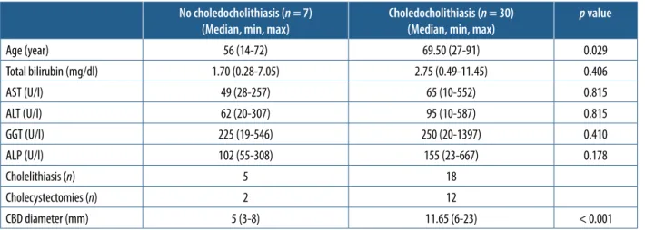
Choledochal pathology was divided into two groups depending on the presence of stones as “there is stone or not”. Extrahepatic bile duct diameters were measured. Gall bladder was examined for the presence of stones. Bio chemical parameters such as alanine aminotransferase (ALT), aspartate aminotransferase (AST), gammaglutam yl transferase (GGT), alkaline phosphatase (ALP), and to tal bilirubin values were recorded.
MRCP was performed with a 3Tesla (Siemens Skyra, Erlangen, Germany) system using respiratorytriggered HASTE technique in axial and coronal planes and with a T2 SPACE sequence in the coronal plane. Images of the T2 SPACE sequence were then reconstructed to maximum intensity projection (MIP) images in the coronal plane with 10 degrees (totally 36 images). Sequence parameters are given in Table 1.
ERCP techniques were performed with a sideviewed Pentax ED 3490 duodenoscope (Tokyo, Japan). Bile ducts were cannulated with a guidewire by sphincterotomy. Images were obtained with nonionic contrast materials. In cases of the presence of a stone, it was retrieved with a balloon catheter.
MannWhitney U test was used to compare demo graphic and laboratory data. Cohen’s κ value was calcu lated for interobserver agreement. Sensitivity, specificity, negative predictive value (NPV), and positive predictive value (PPV) were calculated separately for each observer. The average of both observers was calculated for compar ison with other studies. All statistical analyses were done using SPSS software (IBM SPSS Statistics 20).
Results
Seven patients who had MRCPERCP comparison were diagnosed with tumours and were excluded from the study. Four of the patients who had ERCP failure for oth er reasons were also excluded. The ERCP failure rate was 8.33%. The reasons for failure were as follows: fibrotic pa pilla, papillary oedema, the papilla could not be found in one patient with gastroenterostomy, and papilla was placed at the rim of duodenal diverticula in another patient.
Thirtyseven patients were included in the study pop ulation. The average age of the study population was 63.51 (1491) years. Fifteen of the patients were male and 22 were female. The average age of the male patients was 68.67 (2091) years and of the female patients was 60 (1491)
Table 1. Parameters of sequences used in magnetic resonance cholangio-pancreatography
TR
(msn) (msn)TE Slice thickness (mm) (cm)FOV
Axial HASTE 1600 95 5 37 × 29
Coronal HASTE 1400 108 5 37 × 34
T2 SPACE 4489 702 1 37 × 37
Table 2. Demographic, biochemical, and ultrasonographic data of the cases No choledocholithiasis (n = 7)
(Median, min, max) Choledocholithiasis (n = 30)(Median, min, max) p value
Age (year) 56 (14-72) 69.50 (27-91) 0.029 Total bilirubin (mg/dl) 1.70 (0.28-7.05) 2.75 (0.49-11.45) 0.406 AST (U/l) 49 (28-257) 65 (10-552) 0.815 ALT (U/l) 62 (20-307) 95 (10-587) 0.815 GGT (U/l) 225 (19-546) 250 (20-1397) 0.410 ALP (U/l) 102 (55-308) 155 (23-667) 0.178 Cholelithiasis (n) 5 18 Cholecystectomies (n) 2 12 CBD diameter (mm) 5 (3-8) 11.65 (6-23) < 0.001
years. Demographic features and laboratory data are sum marised in Table 2.
The average time between MRCP and ERCP was 5.46 (115) days. The median bile duct size of the seven pa tients whose MRCP and ERCP results were evaluated as normal was 5 mm (38 mm). The median bile duct size was 11.65 mm (623) in 30 patients with choledocholi thiasis.
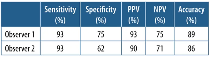
The agreement between the observers was analysed, and Cohen’s κ value was evaluated as 0.84. For two ob servers, the sensitivity of MRCP was 93% whereas the specificity was 75% for the first observer and 62% for the second. Sensitivity, specificity, accuracy, PPV, and NPV values are shown in Table 3.
Discussion
In this study, MRCP was compared with ERCP, and the sensitivity of MRCP in patients with choledocholithiasis was found to be 93% and specificity 69%. The sensitivity of MRCP is reported as being between 27% and 100% in the literature [2,5,6]. In our study, the sensitivity of MRCP that
we found was similar to that of the highlevel sensitivity groups in studies in the literature. Some studies found the specificity of MRCP to be between 40% and 100%. In this perspective, in our study, the sensitivity result seems to be in the medium sensitivity group [1,4]. In a comparison of the specificity value with other series, we found a lower value because the first observer reported suspected stone in the bile ducts of two patients and the second observer in three patients. Both observers interpreted false positive stones in the same cases. The time between MRCP and ERCP of these patients was 35 days. There was a dark focus in the axial HASTE series in the central distal por tion of the bile duct in two patients who had false positive
Table 3. Sensitivity, specificity, positive predictive value (PPV), negative predictive value (NPV), and accuracy values both observers
Sensitivity
(%) Specificity(%) PPV(%) NPV (%) Accuracy(%)
Observer 1 93 75 93 75 89
Observer 2 93 62 90 71 86
Figure 1. A) There was a hypointensity in axial HASTE series in central
por-tion of distal bile duct (choledoc: arrowhead) (gall bladder: open arrow).
B, C) This hypointensity was not seen in coronal HASTE and T2 SPACE MIP
images
A
C
Table 4. Sensitivity, specificity, positive predictive value (PPV), and negative predictive value (NPV) reported in the literature for MRCP in patients with suspected choledocholithiasis
Author Year Scanner (Tesla) n Sensitivity (%) Specificity (%) PPV (%) NPV (%)
Present study 2017 3T 37 93 69 91.5 73 Makmun [1] 2017 1.5 31 81 40 74 50 Badger [2]** 2016 1.5 or 3 47 90 86 97 60 Aydelotte [3] 2015 1.5 36 90 88 97 64 Polistina [4] 2015 1.5 111 77.4 100 100 85 Richard* [5] 2013 NR 70 27 83 36 77 Demartine [6] 2000 1.5 40 100 95.6 92.6 100
*In this study, MRCP was compared with intraoperative cholangiography **In this study, MRCP was compared with intraoperative cholangiography or ERCP NR – not reported
choledocholithiasis as reported by both of the observ ers (Figure 1A). This dark focus was not seen in coronal HASTE and T2 SPACE images (Figure 1BC). It is thought that it could be due to the flow because the location of this false image is in the central distal portion of bile duct and only seen in the axial plane. Millimetric stones and sludge were reported in the ERCP of false negative cases (Table 4).
We also examined the agreement between observers. Cohen’s κ value was calculated as 0.84. This value shows that the agreement between the observers is quite good. Although there are studies concerning interobserver agree ment of MRCP sequences, there is no adequate evidence and discussion about interobserver agreement of MRCP in patients with suspected choledocholithiasis [8,9].
In the first evaluation of patients with suspected chole docholithiasis, blood tests (ALT, AST, GGT, ALP, and total bilirubin) and transabdominal ultrasonography (TAUS) should be performed [10]. Levels of cholestatic biochem ical markers (GGT, ALP, and bilirubin) usually increase gradually, consistent with the duration and severity of bil iary obstruction. However, these biochemical tests are not specific to choledocholithiasis and can increase in some other conditions like pancreatitis in the first line. No statis tical significance was found in levels of biochemical tests in patients with choledocholithiasis and in patients without choledocholithiasis in this study. In the group of patients without choledocholithiasis, one patient had hepatitis and sludge in the gall bladder, one patient had cholecystitis, and two other patients had pancreatitis. The other three patients were investigated because of complaints that de veloped after cholecystectomy and the establishment of large bile duct in ultrasonography. The last patient had increased levels in biochemical tests but normalised in the following tests.
Normally the bile duct diameter is smaller than 6 mm [11]. A mild dilation is reported to be normal due to aging [12]. Biliary obstruction should be suggested when the bile duct diameter is over 8 mm in patients with normal
gallbladder [13]. In the group of patients without choledo cholithiasis in our study, the maximum bile duct diameter was evaluated as 8 mm; this was in the patient with chole cystectomy. TAUS has a low sensitivity in detecting CBD stones. However, it is more reliable in detecting dilation in the bile duct due to choledocholithiasis [14,15].
ERCP is the reference diagnostic method in biliary ob struction [16]. In patients with obstructive jaundice, the ERCP failure rate is about 67% [17]. The rate we found in our study was 8.33%. This rate is slightly higher than re ported. ERCP is recommended in patients with cholelithi asis and with high risk of choledocholithiasis before the op eration. In patients with ERCP failure, EUS and MRCP are recommended [10]. In our opinion, it seems more rational to perform MRCP before ERCP in order to give a broad overview and detect lesions other than choledocholithiasis.
There are some limitations in our study. Nonparamet ric tests were used for comparison of the groups when the number was limited in the group of patients without chole docholithiasis. There was a delay between MRCP and ERCP because it was a retrospective study. The average delay was 5.46 days and in some of the patients it prolonged to 15 days. During this time interval, although it is a low probability, the persistence of stones in the bile duct might have changed, and this situation might have caused false negative results.
Conclusions
As a result, in this study we found a high level of in terobserver agreement in evaluating MRCP. MRCP has a high sensitivity in detecting choledocholithiasis. How ever, the specificity found in this study was comparatively low, and this may be due to artefacts caused by high mag netic field. Further longterm studies are needed.
Conflict of interest
References
1. Makmun D, Fauzi A, Shatri H. Sensitivity and specificity of magne tic resonance cholangiopancreatography versus endoscopic ultraso nography against endoscopic retrograde cholangiopancreatography in diagnosing choledocholithiasis: the Indonesian experience. Clin Endosc 2017; 50: 486490.
2. Badger WR, Borgert AJ, Kallies KJ, Kothari SN. Utility of MRCP in clinical decision making of suspected choledocholithiasis: An insti tutional analysis and literature review. Am J Surg 2017; 214: 251255. 3. Aydelotte JD, Ali J, Huynh PT, et al. Use of magnetic resonance chol angiopancreatography in clinical practice: not as good as we once thought. J Am Coll Surg 2015; 221: 215219.
4. Polistina FA, Frego M, Bisello M, et al. Accuracy of magnetic reso nance cholangiography compared to operative endoscopy in detect ing biliary stones, a single center experience and review of literature. World J Radiol 2015; 28: 7078.
5. Richard F, Boustany M, Britt LD. Accuracy of magnetic resonance cholangiopancreatography for diagnosing stones in the common bile duct in patients with abnormal intraoperative cholangiograms. Am J Surg 2013; 205: 371373.
6. Demartines N, Eisner L, Schnabel K, et al. Evaluation of magnetic resonance cholangiography in the management of bile duct stones. Arch Surg 2000; 135: 148152.
7. Merkle EM, Dale BM, Paulson EK. Abdominal MR imaging at 3T. Magn Reson Imaging Clin N Am 2006; 14: 1726.
8. Nandalur KR, Hussain HK, Weadock WJ, et al. Possible biliary dis ease: diagnostic performance of highspatialresolution isotropic 3D T2weighted MRCP. Radiology 2008; 249: 883890.
9. Sudholt P, Zaehringer C, Urigo C, et al. Comparison of optimized 3DSPACE and 3DTSE sequences at 1.5T MRCP in the diagnosis of choledocholithiasis. Rofo 2015; 187: 467471.
10. Maple JT, BenMenachem T, Anderson MA, et al. The role of endos copy in the evaluation of suspected choledocholithiasis. Gastrointest Endosc 2010; 71: 19.
11. Parulekar SG. Ultrasound evaluation of bile duct size. Radiology 1979; 133: 703707.
12. Bachar GN, Cohen M, Belenky A, et al. Effect of aging on the adult extrahepatic bile duct: a sonographic study. J Ultrasound Med 2003; 22: 879882.
13. Baron RL, Stanley RJ, Lee JKT, et al. A prospective comparison of the evaluation of biliary obstruction using computed tomography and ultrasonography. Radiology 1982; 145: 9198.
14. Lapis JL, Orlando RC, Mittelstaedt CA, et al. Ultrasonography in the diagnosis of obstructive jaundice. Ann Intern Med 1978; 89: 6163. 15. Einstein DM, Lapin SA, Ralls PW, et al. The insensitivity of sonogra phy in the detection of choledocholithiasis. Am J Roentgenol 1984; 142: 725728.
16. Kaltenthaler EC, Walters SJ, Chilcott J, et al. MRCP compared to di agnostic ERCP for diagnosis when biliary obstruction is suspected: a systematic review. BMC Med Imaging 2006; 6: 9.
17. Giovannini M, Bories E, Napoleon B, et al. 855 multicenter rand omized phase II study: percutaneous biliary drainage vs EUS guided biliary drainage: results of the intermediate analysis. Gastrointest Endosc 2015; 81: 174.