Journal of Neurological Sciences [Turkish] 34:(2)# 58; 136-142, 2017
http://www.jns.dergisi.org/text.php3?id=1036 doi: 10.24165/jns.10020.17
Research Article
Correlation of Respiratory Function Tests With Repetitive Stimulation of Long Thoracic Nerve in Myasthenia Gravis
Fatma GENÇ1,Aylin YAMAN1,Burcu YÜKSEL1,Yasemin BİÇER GÖMCELİ1,Gülnihal KUTLU2
1University of Health Sciences Antalya Training and Research Hospital, Department of Neurology,
Antalya, Turkey 2Muğla Sıtkı Koçman University Faculty of Medicine, Department of Neurology and
Clinical Neurophysiology, Muğla, Turkey Summary
Objective: Myasthenia gravis (MG) is an autoimmune disease. Respiratory weakness and
bulbar involvement are the major complications and early diagnosis of these complications is very important for the planning of treatment.
Method and Patients: Twenty-one patients with myasthenia gravis and 21 healthy controls
were included in this study. Repetitive nerve stimulation (3Hz) tests were performed on facial, ulnar and long thoracic nerves. The difference between the first and fourth compound muscle action potential (CMAP) amplitudes was recorded. Respiratory functions were evaluated using forced vital capacity (FVC) and the ratio of forced expiratory volume in 1s (FEV1) to FVC.
Results: 15 male and 6 female patients were included in the patient group. The sensitivity and
specificity of long thoracic nerve compared to facial nerve were 0.86 and 0.66, respectively. There was a significant difference between the decrements recorded from serratus anterior (SA). A strong correlation was observed between the percentage of the decrement recorded from SA and FEV1/FVC ratio.
Conclusions: The use of repetitive stimulation of long thoracic nerve compared to respiratory
function tests might be a well-tolerated and simple way for the early diagnosis of pulmonary dysfunction in MG.
Significance: This well-tolerated method should be recommended in routine
electromyography protocols.
Key words: Myasthenia gravis, repetitive nerve stimulation, long thoracic nerve, respiratory
function tests
Myastenia Graviste Repetetif Uzun Torasik Sinir Uyarımının Klinik ve Solunum Fonksiyon Testleri ile Korelasyonu
Özet
Amaç: Myastenia Gravis (MG) otoimmün bir hastalıktır. Solunum güçsüzlüğü ve bulber
güçsüzlük potansiyel olarak geri dönüşümlü en önemli komplikasyonlar olup erken tespit edilmesi tedavide yol göstericidir. Bu çalışmada MG hastalarında gelişebilecek respiratuar yetmezliğin tespitinde uzun torasik sinir repetetif stimulasyonunun (RNS) solunum fonksiyon testi ile korelasyonu ve güvenilirliğini araştırmayı amaçladık.
Metod: Yirmibir MG hastası ve 21 sağlıklı kontrol grubu çalışmaya dahil edildi. Fasiyal,
ulnar, uzun torasik sinirlerde repetetif sinir uyarımı (3 Hz) yapıldı. Birinci ve dördüncü birleşik kas aksiyon potansiyel (BKAP) amplitüdleri arasındaki fark kaydedildi. Solunum fonksiyonları, zorlu vital kapasite (FVC) ve birinci saniyedeki zorlu ekspirasyon hacminin (FEV1), FVC'ye oranı ölçülerek değerlendirildi.
Bulgular: Hasta grubunda 15 erkek ve 6 kadın vardı. Uzun torasik sinirin fasiyal sinire göre
duyarlılık ve özgüllüğü, 0.86 ve 0.66 idi. Hasta ve kontrol grubu arasında serratus anterior kasından (SA) kaydedilen dekrement yanıtlar arasında anlamlı fark saptandı. SA kasından kaydedilen dekrement yanıtların yüzdesi ile FEV1/FVC oranı arasında güçlü bir korelasyon gözlendi.
Sonuç: Uzun torasik sinirin repetetif uyarımı; solunum fonksiyon testi ile kıyaslandığında iyi
tolere edilebilen ve MG'e bağlı solunum disfonksiyonunun erken teşhisinde basit bir yöntemdir.
Anahtar Kelimeler: Miyastenia Gravis, repetetif sinir uyarımı, uzun torasik sinir, solunum
fonksiyon testi
INTRODUCTION
Myasthenia gravis (MG) is an autoimmune disease of neuromuscular junction characterized by asymmetric muscle weakness and fatigability of extraocular, bulbar, nuchal, or limb muscles, with an incidence of 7-16 per million per year (1,2). Approximately fifteen percent of MG patients have thymoma (2). Electrophysiological testing of neuromuscular junction plays an important role to understand the pathophysiology of the disease (3). Therefore, the most common method used to confirm the diagnosis of neuromuscular junction dysfunction is repetitive stimulation of a motor nerve with recording from a muscle innervated by the same nerve (4). Although proximal muscles (facial muscles, trapezius, deltoid and biceps brachii) have been found to be more sensitive, repetitive nerve stimulation (RNS) studies are usually performed on hand and shoulder muscles (5,6). In this study, we performed repetitive stimulation of long thoracic nerve with recording from serratus anterior (SA), which is one of the accessory respiratory muscles, and aimed at investigating the correlation between respiratory muscle weakness and respiratory function tests through spirometric measurements. These studies
might provide a new perspective for early detection of pulmonary dysfunction in MG patients.
MATERIAL AND METHODS
21 patients with MG who were followed up in our neuromuscular diseases outpatient clinic were included in this study. 21 age and sex matched healthy controls were also included upon their informed consent. Repetitive nerve stimulation (RNS) tests at 3 Hz were performed on facial (orbicularis oculi (OO)), ulnar (abductor digiti minimi (ADM)) and long thoracic (serratus anterior (SA)) nerves. Acetylcholine esterase inhibitors were ceased 12 hours before RNS. Surface stimulation and recordings were performed through Nihon Kohden, NM-312S (Nihon Kohden, Tokyo, Japan) electrodes. Amplifier filter frequencies were set at 10 to 5 kHz with Nihon Kohden Neuropack EMG machine. Ten single square wave pulses of 0.3 ms duration at 3 Hz were delivered in each stimulation run. Surface temperature was maintained at 32 °C - 34 °C. The difference between the first and fourth compound muscle action potential (CMAP) amplitudes was recorded. A decrement greater than 10% was considered as abnormal. Repetitive stimulation of long thoracic nerve was
applied to the patients in supine position. An active electrode was located on the sixth rib on the mid-axillary line. The reference electrode was placed over the medial part of the anterior axillary line on the same rib. Electrodes were fixed to skin by plasters. Stimulations using Nihon Kohden NM-420S bipolar electrodes were applied near the Erb's point, lateral to the clavicular head of the sternomastoid (11). During the stimulation, the patients were advised to hold their breath in mid-expiration. Square wave pulses of 0.3 ms duration were delivered to obtain supramaximal responses. The stimulation was maintained in the range from 15 to 25 mA. The other muscles' decrements were calculated automatically.
Respiratory functions were evaluated through forced vital capacity (FVC) and the ratio of forced expiratory volume in 1s (FEV1) to FVC in all patients. FEV1/FVC ratio and FVC values were calculated according to the age and weight of each individual. The correlation between the clinical findings and electrophysiological or spirometric tests was evaluated in this study.
RESULTS
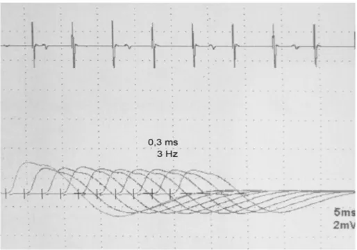
21 patients (15 male, 6 female) with a mean age of 56,9 years (range 27 to 76) and 21 healthy controls (13 male, 8 female) with a mean age of 55,3 (range 19 to 84; 8 women, 13 men) were enrolled. 2 patients were excluded from the study because they refused to undergo spirometric tests. Only one patient had ocular MG while the remaining 20 patients had generalized MG. 8 patients who were hospitalized due to oropharyngeal weakness and dyspnea also had higher decrements, which were not statistically significant. Table-1 shows the demographic and clinical features of the patient group. In the patient group, repetitive facial nerve stimulation was recorded from the OO muscle with a mean amplitude of 1,9 mV and mean decrement of -4.34 %; while the recordings from ADM had a mean amplitude of 10.9 mV and mean decrement of -2.7 %. However, repetitive stimulation of long thoracic nerve recorded from serratus anterior muscle had a mean amplitude of 1.7 mV and mean decrement of -8.91 %. Figure-1 shows an example of RNS recorded from SA muscle.
Figure 1: RNS Example Recorded From SA Muscle
The percent.age of the decrement recorded from OO (p<0.001), ADM (p<0.05) and SA (p<0.001) muscles was calculated, and there were significant differences between the patient and control groups. In female patients, higher decrements were observed in SA recordings (p=0.032) while there were not any significant differences between the other muscles (p>0.05). The sensitivity and specificity of long thoracic nerve compared to facial nerve were 0.86 and 0.66 respectively.
The decrement values were subjected to one-way analysis of variance with a significance set at (F(96,2)=145,55; p=0.00). According to Tukey honest significant difference (TUKEY HSD)
post-hoc test, there was a significant difference in the patient group between OO and ADM decrements (p=0.00) and also SA and ADM decrements (p=0.00), whereas there was not any significant difference between OO and SA decrements (p=0.63).
In the evaluation of spirometric test, a strong correlation was found between the SA decrement recordings and FEV1/FVC ratio r=0.81 (p=0.00) and a medium-strong correlation was found between the decrement recordings from SA and FEV1/FVC r=0.64 (p=0.003). This relation was not observed between respiratory function tests and the percentage of the decrement from OO (Figure-2).
Figure 2:Histogram Graphics of RNS SA, FEV1/FVC and FVC
DISCUSSION
MG is a chronic autoimmune disease characterized by dysfunction in neuromuscular transmission. The entire muscular system including respiratory muscles might be affected mostly in generalized form of the disease. Diaphragm is the main muscle of respiration that is responsible for over two thirds of inspiration during quiet breathing (7). Respiratory muscle weakness including diaphragm and/or intercostal muscles or accessory respiratory muscles can cause respiratory insufficiency and may result in admission to intensive care unit (8). The inspiratory muscles are the diaphragm, external intercostal and accessory muscles (9). The diaphragm innervated by the phrenic nerve is the principal muscle of inspiration. Although respiratory failure in MG is generally due to diaphragm dysfunction and oropharyngeal weakness, long thoracic nerve stimulation was preferred as a simpler and well-tolerated method due to the technical difficulties of phrenic nerve stimulation (10,11). Previous studies have reported that fatigue in intercostal and SA muscles of MG patients results in a greater impairment in neuromuscular transmission when compared with normal subjects (11). Thus, long thoracic nerve stimulation recorded from SA muscle is considered to
be a suitable method to evaluate the respiratory dysfunction.
Weakness of the diaphragm and intercostal muscles is rare (14%) at the early stages of the disease whereas it becomes clinically significant in 50-60% of patients at later stages (7). Myastenic crisis, potentially a fatal complication of MG, occurs in about 20% of patients usually within the first year of illness (12,13). The onset of ventilator insufficiency varies between the beginning of the illness and over 25 years after the onset of symptoms (14). Therefore, acute respiratory failure is a life-threatening condition that might require intubation and mechanical ventilation (15). Early diagnosis of respiratory functions is therefore an important issue to arrange the appropriate therapy for MG.
Although the limitations of respiratory functions tests (RFT) include the lack of cooperation of the patient and facial muscle weakness, these tests are useful parameters for measuring the respiratory muscle strength. Respiratory muscle endurance and respiratory volume with maximal voluntary ventilation can be observed to decrease in patients with generalized MG despite normal spirometry results. After all, these tests can be helpful to predict the need for mechanical
ventilation (11, 16-18). In our study, forced vital capacity (FVC), (FEV1) in one second, (FEV1), FEV1/FVC ratio were measured by using a spirometer. The most common method to detect neuromuscular junction function is repetitive stimulation of a motor nerve with recording from a muscle innervated by the same nerve to compare the decrements between the ulnar, facial and long thoracic nerves (4). Spirometry is not sufficient to identify functional changes in patients with MG (19). We aimed at evaluating the correlation of respiratory function tests with repetitive stimulation of long thoracic nerve.
In previous studies, repetitive stimulation of phrenic nerve was used to detect respiratory muscle weakness in MG. The technical difficulties, lack of tolerance of the patients and artifacts are the limitations of this method (20). In another study, a simpler and well-tolerated method was performed by using the repetitive stimulation of long thoracic nerve recorded from SA muscle, one of the accessory respiratory muscles. This study showed that 8 out of 15 patients who had respiratory symptoms had abnormal repetitive long thoracic nerve stimulation. The remaining 7 patients without symptoms had normal results. For early detection of respiratory muscle weakness, authors recommend the repetitive stimulation of the long thoracic nerve due to its good correlation with respiratory symptoms (11). Similarly in our study, a good correlation was found between RFT and repetitive stimulation of long thoracic nerve. The percentage of the decrement from OO, ADM and SA muscles was calculated and there were significant differences between the patient and control groups. In the patient group, a significant difference was recorded between SA decrements and OO-ADM decrements. Therefore, these results suggest that repetitive stimulation of long thoracic nerve recorded from SA muscle has a diagnostic value in neuromuscular
transmission for the detection of respiratory dysfunction in MG. A good correlation was found between respiratory function tests and repetitive stimulation of long thoracic nerve. Based on these results, it can be suggested that repetitive stimulation of long thoracic nerve is a simple and sensitive method for detecting respiratory muscle weakness and might be useful to predict prognostic value for the course of the disease. This well-tolerated method should be recommended in routine electromyography protocols.
Correspondence to:
Fatma Genç
E-mail: sanivardr@yahoo.com
Received by: 08 January 2017 Revised by: 10 February 2017 Accepted: 06 April 2017
The Online Journal of Neurological Sciences (Turkish) 1984-2017
This e-journal is run by Ege University Faculty of Medicine,
Dept. of Neurological Surgery, Bornova, Izmir-35100TR
as part of the Ege Neurological Surgery World Wide Web service.
Comments and feedback: E-mail: editor@jns.dergisi.org URL: http://www.jns.dergisi.org
Journal of Neurological Sciences (Turkish) Abbr: J. Neurol. Sci.[Turk]
REFERENCES
1. Heldal AT, Eide GE, Romi F, Owe JF, Gilhus NE. Repeated Acetylcholine Receptor Antibody-Concentrations and Association to Clinical Myasthenia Gravis Development. PLoS One 2014;9:e114060.
2. Jacob S, Viegas S, Lashley D, Hilton-Jones D. Myasthenia gravis and other neuromuscular junction disorders. Pract Neurol 2009;9:364-371. 3. Plomp JJ, Morsch M, Phillips WD, Verschuuren JJ.
Electrophysiological analysis of neuromuscular synaptic function in myasthenia gravis patients and animal models. Exp Neurol 2015; 270:41-54. 4. Ajith Cherian, Neeraj N. Baheti, Thomas Iype.
Electrophysiological study in neuromuscular junction disorders. Ann Indian Acad Neurol 2013;16:34-41.
5. Howard JF, Sanders DB, Massey JM. The electrodiagnosis of myasthenia gravis and the Lambert-Eaton myasthenic syndrome. Neurol Clin 1994;12:305-330.
6. Costa J, Evangelista T, Conceição I, de Carvalho M. Repetitive nerve stimulation in myasthenia gravis relative sensitivity of different muscles. Clin Neurophysiol 2004;115:2776-2782.
7. Simpson JA. Myasthenia gravis and myasthenic syndromes. In: Walton JN, ed. Disorders of voluntary muscle, 4th edn. Edinburgh: Churchill Livingstone, 1981:585-624.
8. Pfeffer G, Povitz M, Gibson GJ, Chinnery PF. Diagnosis of muscle diseases presenting with early respiratory failure. J Neurol 2015;262:1101-1114. 9. Roussos C, Macklem PT. The Respiratory Muscles.
The N Eng J Med 1982; 307:786-797.
10. Evoli A, Alboini PE, Bisonni A, Mastrorosa A, Bartoccioni E. Management challenges in muscle-specific tyrosine kinase myasthenia gravis. Ann N Y Acad Sci 2012;1274:86-91.
11. Y L Lo, T H Leoh, Y F Dan, Y E Tan, S Nurjannah, P Ratnagopal. Repetitive stimulation of the long thoracic nerve in myasthenia gravis: clinical and electrophysiological correlations. J Neurol Neurosurg Psychiatry 2003;74:379-381.
12. Vern C Juel, Janice M Massey. Myasthenia gravis. Orphanet J Rare Dis. 2007; 2: 44.
13. Thomas CE, Mayer SA, Gungor Y, et al. Swarup R, Webster EA, Chang I, Brannagan TH, Fink ME, Rowland LP. Myasthenic crisis: Clinical features, mortality, complications, and risk factors for prolonged intubation. Neurology. 1997;48:1253-1260.
14. Ferguson P, Murphy R, Lascelles R. Ventilatory failure in myasthenia gravis. J Neurol Neurosurg Psychiatry 1982;45:217-22.
15. Trouth AJ, Dabi A, Solieman N, Kurukumbi M, Kalyanam J. Myasthenia Gravis: A Review. Autoimmune Dis 2012;2012: 874680.
16. Evoli A, Batocchi AP, LoMonaco M, et al. Clinical heterogeneity of seronegative myasthenia gravis. Neuromuscul Disord 1996;6:155-61.
17. Cabrera Serrano M, Rabinstein AA. Usefulness of pulmonary function tests and blood gases in acute neuromuscular respiratory failure. Eur J Neurol 2012;19:452-456.
18. Heliopoulos I, Patlakas G, Vadikolias K, et al. Maximal voluntary ventilation in myasthenia gravis. Muscle Nerve 2003; 27:715-719.
19. A Mier, C Brophy, J Moxham, M Green. Repetitive stimulation of phrenic nerves in myasthenia gravis. Thorax 1992;47:640-644.
20. Zifko UA, Nicolle MW, Grisold W, Bolton CF. Repetitive phrenic nerve stimulation in myasthenia gravis. Neurology. 1999;53:1083-1087.

