ABSTRACT
This work is licensed under a Creative Commons Attribution-NonCommercial 4.0 International License.
Zeynep Gizem Kaya İslamoğlu1 , Abdullah Demirbaş1 , Mehmet Unal1 , Duygu Fındık2
Nail Digital Dermoscopy in Onychomycosis:
A Correlation with Clinical Type, Gender, and
Culture Examination
Objective: Onychomycosis (OM) is a common disease that covers both tinea unguium and those remaining cases caused by yeasts, mainly of the Candida and various non-dermatophyte molds. Diagnosis is usually confirmed with direct microscopy and fungal culture. Nail dermoscopy is a non-invasive tool to diagnose various nail disorders and also to avoid time-consuming investigations. The aim of the present study was to determine the dermoscoping findings in OM and to correlate this with clinical type, gender, and culture results.
Materials and Methods: This was a cross-sectional study of 100 patients diagnosed with OM according to clinical findings and direct microscopic examination. Nail dermoscopy was performed using a FotoFinder Digital Dermoscope, and images were recorded. A part of the samples was cultured in all patients.
Results: The most frequent clinical type was distal lateral subungual onychomycosis (80.0%). The culture was negative in 72.0% of the samples. In the positive group, 48% of Trichophyton rubrum was cultured. The most common dermoscopic findings were longitudinal stria, ruin appearance, and longitudinal leukonychia. In culture-negative samples, irregular termi-nation was most commonly seen. Ruin appearance, brown discoloration, hematoma, and transverse leukonychia, such as brushing, were compatible with total dystrophic OM.
Conclusion: Determinative dermoscopic findings for OM, clinical types, and fungus forms were identified. These signs can avoid unnecessary mycology in selected cases.
Keywords: Onychomycosis, dermoscopy, toenails, culture
INTRODUCTION
Onychomycosis (OM) is one of the most common nail infective disorders, accounting for nearly 50% of patients who have nail disorders. It is a term that covers all fungal infections of the nail. Approximately 90% of most cases are caused by dermatophytes, especially Trichophyton rubrum, Trichophyton mentagrophytes, and Epidermophy-ton floccosum. The other 10% are developed by yeast or non-dermatophyte-type fungus. OM primarily affects
adults between ages 30 and 60 years (1). Generally, the toenails are kept more than the fingernails. The nail bed, matrix, and nail plate that formed the nail unit may be involved particularly in OM, but the infection may have affected them all together (2). OM can be mimicking several diseases, such as psoriasis, lichen planus, alopecia areata, viral warts, chronic paronychia, traumatic onycholysis, and nail apparatus tumors (3). For this reason, diag-nosing OM is extremely important. Direct microscopic examination with potassium hydroxide (KOH), fungal cul-ture, histopathological evaluation, immunofluorescence examination with calcofluor white dye, enzyme analysis, and polymerase chain reaction can be used for diagnosing OM. The most commonly used conventional methods for diagnosis are direct microscopy and culture. However, the sensitivity and specificity of KOH examination and culture can vary from center to center (4). The other methods increase the sensitivity, but they are time-consum-ing, expensive, or invasive options for diagnosing OM (5, 6). Dermoscopy is a non-invasive diagnostic tool that is widely used for the diagnosis of melanocytic and non-melanocytic lesions and inflammatory and infectious diseases (7). Nail dermoscopy, also known as “onychoscopy,” is a technique that aids clinical examination in the diagnosis of nail pigmentations; melanocytic and non-melanocytic tumors; and inflammatory, traumatic, and infectious dis-eases (8). It is substantial for avoiding unnecessary nail biopsies (9).
There are restricted studies guided by the diagnosis of OM with dermoscopy. The aim of the present study was to determine the most frequent dermoscopic patterns associated with OM and the correlation between fungus cultures and dermoscopic patterns in patients with a clinical diagnosis of OM.
MATERIALS and METHODS
This was a cross-sectional study. The study was approved by the Institutional Review Board (no. 2018/305).
Cite this article as:
Kaya İslamoğlu ZG, Demirbaş A, Unal M, Fındık D. Nail Digital Dermoscopy in Onychomycosis: A Correlation with Clinical Type, Gender, and Culture Examination. Erciyes Med J 2019; 41(3): 288–94.
1Department of Dermatology, Selcuk University Faculty of Medicine, Konya, Turkey
2Department of Medical Microbiology, Selcuk University Faculty of Medicine, Konya, Turkey
Submitted
28.02.2019
Accepted
10.06.2019
Available Online Date
07.08.2019
Correspondence
Zeynep Gizem Kaya İslamoğlu, Department of Dermatology, Selcuk University, Faculty of Medicine, Konya, Turkey Phone: +90 506 691 00 47 e-mail: [email protected]
©Copyright 2019 by Erciyes University Faculty of Medicine - Available online at www.erciyesmedj.com
Informed consent was obtained from all participants of the study. Patients were admitted to the dermatology outpatient clinic of Sel-cuk University, Faculty of Medicine Hospital with a complaint of nail disturbance between June 2018 and November 2018. A total of 100 patients clinically and according to direct microscopic ex-amination diagnosed with OM were included in the present study. KOH negative cases were excluded from the study. A part of the sample from direct microscopic examination (KOH 40%) was sys-tematically performed for culture. Patients who used oral or topi-cal antifungal therapy in the last 3 months; had diseases, such as psoriasis, lichen, eczema, autoimmune bullous diseases, and gen-odermatoses; and with nail involvement were excluded from the study. Age, gender, type of OM, limbs held, and number of nails were noted.
In all cases, one of the affected nails was examined by digital dermo-scope (FotoFinder Dermodermo-scope; FotoFinder Systems GmbH, Bad Birnbach, Germany) with a magnification of 20×. Higher magnifi-cation of up to 70× and unlimited manual magnifimagnifi-cation were used if necessary. The linkage fluid used was ultrasound gel in most of the cases. The photos were taken using Medicam 800 HD by FotoFin-der VideoFotoFin-dermoscope and recorded. Dermoscopy was performed to include the nail plate, nail plate free end, hyponeidia, and nail folds. A single observer analyzed each of the dermoscopy images to define the most common patterns and compare them with the patterns described by Piraccini et al. (10) and Kaynak et al. (11). Statistical analysis: Data obtained in the study were analyzed using SPSS (Statistical Package for Social Sciences) for Windows 22.0 program (SPSS Inc., Chicago, IL, USA). Number, percentage, mean, and standard deviation were used as descriptive statisti-cal methods in the evaluation of the data. The t-test was used to compare quantitative continuous data between two independent groups. The chi-square analysis was used to examine the relation-ship between group variables. The findings were evaluated at 95% confidence interval and 5% significance level.
RESULTS
The study included 100 patients with clinical suspicion and positive KOH examination of OM. Of the 100 patients, 66 (66.0%) were males, and 34 (34.0%) were females. The mean age of the patients was 49.52±13.62 years, and the mean age of the control group was 47.18±14.47 years. No significant difference was found be-tween age and groups (p>0.05) (Table 1).
All patients had OM in the toenails. Of the 100 patients, 80 (80.0%) were clinically classified as distal lateral subungual OM (DLSO), 18 (18.0%) as total dystrophic OM (TDO), and 2 (2.0%) as white superficial OM (WSO).
The culture was negative in 42 (42.0%) samples. In 48 (48%) pa-tients, T. rubrum was the causative agent, and in 10 (10%) patients, Trichophyton tonsurans was the causative agent.
In dermoscopy, longitudinal stria was seen as the most common pattern (66.0%) (Fig. 1). The second most common pattern was ruin appearance, meaning subungual hyperkeratosis (54.0%) (Fig. 2) (11). The incidence and distribution of other patterns are shown in Table 2 (Fig. 3–6).
The dermoscopic patterns were correlated with the clinical pat-tern of OM. A significant relationship was found between ruin Table 1. The mean age of the groups
Groups Patients Controls t p
(n=100) (n=120) Mean SD Mean SD
Age 49.520 13.626 47.183 14.473 1.224 0.222
SD: Standard deviation
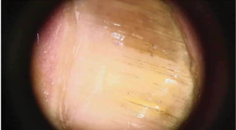
Figure 1. Longitudinal striae which is whitish to brown in the onycholytic nail plate
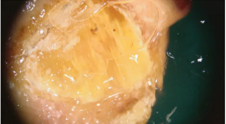
Figure 2. Ruin appearancewhich means subungual hyperk-eratosis and hematoma
Figure 3. Longitudinal leukonychia which is a white color change that extends parallel to the grooves in the nail plate
appearance and clinical type (X2=6.960, p=0.031 <0.05). Ruin appearance was presented in 40 (50.0%) patients with DLSO type and 14 (77.8%) patients with TDO type. Furthermore, there was significance with punctate leukonychia (X2=11.748, p=0.003 <0.05). It was seen in 10 (12.5%) patients with DLSO, 2 (10.0%) with WSO, and 4 (22.2%) with TDO. Brown discoloration was most commonly seen in TDO (55.6%). Punctate hemorrhage was found to be related with DLSO (27.5%). Hematoma was seen only in TDO (1.1%). Finally, transverse leukonychia, such as brushing, was clinically compatible with TDO (55.6%). The distribution of patterns according to clinical type is shown in Table 3.
Then, the dermoscopic patterns were correlated with gender. Punctate leukonychia pattern was seen especially in females (29.4%) (X2=6.895, p=0.011 <0.05), black discoloration pattern in males (15.2%) (X2=5.724, p=0.012 <0.05), and transverse leukonychia, such as brushing, in males (30.3%) (X2=4.228, p=0.032 <0.05) (Table 4).
Finally, the dermoscopic patterns were tried to be correlated with the culture results. Distal irregular termination was most commonly seen in culture-negative samples (33.3%) (X2=8.750, p=0.013 <0.05), longitudinal leukonychia in T. rubrum
posi-tive samples (41.7%) (X2=8.163, p=0.017 <0.05), and black discoloration in culture-negative samples (19.0%), and it was not seen in T. tonsurans positive patients (100.0%) (X2=6.746, p=0.034 <0.05). Punctate hemorrhage was almost seen in
T. tonsurans positive cultures (60.0%) (X2=9.424, p=0.009
<0.05) (Table 5).
Table 2. Distribution and percentage of clinical type, culture results, and dermoscopic patterns in patients
Groups Frequency Percentage
n %
Clinical pattern DLSO 80 80.0
WSO 2 2.0 TDO 18 18.0 Total 100 100.0 Culture T. rubrum 48 48.0 T. tonsurans 10 10.0 Negative 42 42.0 Total 100 100.0
Dermoscopic Ruin appearance 54 54.0 patterns Longitudinal stria 66 66.0
Jagged edge with spikes 6 6.0 Distal irregular termination 20 20.0 Homogenous leukonychia 28 28.0 Punctate leukonychia 16 16.0 Longitudinal leukonychia 30 30.0 Black discoloration 10 10.0 Brown discoloration 22 22.0 Splinter hemorrhage 8 8.0 Punctate hemorrhages 22 22.0 Hematoma 2 2.0 Transverse leukonychia, such as brushing 2 2.0
DLSO: Distal lateral subungual OM; WSO: White superficial OM; TDO: Total dystrophic OM
Figure 4. Punctate hemorrhages and transvers leukonychia such as brushing
Figure 5. Jagged edge with spikes of the whitish longitudi-nal lines that seen in proximal side of the onycholytic area
Figure 6. Distal irregular termination which is the end of the thickened nail plate ending with an irregular
DISCUSSION
Our data demonstrate that dermoscopic patterns of OM showed the following frequencies: longitudinal stria, ruin appearance,
longitudinal leukonychia, homogenous leukonychia, and trans-verse leukonychia, such as brushing. Longitudinal stria was more frequently observed in patients with DLSO, homogenous Table 3. The distribution of patterns according to clinical type
DLSO WSO TDO p
n % n % n % Ruin appearance Positive 40 50.0 0 0.0 14 77.8 X2=6.960 Negative 40 50.0 2 100.0 4 22.2 p=0.031 Longitudinal stria Positive 56 70.0 0 0.0 10 55.6 X2=5.328 Negative 24 30.0 2 100.0 8 44.4 p=0.070
Jagged edge with spikes
Positive 6 7.5 0 0.0 0 0.0 X2=1.596
Negative 74 92.5 2 100.0 18 100.0 p=0.450
Distal irregular termination
Positive 14 17.5 0 0.0 6 33.3 X2=2.813 Negative 66 82.5 2 100.0 12 66.7 p=0.245 Homogenous leukonychia Positive 20 25.0 2 100.0 6 33.3 X2=5.754 Negative 60 75.0 0 0.0 12 66.7 p=0.056 Punctate leukonychia Positive 10 12.5 2 100.0 4 22.2 X2=11.748 Negative 70 87.5 0 0.0 14 77.8 p=0.003 Longitudinal leukonychia Positive 22 27.5 0 0.0 8 44.4 X2=2.884 Negative 58 72.5 2 100.0 10 55.6 p=0.237 Black discoloration Positive 6 7.5 0 0.0 4 22.2 X2=3.765 Negative 74 92.5 2 100.0 14 77.8 p=0.152 Brown discoloration Positive 12 15.0 0 0.0 10 55.6 X2=14.659 Negative 68 85.0 2 100.0 8 44.4 p=0.001 Splinter hemorrhage Positive 8 10.0 0 0.0 0 0.0 X2=2.174 Negative 72 90.0 2 100.0 18 100.0 p=0.337 Punctate hemorrhages Positive 22 27.5 0 0.0 0 0.0 X2=7.051 Negative 58 72.5 2 100.0 18 100.0 p=0.029 Hematoma Positive 0 0.0 0 0.0 2 11.1 X2=9.297 Negative 80 100.0 2 100.0 16 88.9 p=0.010
Transverse leukonychia, such as brushing
Positive 14 17.5 0 0.0 10 55.6 X2=12.311
Negative 66 82.5 2 100.0 8 44.4 p=0.002
leukonychia in WSO, and ruin appearance and brown discol-oration in TDO. In addition to other similar studies, the cor-relation between gender and culture results with dermoscopic patterns was also investigated.
Dermoscopy plays an important role to ease the diagnosis of dermatological disorders as it is a non-invasive procedure (7). The use of a dermoscope in OM has been first performed by Piraccini et al. (10). They studied the difference between DLSO and trau-matic onycholysis. According to their study, two patterns, jagged proximal edge with spikes of the onycholytic area and longitudi-nal striae, had been presented in DLSO and only one pattern, linear edge without spikes, in traumatic onycholysis. In our study, jagged proximal edge with spikes had been presented in 6% of patients with OM, and all of them had DLSO as clinical type; longitudinal striae were 66.0% and 70.0% in DLSO and 55.6% in TDO. The rate of longitudinal striae was similar, but the rate of jagged edge with spikes was lower in our study. In culture results, longitudinal stria was the most frequent in T. tonsurans positive
samples (80.0%).
Nakamura et al. performed dermoscopy in 500 cases of nail dys-trophies and found chromonychia, onycholysis, opacity, and longi-tudinal stripes in patients with OM (1). In another study with ony-choscopy in 237 patients with nail disorder, OM showed jagged pattern of the proximal margin of the onycholytic area, spikes in the proximal fold, white-yellow longitudinal striae in the ony-cholytic nail plate, and distal irregular termination pattern (12). Similar to our study, Jesus-Silva et al. found that TDO (56.13%) is the most common clinical pattern in OM, and that longitudinal striae pattern (32.9%) is the most common dermoscopic pattern. In addition, they described a pattern as distal irregular termination in TDO and DLSO samples (13). It means “the end of the thick-ened nail plate ending with an irregular and crumbly appearance” (11). Its frequency was found in 20% of patients with OM, and it was more common in DLSO type (17.5%) and culture-negative samples (33.3%).
In a case control study, spikes, longitudinal striations, and color changes were statistically significantly higher in patients than in controls. They found that the sensitivity of spikes in OM was 100% and that of longitudinal striations was 82.5%, and color changes were not only seen in OM but also seen in healthy controls (14). This was consistent with what Piraccini et al. stated (10). Kaynak et al. diagnosed 205 patients with DLSO and found that longitudinal striae pattern is observed in 79.5% of the samples, and that jagged edge with spikes pattern is observed in 63.4% of the samples. At the end of the study, they suggested that ruin appearance and punctate leukonychia are identified as dermoscopic patterns with high sensitivity only for DLSO diagnosis (11). In another study by Nargis et al., longitudinal striae and jagged proximal edges were seen in all patients, intermittent spiked pattern was seen in 78.3% of patients, and chromonychia and distal irregular termination were noticed in 38.3% and 11.7% patients, respectively (15). Nail unit pathology reported that 52 patients with abnormal great toe-nails were compared with the dermoscopic features detected by nail unit dermoscopy in Bodman’s study. The specific dermoscopic findings of short spikes (p<0.001), long striae (p<0.001), aurora borealis (p<0.001), irregular termination (p=0.003), dermatophy-toma (p=0.011), transverse onycholysis (p=0.018), and dry scale (p=0.04) patterns were all significantly associated with pathology test results consistent with OM. Transverse onycholysis (p=0.018) was significantly associated with negative pathology results consis-tent with the diagnosis of nail dystrophy (16).
Table 4. The distribution of dermoscopic patterns to gender Males Females p n % n % Ruin appearance Positive 40 60.6 14 41.2 X2=3.410 Negative 26 39.4 20 58.8 p=0.051 Longitudinal stria Positive 46 69.7 20 58.8 X2=1.182 Negative 20 30.3 14 41.2 p=0.193
Jagged edge with spikes
Positive 2 3.0 4 11.8 X2=3.035
Negative 64 97.0 30 88.2 p=0.100
Distal irregular termination
Positive 16 24.2 4 11.8 X2=2.184 Negative 50 75.8 30 88.2 p=0.110 Homogenous leukonychia Positive 16 24.2 12 35.3 X2=1.360 Negative 50 75.8 22 64.7 p=0.176 Punctate leukonychia Positive 6 9.1 10 29.4 X2=6.895 Negative 60 90.9 24 70.6 p=0.011 Longitudinal leukonychia Positive 22 33.3 8 23.5 X2=1.027 Negative 44 66.7 26 76.5 p=0.218 Black discoloration Positive 10 15.2 0 0.0 X2=5.724 Negative 56 84.8 34 100.0 p=0.012 Brown discoloration Positive 16 24.2 6 17.6 X2=0.569 Negative 50 75.8 28 82.4 p=0.313 Splinter hemorrhage Positive 4 6.1 4 11.8 X2=0.992 Negative 62 93.9 30 88.2 p=0.266 Punctate hemorrhages Positive 14 21.2 8 23.5 X2=0.070 Negative 52 78.8 26 76.5 p=0.490 Hematoma Positive 2 3.0 0 0.0 X2=1.051 Negative 64 97.0 34 100.0 p=0.433 Transverse leukonychia, such as brushing Pozitif 20 30.3 4 11.8 X2=4.228 Negative 46 69.7 30 88.2 p=0.032
In our study, longitudinal stria and punctate hemorrhage were iden-tified as dermoscopic patterns with high sensitivity for DLSO; ruin appearance, longitudinal leukonychia brown discoloration, and transverse leukonychia, such as brushing, for TDO; and homoge-nous leukonychia for WSO. This finding suggests that if these
pat-terns are detected in cases with uncertainty of OM, they can guide us. Again, in the same way, jagged edge with spikes and longitudi-nal leukonychia can be strongly associated with fungi and punctate hemorrhage pattern of samples showing high T. tonsurans
repro-duction probability in culture. Table 5. The distribution of dermoscopic patterns by type of culture
T. rubrum T. tonsurans Culture (-) p
n % n % n % Ruin appearance Positive 28 58,3 4 40,0 22 52,4 X2=1,196 Negative 20 41,7 6 60,0 20 47,6 p=0,550 Longitudinal stria Positive 26 54,2 8 80,0 32 76,2 X2=5,812 Negative 22 45,8 2 20,0 10 23,8 p=0,055
Jagged edge with spikes
Positive 6 12,5 0 0,0 0 0,0 X2=6,915
Negative 42 87,5 10 100,0 42 100,0 p=0,032
Distal irregular termination
Positive 4 8,3 2 20,0 14 33,3 X2=8,750 Negative 44 91,7 8 80,0 28 66,7 p=0,013 Homogenous leukonychia Positive 14 29,2 2 20,0 12 28,6 X2=0,357 Negative 34 70,8 8 80,0 30 71,4 p=0,837 Punctate leukonychia Positive 6 12,5 2 20,0 8 19,0 X2=0,847 Negative 42 87,5 8 80,0 34 81,0 p=0,655 Longitudinal leukonychia Positive 20 41,7 0 0,0 10 23,8 X2=8,163 Negative 28 58,3 10 100,0 32 76,2 p=0,017 Black discoloration Positive 2 4,2 0 0,0 8 19,0 X2=6,746 Negative 46 95,8 10 100,0 34 81,0 p=0,034 Brown discoloration Positive 12 25,0 0 0,0 10 23,8 X2=3,152 Negative 36 75,0 10 100,0 32 76,2 p=0,207 Splinter hemorrhage Positive 4 8,3 2 20,0 2 4,8 X2=2,562 Negative 44 91,7 8 80,0 40 95,2 p=0,278 Punctate hemorrhages Positive 8 16,7 6 60,0 8 19,0 X2=9,424 Negative 40 83,3 4 40,0 34 81,0 p=0,009 Hematoma Positive 2 4,2 0 0,0 0 0,0 X2=2,211 Negative 46 95,8 10 100,0 42 100,0 p=0,331
Transverse leukonychia, such as brushing
Positive 10 20,8 2 20,0 12 28,6 X2=0,833
The limitations of our study include a small sample size of the study group selection that does not reflect the general population at a single clinic. Second, there was no control group to compare. In addition to this, our study was a prospective study, used digital dermoscopy in determining dermoscopic patterns, and in contrast to other studies, dermoscopic patterns were also correlated with culture results and gender.
In summary, the diagnosis of OM cannot be built trusting on the clinical appearance alone. There is a need for a supportive diag-nostic tool. Nail dermoscopy is an easy, quick, non-invasive and cost-effective tool to showing the OM changes when mycology is not available. Further studies with large samples will confirm these dermoscopic patterns in OM and will compare these findings with other nail diseases.
Ethics Committee Approval: Selçuk University Faculty of Medicine Non-interventional Clinical Research Ethics Committee. Number: 2018/305 Date: 12.09.2018.
Informed Consent: Written informed consent was obtained from patients who participated in this study.
Peer-review: Externally peer-reviewed.
Author Contributions: Concept – ZGKİ, AD; Design – ZGKİ, AD; Super-vision – ZGKİ, AD, MU, DF; Resource – ZGKKİ, AD, MU, DF; Materials – ZGKİ, AD, DF; Data Collection and/or Processing – ZGKİ, AD; Analysis and/or Interpretation – ZGKİ; Literature Search – ZGKİ, AD; Writing – ZGKİ; Critical Reviews – ZGKİ.
Conflict of Interest: The authors have no conflict of interest to declare.
Financial Disclosure: The authors declared that this study has received no financial support.
REFERENCES
1. Nakamura RC, Costa MC. Dermatoscopic findings in the most fre-quent onychopathies: descriptive analysis of 500 cases. Int J Dermatol 2012; 51(4): 483–5. [CrossRef]
2. Rinaldi MG. Dermatophytosis: epidemiological and microbiological up-date. J Am Acad Dermatol 2000; 43(5 Suppl): S120–4. [CrossRef]
3. Allevato MA. Diseases mimicking onychomycosis. Clin Dermatol 2010; 28(2): 164–77. [CrossRef]
4. Weinberg JM, Koestenblatt EK, Tutrone WD, Tishler HR, Najarian L. Comparison of diagnostic methods in the evaluation of onychomyco-sis. J Am Acad Dermatol 2003; 49(2): 193–7. [CrossRef]
5. Richert B, Lateur N, Theunis A, Andre J. New tools in nail disorders. Semin Cutan Med Surg 2009; 28(1): 44–8. [CrossRef]
6. Prakash R, Prashanth HV, Ragunatha S, Kapoor M, Anitha TK, Kr-ishnamurthy V. Comparative study of efficacy, rapidity of detection, and cost-effectiveness of potassium hydroxide, calcofluor white, and Chicago sky blue stains in the diagnosis of dermatophytoses. Int J Der-matol 2016; 55(4): e172–5. [CrossRef]
7. Campos-do-Carmo G, Ramos-e-Silva M. Dermoscopy: basic concepts. Int J Dermatol 2008; 47(7): 712–9. [CrossRef]
8. Di Chiacchio ND, Farias DC, Piraccini BM, Hirata SH, Richert B, Za-iac M, et al. Consensus on melanonychia nail plate dermoscopy. An Bras Dermatol 2013; 88(2): 309–13. [CrossRef]
9. Wang YJ, Sun PL. Fungal melanonychia caused by Trichophyton rubrum and the value of dermoscopy. Cutis 2014; 94(3): E5–6. 10. Piraccini BM, Balestri R, Starace M, Rech G. Nail digital dermoscopy
(onychoscopy) in the diagnosis of onychomycosis. J Eur Acad Derma-tol Venereol 2013; 27(4): 509–13. [CrossRef]
11. Kaynak E, Göktay F, Güneş P, Sayman E, Turan D, Baygül A, et al. The role of dermoscopy in the diagnosis of distal lateral subungual onychomycosis. Arch Dermatol Res 2018; 310(1): 57–69. [CrossRef]
12. Bhat YJ, Mir MA, Keen A, Hassan I. Onychoscopy: an observational study in 237 patients from the Kashmir Valley of North India. Dermatol Pract Concept 2018; 8(4): 283–91. [CrossRef]
13. Jesús-Silva MA, Fernández-Martínez R, Roldán-Marín R, Arenas R. Dermoscopic patterns in patients with a clinical diagnosis of ony-chomycosis-results of a prospective study including data of potassium hydroxide (KOH) and culture examination. Dermatol Pract Concept 2015; 5(2): 39–44.
14. El-Hoshy KH, Abdel Hay RM, El-Sherif RH, Salah Eldin M, Moussa MF. Nail dermoscopy is a helpful tool in the diagnosis of onychomycosis: A case control study. Eur J Dermatol 2015; 25(5): 494–5. [CrossRef]
15. Nargis T, Pinto M, Shenoy MM, Hegde S. Dermoscopic Features of Distal Lateral Subungual Onychomycosis. Indian Dermatol Online J 2018; 9(1): 16–9.
16. Bodman MA. Point-of-Care Diagnosis of Onychomycosis by Der-moscopy. J Am Podiatr Med Assoc 2017; 107(5): 413–8. [CrossRef]