Received: 16.02.2019 Accepted: 04.05.2019 Published Online: 25.07.2019
Ferhat CUCE
1, Gokalp TULUM
2, Ozgur DANDIN
3, Tuncer ERGIN
1, Omer KARADAS
4, Onur OSMAN
21Gulhane Education and Training Hospital, Department of Radiology, Ankara, Turkey
2Istanbul Arel University, Department of Electrical and Electronics Engineering, Istanbul, Turkey 3Gulhane Education and Training Hospital, Department of General Surgery, Ankara, Turkey 4Gulhane Education and Training Hospital, Department of Neurology, Ankara, Turkey
This study has been presented as an oral presentation at the Balkan Radiology Congress between 12 and 14 October in 2017 at Budapest, Hungary.
A New Practical Intracerebral Hematoma Volume Calculation
Method and Comparison to simple ABC/2
AIM: To find a more practical and effective formula than simple ABC/2 (sABC/2) to calculate the hematoma volume in patients with
subdural and parenchymal haemorrhage.
MATERIAL and METHODS: We reviewed the records of 157 patients who underwent brain computed tomography examinations
for stroke from January to October 2017. Our method, sABC/2 formula, and the planimetric method (the gold standard) were used for measuring the volumes of hematoma.
RESULTS: The concordance in brain hematoma volumes calculated by sABC/2 and the proposed method as compared to
planimetry were 0.92 and 0.93, respectively (p<0.05). The proposed method calculates the subdural hematoma volumes much better than the conventional one, and the root mean square error (RMSE) values were 32.17 and 20.62 ml for sABC/2 and our new method, respectively, whereas the RMSE values for parenchymal hematomas were 25.01 and 20.46 ml for sABC/2 and our new method, respectively.
CONCLUSION: Our new formula for calculating the volume of subdural and parenchymal hematomas is as practical as sABC/2 and
allows the clinician to apply the method bedside.
KEYWORDS: Brain, Cerebral hemorrhage, Computed tomography, Volume, Calculation, ABC/2 Corresponding author: Omer KARADAS
[email protected]Original Investigation
█
INTRODUCTION
T
he most devastating type of stroke is the intracerebral haemorrhage (ICH), and 30%–50% of the patients with ICH die within 30 days of onset. The cerebral hematoma volume is an independent predictor of clinical outcomes and one of the scoring parameters used to predict monthly morbidity and mortality of a patient (8). Thus, the calculation of accurate hematoma volume at admission and its progression during hospitalisation is crucial for clinical prognosis.In neuroradiology, the most common and practical formula to calculate hematoma volume is simple ABC/2 (sABC/2) (2,4,6,8,11,13,15,16,18). The planimetric method, which depends on a software, is the gold standard; however, in practice, it is not yet available. The present study is about the use of sABC/2, a practical formula for clinicians and radiologists in the computation of acute parenchymal hematoma (PH), and subdural hematoma (SH) volumes and the derivation of a more effective formula.
Ferhat CUCE : 0000-0003-1831-3868 Gokalp TULUM : 0000-0003-1906-0401 Ozgur DANDIN : 0000-0002-7386-6871 Tuncer ERGIN : 0000-0002-2407-8844 Omer KARADAS : 0000-0003-0457-3722 Onur OSMAN : 0000-0001-7675-7999 ABSTRACT DOI: 10.5137/1019-5149.JTN.25996-19.2
█
MATERIAL and METHODS
This prospective study was approved by the hospital’s institutional review board (no: 1491-987-10/1539). The data of consecutive patients with acute cerebral hematoma, presenting to the emergency department between January and October 2017, were collected. A written informed consent was waived by the local institutional review board.
Study Population
Brain computed tomography (CT) images of 157 patients, who were admitted to the emergency department with the diagnosis of acute haemorrhagic stroke, were enrolled in the study. All patients underwent a brain CT without contrast agent within 24 hours after the onset of symptoms. Diagnosis was made by two radiologists (F.C, T.E.) and confirmed by a neurologist (O.K).
A total of 79 patients with <18 years of age, brain tumour and metastasis, traumatic haemorrhage, arteriovenous malformation or image motion artefacts were excluded from the study. Finally, 78 patients with 90 hematomas were included in the study for further statistical analysis.
CT Technique
All examinations were performed on 64-slice CT scanner (Brilliance 64; Philips Healthcare, Cleveland, Ohio, USA) providing 4-cm coverage. Exposure parameters were 120 kV and 350 mAs. The collimation was 64x0.625 mm and the images were obtained by axial acquisition. Matrix size was 512 × 512, and the field of view was 250 mm. In addition, the images were reconstructed 3.0 mm thick on a transverse plane.
Image Evaluation and Hematoma Volume Calculation Methods
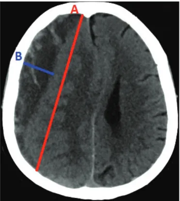
A total of 78 brain CT examination data sets were acquired in Digital Imaging and Communications in Medicine (DICOM) format. Appropriate image settings were performed for the CT, according to the hematoma type. PHs were evaluated using standard window widths (WW) and window levels (WL) to assess brain parenchyma (WW: 80, WL: 30). The SHs were evaluated with adjusted window settings to minimise the underestimation of hematoma thickness due to the proximity to the bone (WW: 250, WL: 100). The three lengths required for the sABC/2 formula were hand-traced by the radiologist. The two longest diameters [anteroposterior (A) and transverse (B) diameters], perpendicular to each other, were calculated at the level of the widest dimension on the axial image. For the calculation of craniocaudal length (C), the number of the slices between the start and end of the hematoma from the bottom to top, was calculated and multiplied by the slice thickness of 3 mm. The units of all the lengths were in millimetres and those of the volume in millilitres. Finally, hematoma volume was calculated by the sABC/2 formula (Figure 1). The formulas, such as C-weighted ABC/2 and 2/3sh in the literature, using the area of the hematoma, are not practical (8,10,17). sABC/2 does not require area calculation and thus is considered as ‘simple’.
The determination of hematoma shape and volume was performed. The shape of the PH was categorised into regular (round, ellipsoid and regular with convex margin) and irregular. SHs were not classified according to shape.
The hematoma volumes were calculated with sABC/2 and the proposed formula by a calculator as well as with the planimetric technique using the ManSeg 2.6 software. The planimetric analysis was performed using a semi-automated segmentation and volume calculation programme ManSeg 2.6 (1). Moreover, a joint decision of the observers was made in cases of disagreement, thus avoiding bias.
The Two Step Method for Cerebral Hematoma Volume Calculation: TSM
When the correlation coefficient (r) between volume and three lengths (anteroposterior, transverse and craniocaudal diameters) of hematoma were investigated, it was observed that there was a significant relationship between volume
Figure 1: An acute onset chronic subdural hematoma case: The
hematoma volume was calculated with both ABC/2 and our proposed formula. The diameter of C was 51 mm [as the number of the slice between the bottom and up (17 slice) was multiplied with the slice thickness of 3 mm], the diameter of A and B was 101.4 mm and 19.4 mm, respectively.
sABC/2=101.4(A) ×19.4 (B) ×51(C)/2=50.16 ml
Proposed formula: 0.3ABC+30 = 0.3 × 101.4 (A) × 19.4 (B) ×51 (C)+30=60.09 ml
[The result of the planimetric volume analysis (gold standart) of the case was 58.95 ml. While sABC/2 methods underestimated volume of hematoma with differences of 8.79 ml. The proposed method TSM has only -1.14 ml of the volume difference.]
and anteroposterior diameter (r=0.80 for p<0.05). Therefore we proposed two different linear functions to calculate the hematoma volume according to the anteroposterior diameter of the hematoma. In this work, the proposed linear functions and the related threshold diameters were determined in two steps.
While 30 ml was taken into consideration as a grading score criteria for a hematoma volume, we firstly selected 45 ml as a soft margin and applied least squares approximation to hematomas that are lower and higher than 45 ml separately and determined two different linear functions. Second, we determined types of hematoma as regular parenchymal, irregular parenchymal, and crescent subdural hematoma. For each category, the anteroposterior diameter, which has the minimum error probability, determined as a threshold. In order to designate the relevant diameter for the threshold, we increased the threshold value 5 mm in each step and calculated the conditional probability. We obtained 50 mm diameter as the threshold for irregular and regular parenchymal hematomas (Volume of all irregular and regular parenchymal hematomas less than 50 mm in diameter were smaller than 45 ml. Also volume of all irregular and regular parenchymal hematomas greater than 50 mm in diameter were large than 45 ml) and 70 mm diameter as threshold for crescent subdural hematoma (87.5% of crescent subdural hematoma had less than 70 mm in diameter were smaller than 45 ml. Also the volume of all crescent subdural hematoma greater than 70 mm in diameter were large than 45 ml.) Two linear equations for volume estimation of hematoma were given as,
V= 0.45ABC + 1
If anteroposterior diameter < 50 mm for regular parenchymal hematoma If anteroposterior diameter < 50 mm for irregular parenchymal hematoma If anteroposterior diameter < 70 mm for crescent subdural hematoma
V= 0.3ABC + 30
If anteroposterior diameter > 50 mm for regular parenchymal hematoma If anteroposterior diameter > 50 mm for irregular parenchymal hematoma If anteroposterior diameter > 70 mm for crescent subdural hematoma
Statistical Analysis
Agreement between planimetry and TSM was evaluated using Bland–Altman plots. Sensitivity, specificity, and AUC were calculated. Root mean square errors of the methods were obtained. Concordance between planimetry and volumes obtained by other estimation methods were assessed using t - test. Bland–Altman plots were generated for sABC/2 and TSM methods in comparison to the planimetric method using both original and log-transformed units.
█
RESULTS
Total 90 cerebral hematomas from 78 patients (29 females, 49 males) were evaluated for this study. The patients’ mean age with standard deviation (SD) was 52.35 ± 22.47. The type and number of hematomas are given in Table I.
The results of hematoma volumes calculated by both sABC/2 and our proposed formula:
While sABC/2 methods overestimated planimetry with mean differences of -8.51 ml the proposed method TSM has the only -0.31 ml of the mean difference.
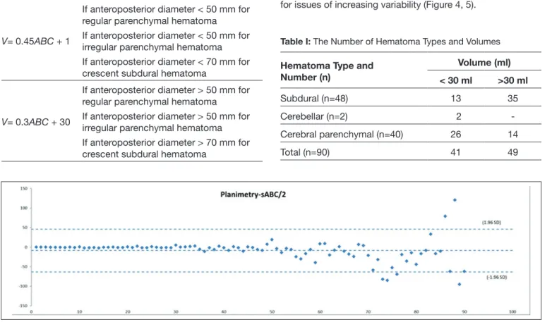
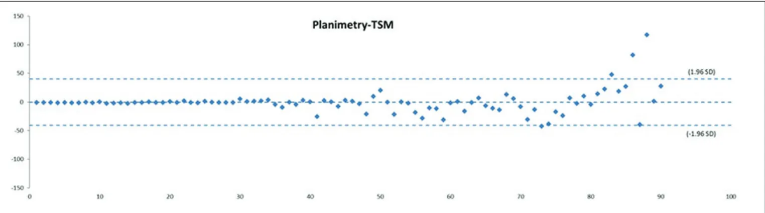
Bland–Altman analysis illustrated that all methods were concordant with the planimetric method and no evidence of a linear trend (Figure 2, 3).
Variability in the concordance between planimetry and the two methods appeared to increase as volumes increased. Therefore, geometric Bland–Altman plots were drawn in the semi-log domain to evaluate concordance while compensating for issues of increasing variability (Figure 4, 5).
Table I: The Number of Hematoma Types and Volumes Hematoma Type and
Number (n) Volume (ml) < 30 ml >30 ml Subdural (n=48) 13 35 Cerebellar (n=2) 2 -Cerebral parenchymal (n=40) 26 14 Total (n=90) 41 49
Concordance between planimetry, sABC/2 and TSM were all high and obtained as 0.92 and 0.93 respectively as seen in Table IV.
Sensitivity and specificity values were obtained for differentiating a cerebral hematoma volume of 30 ml. When considering planimetry as the gold standard, sABC/2 had a sensitivity of 95.6% at a specificity of 95.4% and AUC: .99, TSM had a sensitivity of 91.3% at a specificity of 95.4% and AUC: 0.99.
Planimetry to sABC/2 and TSM ratios were all very close to 1, which means that these methods only slightly overestimated and underestimated planimetry, respectively.
RMSE is another important metric for comparing methods. Table II and Table III demonstrate the RMSE values for hematomas less than 45 ml and greater than 45 ml respectively. For small ICH volumes, 0.45ABC+1 and for large volumes 0.3ABC+30 perform better approximation than sABC/2 for all types of hematomas.
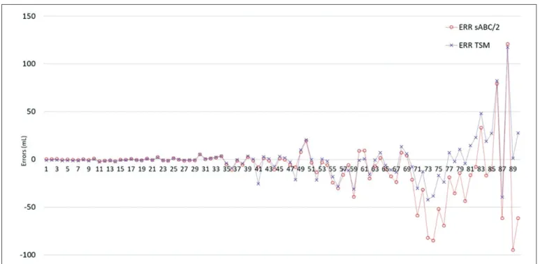
Volume errors of cerebral hematoma for all cases are shown in Figure 6.
Figure 3: Arithmetic mean: Planimetry - TSM (Two step method).
Figure 4: Geometric mean: Planimetry / sABC/2.
█
DISCUSSION
In the present study, the hematoma volumes calculated by sABC/2 and our new formula were found to be close to the values obtained by the planimetric method, and both the formulas were found to be highly correlated between different users.
Various grading scores have been used to predict the prognosis of spontaneous PH (5). The ICH score is most Figure 6: Errors of sABC/2 and two step method (TSM).
Table II: Root Mean Square Errors of ICH Volume for Less than 45 ml
1ICH Type Formula and
2RMSE
ABC/2 (ml) 0.3ABC + 30(ml) 0.45ABC + 1(ml)
Subdural 13.93 21.43 9.38
Irregular parenchymal 8.53 25.46 8.07
Regular parenchymal 3.94 26.02 3.85
1ICH (intracerebral hemorrhage), 2RMSE( Root mean square error).
Table III: Root Mean Square Errors of ICH Volume for Greater than 45 ml
1ICH Type Formula and
2RMSE
ABC/2 (ml) 0.3ABC + 30(ml) 0.45ABC + 1(ml)
Subdural 43.74 27.88 34.95
Irregular parenchymal 73.05 62.27 70.22
Regular parenchymal 27.46 16.09 17.74
1ICH (intracerebral hemorrhage), 2RMSE (Root mean square error).
Table IV: ICH Concordance for sABC/2 and TMS as Compared
to Planimetry
Method 1ICH 95% 2CI
sABC/2 0.92 (.88-.95)
3TSM 0.93 (.89-.95)
1ICH (intracerebral hemorrhage), 2CI (Confidential Interval), 3TSM (Two
with different experiences work together. Therefore, the volume formula shall be easy to use. ABC/2 is an ellipsoid volume formula and commonly used in radiology. Khan et al. reported a highdegree of coherence amongst the readers, comprising medical students, neurology residents and vascular neurologists (ICC: 0.96), with regard to sABC/2 (8). However, in a study about the reliability of sABC/2 amongst different readers, Hussein et al. found that the planimetric method correlation was lower for various readers with little experience at the incident scene in comparison to an experienced single reader (6). Divani et al. revealed that two radiologists performed volume calculations at an imaging laboratory, and found sABC/2 and planimetric method correlation to be high (0.91–1.0) amongst the readers, reasoning a high applicability rate of this formula (2). In the present study, a high degree of correlation between our method and sABC/2 was evident as well as between the two radiologists was observed (0.98). The present method is less practitioner-dependent. Furthermore, the degree of correlation between the planimetric method, proposed formula and sABC/2 were calculated and found to be equal.
The limitations of this study include a small sample size and thus our new formula needs to be tested with larger groups of patients. Furthermore, although the two formulas mentioned in the study may be easily applicable, they were used by two radiologists in the reporting room, rather than a practitioner or a clinician at the place of incident.
█
CONCLUSION
Our new formula has a high accuracy rate for both SHs and PHs and may prove to be simpler to use, fast and deliver the most accurate measurement, which would help the clinician in bedside management of the patients.
█
REFERENCES
1. Cuce F, Dandin O, Teomete U, Osman O, Ergin T, Tulum G, Aydin OU: Semi-automated calculation of intracranial haematoma volume with a new method. Injury 47(6):1361-1363, 2016
2. Divani AA, Majidi S, Luo X, Souslian FG, Zhang J, Abosch A, Tummala RP: The ABCs of accurate volumetric measurement of cerebral hematoma. Stroke 42:1569-1574, 2011
3. Freeman WD, Barrett KM, Bestic JM, Meschia JF, Broderick DF, Brott TG: Computer-assisted volumetric analysis com-pared with ABC/2 method for assessing warfarin-related intracranial hemorrhage volumes. Neurocrit Care 9:307-312, 2008
4. Gebel JM, Sila CA, Sloan MA, Granger CB, Weisenberger JP, Green CL, Topol EJ, Mahaffey KW: Comparison of the ABC/2 estimation technique to computer-assisted volumetric analysis of intraparenchymal and subdural hematomas complicating the GUSTO-1 trial. Stroke 29:1799-1801, 1998 5. Hemphill JC, Bonovich DC, Besmertis L, Manley GT, Johnston
SC: The ICH score: A simple, reliable grading scale for intracerebral hemorrhage. Stroke 32:891-897, 2001
commonly used and can be easily calculated based on neurological examination and CT findings (5). The hematoma volume is one of the scoring criteria of ICH score and assessed as being lower or higher than 30 ml (1). Khan et al. found that in patients with acute PH, the sensitivity and specificity value of sABC/2 at distinguishing the volume over 30 ml was 93.3% and 91.1%, respectively (8). Likewise, as mentioned in the literature (9,14), Hussein et al. found that the error rate of sABC/2 was 10% in haemorrhages <20 cm³, whereas it was 37% in those >40 cm³ (6). Wang et al. suggested the error rate of sABC/2 as 9.9% in a haemorrhage volume of <20 ml, 16.7% in 20–40 ml and 37.1% in >40 ml (14). This error rate, which increases parallel to the volume, generally tends to be overestimated. In the present study, sABC/2 overestimated planimetry, with mean differences of−8.51 ml, whereas TSM method overestimated planimetry with only −0.31 ml of the mean difference. For differentiating a hematoma volume of >30 ml, sABC/2 had a sensitivity of 95.6% at a specificity of 95.4% and AUC 0.99, whereas TSM had a sensitivity of 91.3% at a specificity of 95.4% and AUC 0.99, thus being close to each other. It should be noticed that the values of sensitivity and specificity are dependent on the number of cases that have approximately 30 ml of volumes.
One of the difficulties in volume calculation is the shape of the hematoma. Irregular and discrete shapes were calculated using an ellipsoid volume formula. Xu et al. suggested that erroneous outcomes were more likely because of the hematoma shape compared to the hematoma volume (16). Erroneous results generally tend to be overestimated, and such erroneous results in hematoma with regular shape are by 3.33 cm³ (9.76%) and in irregular-shaped hematoma by 7.19 cm³ (18.37%). Huttner et al. reported the over-measured volume by sABC/2 to be 32.1% in an irregular-shaped hematoma (7). Similar to the previous studies (3,12), Khan et al. found that sABC/2 was much better than another formula, C-weighted ABC/2, for measuring irregular-shaped hematoma (8). The planimetric correlation of our new formula and sABC/2 in irregular-shaped hematoma was 72.7%.
For SHs, there is no scoring system to determine the treatment or prognosis. Although the indirect findings, such as midline shift, could have been used in surgical decision making, some of the studies have been done to calculate the volume of subdural hematomas in the literature. Gebel et al. reported that the correlation of sABC/2 with the planimetric method for acute SH volume was high (average volume, 91.0 cm³ and 82.4 cm³ with the sABC/2 and planimetric method, respectively), and it could be practically used at the bedside (4). In chronic SH, Sucu et al. found that the correlation of all the five different sABC/2 types, which they used at different measurement points, was high (13). For parenchymal cases, both the methods we used had approximately similar error values; however, the proposed method had a correlation of 91% with planimetric volume, which was slightly better than sABC/2 for subdural cases.
The initial diagnosis of ICH is generally made at the emergency room, and the emergency teams from different departments
13. Sucu HK, Gokmen M, Gelal F: The value of XYZ/2 technique compared with computer-assisted volumetric analysis to estimate the volume of chronic subdural hematoma. Stroke 36:998-1000, 2005
14. Wang CW, Juan CJ, Liu YJ, Hsu HH, Liu HS, Chen CY, Hsueh CJ, Lo CP, Kao HW, Huang GS: Volume dependent overestimation of spontaneous intracerebral hematoma volume by the ABC/2 formula. Acta Radiol 50:306–311, 2009 15. Webb AJS, Ullman NL, Morgan TC, Muschelli J, Kornbluth
J, Awad IA, Mayo S, Rosenblum M, Ziai W, Zuccarrello M, Aldrich F, John S, Harnof S, Lopez G, Broaddus WC, Wijman C, Vespa P, Bullock R, Haines SJ, Cruz-Flores S, Tuhrim S, Hill MD, Narayan R, Hanley DF; MISTIE and CLEAR Investigators: MISTIE and CLEAR Investigators. Accuracy of the ABC/2 Score for intracerebral hemorrhage: Systematic review and analysis of MISTIE, CLEAR-IVH, and CLEAR III. Stroke 46:2470-2476, 2015
16. Xu X, Chen X, Zhang J, Zheng Y, Sun G, Yu X, Xu B: Comparison of the Tada formula with software slicer: Precise and low-cost method for volume assessment of intracerebral hematoma. Stroke 45:3433-3435, 2014
17. Yan P, Yan L, Hu T, Zhang Z, Feng J, Zhao H: Assessment of the accuracy of ABC/2 variations in traumatic epidural hematoma volume estimation: A retrospective study. PeerJ 4:e1921, 2016
18. Zhao KJ, Zhang RY, Sun QF, Wang XQ, Gu XY, Qiang Q, Gao C, Shen JK: Comparisons of 2/3Sh estimation technique to computer-assisted planimetric analysis in epidural, subdural and intracerebral hematomas. Neurol Res 32(9):910-917, 2010
6. Hussein HM, Tariq NA, Palesch YY, Qureshi AI; ATACH Investigators: ATACH investigators. Reliability of hematoma volume measurement at local sites in a multicenter acute intracerebral hemorrhage clinical trial. Stroke 44:237-239, 2013
7. Huttner HB, Steiner T, Hartmann M, Köhrmann M, Juettler E, Mueller S, Wikner J, Meyding-Lamade U, Schramm P, Schwab S, Schellinger PD: Comparison of ABC/2 estimation technique to computer-assisted planimetric analysis in war-farin-related intracerebral parenchymal hemorrhage. Stroke 37:404–408, 2006
8. Khan M, Baird GL, Elias R, Rodriguez-Srednicki J, Yaghi S, Yan S, Collins S, Thompson BB, Wendell LC, Potter NS, Fehnel C, Saad A, Silver B: Comparison of intracerebral hemorrhage volume calculation methods and their impact on scoring tools. J Neuroimaging 27:144-148, 2017
9. Kothari RU, Brott T, Broderick JP, Barsan WG, Sauerbeck LR, Zuccarello M, Khoury J: The ABCs of measuring intracerebral hemorrhage volumes. Stroke 27:1304–1305, 1996
10. Maeda AK, Aguiar LR, Martins C, Bichinho GL, Gariba MA: Hematoma volumes of spontaneous intracerebral hemorrhage: The ellipse (ABC/2) method yielded volumes smaller than those measured using the planimetric method. Arq Neuropsiquiatr 71:540-544, 2013
11. Morgan T, Zuccarello M, Narayan R, Keyl P, Lane K, Hanley D: Preliminary findings of the minimally-invasive surgery plus rtPA for intracerebral hemorrhage evacuation (MISTIE) clinical trial. Acta Neurochir Suppl 105:147-151, 2008
12. Sheth KN, Cushing TA, Wendell L, Lev MH, Romero JM, Schwab K, Smith EE, Greenberg SM, Rosand J, Goldstein JN: Comparison of hematoma shape and volume estimates in warfarin versus non-warfarin related intracerebral hemorrhage. Neurocrit Care 12:30-34, 2010