1 Erciyes University, Faculty of Medicine, Department of Biochemistry, Kayseri, Turkey 2 Erciyes University, Faculty of Pharmacy, Department of Biochemistry, Kayseri, Turkey 3 Batman University, School of Health, Department of Nutrition and Dietetics, Batman, Turkey
4 Erciyes University, Faculty of Medicine, Department of Immunology, Turkey, Turkey 5 Erciyes University, Hematology-Oncology Hospital, Department of Hematology, Kayseri, Turkey
Yazışma Adresi /Correspondence: Behzat Çimen,
Erciyes University, Faculty of Pharmacy, Department of Biochemistry, Kayseri, Turkey Email: [email protected] ORIGINAL ARTICLE / ÖZGÜN ARAŞTIRMA
Determination of P-Glycoprotein Expression by Flow Cytometry in Hematological
Malignancies
Hematolojik Malignansilerde P-Glikoprotein Ekspresyonunun Akım Sitometri İle
Belirlenmesi
Berkay Saraymen1,4, Behzat Çimen2, İhsan Çetin3, Mustafa Yavuz Köker4, Aysun Çetin1, Bülent Eser5
ÖZET
Amaç: Duyarlılık açısından sınırlı olan immünolojik
yöntem-ler kullanılarak özellikle normal dokulardaki P-glikoprotein ekpresyonunun belirlenmesi sorun oluşturmaktadır. Bu çalış-mada hematolojik malignansi tanısı almış ve tedaviye yanıt vermeyen hastalarda P-glikoprotein ve CD34 ekpresyonu-nun değerlendirilmesi için, P-glikoprotein and CD34 ekpres-yonunun akım sitometri ile belirlenmesini amaçladık.
Yöntemler: Erciyes Üniversitesi Mehmet-Kemal Dedeman
Hematoloji-Onkoloji Hastanesine başvuran akut miyeloblas-tik lösemi ile akut lenfoblasmiyeloblas-tik lösemi tanısı almış 50 hasta ve yirmi kontrol çalışmaya dâhil edildi. Hastaların kemik iliği örnekleri ile kontrol grubundan alınan periferik kan örnekle-rinden elde adilen süspanse hücreler, P-glikoprotein fikoerit-rin ile ve CD34 FITC ya da PerCP Cy 5.5 antikorları ile işa-retlendi ve daha sonra yüzey ekspresyonları akım sitometri ile ölçüldü.
Bulgular: Çalışmamızda 30 akut miyeloblastik lösemi
has-tasının 6’sında (%20) P-glikoprotein ve CD34 ekspresyonu tespit edilirken, 20 akut lenfoblastik lösemi hastasının 6’sında (%30) P-glikoprotein, 5’inde (%25) CD34 ekspresyonu tespit edildi. Akut miyeloblastik lösemi ve akut lenfoblastik lösemi kemik iliği örneklerinde P-glikoprotein ve CD34 ekspresyon-ları arasında anlamlı bir ilişki olduğu görüldü.
Sonuç: Bulgular, akım sitometri yöntemi ile P-glikoprotein
ekspresyonunun akut miyeloblastik lösemi ve akut lenfob-lastik lösemi kemik iliği örneklerinde moleküler yöntemlerden daha hızlı, güvenilir ve doğru bir şekilde ölçülebildiğini, P-gli-koprotein ile CD34 ekspresyonları arasında anlamlı bir ilişki olabileceğini göstermiştir. Yüzeyinde CD34 eksprese eden blast hücrelerinin aynı zamanda P-glikoprotein eksprese et-meleri hücrelerdeki çoklu ilaç direnci geninin daha çok olgun-laşmamış hücrelerde aktif olduğunu göstermektedir.
Anahtar kelimeler: Çoklu ilaç direnci-1 geni, AML, ALL,
P-glikoprotein, akım sitometri
ABSTRACT
Objective: Determination the expression of P-glycoprotein is
especially problematic for normal tissues because immuno-logical methods are limited in terms of sensitivity. We aimed to determine the expression of P-glycoprotein and CD34 by flow cytometry, and to evaluate the level of expression of P-glycoprotein and CD34 with unresponsive to treatment in pa-tients diagnosed with hematologic malignancy.
Methods: Our study included fifty patients diagnosed with
acute myeloblastic leukemia and acute lymphoblastic leuke-mia, and twenty healthy controls who were admitted to Erci-yes University Hematology-Oncology Hospital. The suspend-ed cells from bone marrow samples of patients and the pe-ripheral blood samples of healthy people were marked with P-glycoprotein phycoerythrin and CD34 FITC or PerCP Cy 5.5; and then surface expression was measured by means of flow cytometry.
Results: In 6 of 30 acute myeloblastic leukemia patients
P-glycoprotein and CD34 expression, in 6 of 20 acute lympho-blastic leukemia patients P-glycoprotein, in 5 of them CD34 expression were determined. A significant relation between P-glycoprotein and CD34 expressions in acute myeloblas-tic leukemia and acute lymphoblasmyeloblas-tic leukemia bone marrow samples was reported.
Conclusion: Our data indicate that flow cytometry is more
reliable, precise and faster than molecular methods for mea-suring P-glycoprotein expression and suggests the pos-sibility of a significant relationship between P-glycoprotein and CD34 expressions in acute myeloblastic leukemia and acute lymphoblastic leukemia bone marrow samples. The blast cells expressing CD34 on their surface along with P-glycoprotein simultaneously show that multi drug resistance 1 gene is mostly active in immature cells.
Key words: MDR-1 gene, AML, ALL, P-glycoprotein, flow
INTRODUCTION
Considerable progress has been made in the therapy of acute lymphocytic leukemia (ALL) and despite complete remission exceeding 90% in contemporary treatment series, overall survival rates of adults with ALL are within the range of 30-40% [1-3]. Similar-ly, there has been great progress in the treatment of acute myeloid leukemia (AML), and 70 to 80% of newly diagnosed patients have achieved complete remission. However, often associated with multiple drug resistance (MDR), relapse occurs in more than 50% of the patients [4].
P-glycoprotein (P-gp) expression, which causes the formation of MDR in the cell, can be assessed by various laboratory methods [5]. Determination expression of P-gp is especially problematic for normal tissues since immunological methods are limited in terms of sensitivity. The use of western blot methodologies which require large numbers of tumor cells to obtain sufficient protein for a reliable result is insufficient. A more focused approach us-ing flow cytometric methodologies offers potential for the solution to this problem. Flow cytometry is a method that can be applied quickly and easily be-fore and after chemotherapy in hematologic malig-nancies [6-10].
P-gp is believed to function as an energy-de-pendent drug efflux pump for various lipophilic xenobiotics, resulting in reduced intracellular ac-cumulation [11]. CD34 is a member of the single-pass transmembrane sialomucin protein family and a cell-cell adhesion factor expressed in some cell types; and recent reports have demonstrated that CD34 can alter the cell adhesion, migration, and engraftment potential of hematopoietic progenitor cells in bone marrow (BM) niche [12-15]. During the differentiation of leukemia cells, while the ex-pression of P-gp decreases, exex-pression of CD34 in-creases in AML malignant cells [16-18]. However, whether CD34 levels correlate with the expression or function of P-gp remains unclear [19].
Therefore, this study was designed to deter-mine the expression of P-gp and CD34 by flow cy-tometry, and to find out the expression of P-gp and CD34 with unresponsive treatment in patients with AML and ALL.
METHODS Participants
The study group consisted of 50 patients who were present at Erciyes University, Mehmet Kemal Dede-man Hematology-Oncology Hospital. The patients were diagnosed with AML (16 females, 14 males) and diagnosed with ALL (10 females, 10 males). Twenty healthy controls (10 females, 10 males) were also included in the study. After they were asked for their consent, BM samples were obtained from 30 patients, and blood samples were obtained from both patients and healthy controls group. The study was approved by Ethics Committee of Erciyes University Faculty of Medicine (Decision number: 2010/65, Decision Date: 08.07.2010). The study was carried out in accordance with the principles of the Helsinki Convention on Human Rights [20].
Preparation of Samples
Peripheral blood samples were collected in glass tubes (sample tubes with acidum citrose-dextrose solution A; Becton Dickinson, San Jose, CA, USA); and leukemic peripheral blood mononuclear cells were isolated by density gradient separation (Ficoll-Hypaque specific density 1.077; NycoMed, Oslo, Norway) immediately after sampling.
Cells were frozen without delay and stored fro-zen in liquid nitrogen. BM aspirate samples were washed once in pre-warmed phosphate buffered saline. Lymphocytes, monocytes, and granulocytes must exist in the cell environment to make optimal analysis possible by flow cytometry. Therefore, the erythrocytes were removed from peripheral blood and BM samples. To achieve this, 1.5 mL BD cell lysis solution was added to each of the mixture in the tubes Falcon; and mixtures were incubated in darkness for 8 minutes after they were quickly vor-texed. Each tube was centrifuged for 5 minutes at 1500 rpm after the incubation for 8 minutes. Super-natant was discarded after centrifugation. 2 mL of BD cell wash solution was added to pellet on the bottom of the tubes and tubes were centrifuged for 5 minutes at 1500 rpm for the second time. Superna-tant was discarded after centrifugation. Pellets were vortexed quickly, and 500 μL of BD cell washing solution was added into the tubes [21].
Cell Analysis with Flow Cytometry
Initially, 10µL of phycoerythrin Mouse anti-human P-gp kit, CD45 FITC kit and CD34 FITC or PerCP Cy 5.5 kit were added to 5 ml Falcon tubes. 100 µL BM or peripheral blood sample were added onto Antibodies. Considering the possibility of clots in BM, samples were filtered from 40μm diameter millipore and risk of contamination was removed. Only 100 µL BM or peripheral blood sample were added to another 5 mL Falcon tube. The second tube was used for control purpose. Vortexing was ap-plied to the cells to form antigen-antibody complex and to mark a fluorescently labeled antibody; and they were incubated in the darkness for 15 minutes. Suspension cells were analyzed by flow cytometry [FACSCalibur (BD Bioscience)]. The analysis was made by counting 20000 cells on the device’s Cel-lquest program. The unfixed cells were run on a BD FACSCalibur flow cytometer with forward scatter and side scatter gate set around the blast population by using pulse processing of the forward scattered light signal to gate out doublets. Phycoerythrin la-beled Mouse IgG2b, kappa isotype control kit was used for positive and negative control in the study [21].
Statistical analysis
Statistical analysis were performed using statistics programs with SPSS software version 15.0. Statisti-cal comparisons were performed using chi-square exact method. Data were expressed as mean ± SD for continuous variables. Statistical significance was set as p<0.05.
RESULTS
The control group consisted of 20 individuals of without hematologic malignancy (10 females, 10 males, mean age 48.2±16.5 years). The AML group
consisted of 30 individuals (16 females, 14 males, mean age 46.1±16.6 years). The ALL group consist-ed of 20 individuals (10 females, 10 males, mean age 31.3±13.2 years). The three groups were almost similar in terms of age and gender.
Flow cytometric analysis of CD34 and P-gp expression
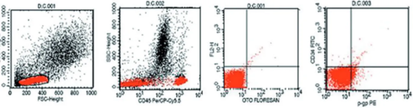
Expression of P-gp and CD34 was not observed in the samples of control group. Flow cytometric im-ages of one subject in the control group are shown in Figure 1.
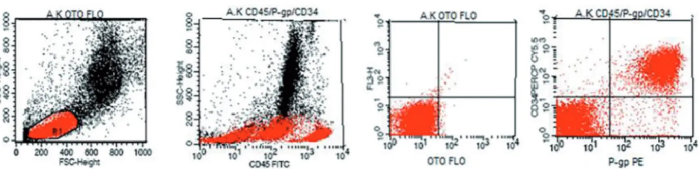
When we analyzed the BM of AML group us-ing flow cytometry, we observed strong expression of P-gp and CD34 in 6 of 30 (20%) AML patients. P-gp and CD34 expression was not observed in 24 of 30 (80%) AML patients (Table 1). Flow cyto-metric images of one subject in the AML group are shown in Figure 2.
Table 1. Comparison of AML and ALL patients in terms of the P-gp and CD34 expression
AML
P-gp Expression P-gp ExpressionALL
CD34
Expression (n) P-gp (+) (n) P-gp (-) (n) P-gp (+) (n) P-gp (-)
CD34 (+) (6) 20% (0) 0% (5) 25% (0) 0%
CD34 (-) (0) 0% (24)80% (1) 5% (14) 70%
AML: p=0.001, ALL: p=0.001, n: number of patients
When we analyzed BM of ALL group using flow cytometry, we observed strong expression of P-gp and CD34 in 5 of 20 (25%) ALL patients. We found that there was strong expression of P-gp but there was not CD34 expression in one of 20 (5%) ALL patients. P-gp and CD34 expression was not observed in 14 of 20 (70%) ALL group (Table 1). Flow cytometric images of one subject in the ALL group are shown in Figure 3.
Figure 2. Expression of P-gp and CD34 in a participant of AML group
Figure 3. Expression of P-gp and CD34 in a participant of ALL group
The distribution of P-gp and CD34 expression by gender in AML and ALL groups
P-gp expression was observed 12.5% in females and 28.6% in males of AML patients. P-gp expres-sion was observed 20% in females and 40% in males of ALL patients. CD34 expression was
ob-served 12.5% in females and 28.6% in males of AML patients. CD34 expression was observed 20% in females and 30% in males of ALL patients. There was not any significant difference between male and female expressions in terms of P-gp and CD34 (Table 2).
AML Group P-gp Expression ALL Group P-gp Expression
Gender (n) (n) P-gp (+) (n) P-gp (-) Gender (n) (n) P-gp (+) (n) P-gp (-) Female (16) (2) 12.5% (14) 87.5% Female (10) (2) 20% (8) 80% Male (14) (4) 28.6% (10) 71.4% Male (10) (4) 40% (6) 60% CD34 Expression CD34 Expression Gender (n) (n) CD34 (+) (n) CD34 (-) Gender (n) (n) CD34 (+) (n) CD34 (-) Female (16) (2) 12.5% (14) 87.5% Female (10) (2) 20% (8) 80% Male(14) (4) 28.6% (10) 71.4% Male (10) (3) 30% (7) 70%
AML: p=0.378, ALL: p=0.628, (n): Number of patients Table 2. The distribution
of P-gp expression by gender in AML and ALL groups
DISCUSSION
Our results provide evidence of MDR as shown by positive expression of P-gp and CD34 in patients with AML and ALL. On the other hand, very low P-gp expression is observed in stem cells of normal
hematopoiesis, which is consistent with previous studies [22,23].
Many methods using the determination of P-gp, responsible for cellular MDR, have been developed that analyze the level of protein, mRNA levels and
the functional levels [24-26]. However, as a result of research conducted in several centers, Chevillard et al. recommended the use of flow cytometry for analyses of MDR in protein levels and the use of real time polymerase chain reaction method in the mRNA levels [27]. In our study, MDR-1 expres-sions were measured with flow cytometry using 17F9 specific monoclonal antibody for P-gp mem-brane surface epitopes. Both the threshold value of P-gp and the functional status of the synthesized P-gp play a role in the appearance of clinical resis-tance phenotype [28]. Therefore, it is necessary to determine the threshold value for P-gp positivity in the method used in the determination of P-gp. In the present study, 102 and a higher level of fluorescence intensity was evaluated as positive on forward scat-tered light-side scatscat-tered light screen in flow cytom-etry.
We found that CD34 and P-gp expression lev-els are consistent with each other in patients with AML. We also showed that positive expression of P-gp and CD34 in 6 (20%) of 30 patients with AML and in 5 (25%) of 20 patients with ALL receiving chemotherapy.
The positive expressions of P-gp were evaluated for the presence of drug resistance. The increase of P-gp expression accelerates the elimination of drugs which are used in the treatment of ALL and AML. Thus, cancer cells are protected from the cytotoxic effects of chemotherapeutic drugs. Kuwazuru et al. showed that 9 of 17 patients with AML and 4 of 11 patients with ALL had P-gp positive results at the initial presentation, and most P-gp positive pa-tients did not respond to chemotherapy [29]. Pall et al. observed a significant correlation between CD34 expression and activity of P170 in AML patients. Senent et al. [30] conducted a study in patients with 82 AML by immunocytochemical method using the monoclonal antibody C219, and showed that CD34 expression were positive in 8 of P-gp positive 13 cases (62%). The researchers emphasized that P-glycoprotein expression is a reliable marker of pa-tients first diagnosed with AML. Consistent with our study, Gruber et al. [31] stated that expression of the MDR-1 gene could be an important factor con-tributing to drug resistance in acute leukemia. Simi-larly, Boekhorst et al. [32] reported that expression of P-170 encoded by the MDR-1 should be related
to the MDR. In the present study, we also found that 24 patients were seen in the remission process. P-gp and CD34 expressions were not observed in individuals entering remission process and controls group. These findings show that the treatments were successful. El-Ghaffar et al. [33] conducted a study on patients with acute leukemia (14 AML cases and 6 ALL cases); and showed that the functional activ-ity of MDR-1 P-gp was 71.4% in AML and 33.3% in ALL patients compared with 16.6% in normal lymphocytes. They suggested that P-gp/170 is ex-pressed to a higher degree in leukemic cells and this is greater in relapse when compared to de novo cases and more in AML than ALL blasts.
In light of these findins, the results suggest the expression of P-gp is closely related to clini-cal drug resistance in acute leukemia. We also in-vestigated the expression of P-170 encoded by the MDR-1 with flow cytometry; and we observed that P-gp was positive in patients with recurrence. The observation of MDR at a higher rate in ALL pa-tients compared to papa-tients with AML suggests that it may have occurred due to different morphologi-cal and phenotypic characteristics of ALL patients. There are several limitations for our study. Firstly, the small number of patients may be perceived as a limitation of the study. However, the major limita-tion of the study is that there was no way to evalu-ate the effect of individual chemotherapeutic agents that was used.
In conclusion, in terms of MDR, examination of patients diagnosed with hematologic malignancy especially in recurrent cases will shed light on che-motherapy resistance encountered in these patients. For measurement of MDR by flow cytometry, the most important advantages are the ones that make it possible to easily identify the cell clones of gated leukemic (blastic) and to make them specifically be analyzed in terms of P-gp. P-glycoprotein expres-sion can be measured quickly by flow cytometry; and this may pave the way to more rapid implemen-tation of alternative treatment regimens in especial-ly in recurrent cases.
Declaration of Conflicting Interests: The
au-thors declare that they have no conflict of interest.
Financial Disclosure: No financial support
REFERENCES
1. Faderl S, O’Brien S, Pui CH, et al. Adult acute lymphoblastic leukemia: concepts and strategies. Cancer 2010;116:1165-1176.
2. Ribera JM. Advances in acute lymphoblastic leukemia in adults. Curr Opin Oncol 2011;23:692-699.
3. Ayyıldız O, Işıkdoğan A, Çelik M, et al. Invasive pulmonary aspergillosis in a patient with acute lymphoblastic leuke-mia. Dicle Medical Journal 2004;31:57-61.
4. Döhner H, Estey EH, Amadori S, et al. European Leukemi-aNet. Diagnosis and management of acute myeloid leuke-mia in adults: recommendations from an international ex-pert panel, on behalf of the European Leukemia Net. Blood 2010;115:453-474.
5. Consoli U, Santonocito A, Stagno F, et al. Multidrug resis-tance mechanisms in chronic lymphocytic leukaemia. Br J Haematol 2002;116:774-780.
6. Gross HJ, Verwer B, Houck D, et al. Detection of rare cells at a frequency of one per million by flow cytometry. Cytom-etry 1993;14:519-526.
7. Gross HJ, Verwer B, Houck D, et al. Model study detecting breast cancer cells in peripheral blood mononuclear cells at frequencies as low as 10(-7), Proc Natl Acad Sci USA 1995;92:537-541.
8. Rosenblatt JI, Hokanson JA, McLaughlin SR, et al. Theo-retical basis for sampling statistics useful for detecting and isolating rare cells using flow cytometry and cell sorting. Cytometry 1997;27:233-238.
9. Rehsem MA, Corpuzm S, Heimfeldm S, et al. Use of fluores-cence threshold triggering and high-speed flow cytometry for rare event detection. Cytometry 1995;22:317-322. 10. Bay A, Boşnak V, Coşkun E, et al. Severe Rotavirus
gas-troenteritis in a patient with infant leukemia. Dicle Medical Journal 2011;38:101-103.
11. Drach D, Zhao S, Drach J, et al. Subpopulations of normal peripheral blood and bone marrow cells express a functional multidrug resistant phenotype. Blood 1992;80:2729-2734. 12. Fackler MJ, Krause DS, Smith OM, et al. Full-length but
not truncated CD34 inhibits hematopoietic cell differentia-tion of M1 cells. Blood 1995;85:3040-3047.
13. Healy L, May G, Gale K, et al. The stem cell antigen CD34 functions as a regulator of hemopoietic cell adhesion. Proc Natl Acad Sci USA 1995;92:1220-1224.
14. Cheng J, Baumhueter S, Cacalano G, et al. Hematopoi-etic defects in mice lacking the sialomucin CD34. Blood 1996;87:479-490.
15. Salati S, Zini R, Bianchi E, et al. Role of CD34 antigen in myeloid differentiation of human hematopoietic progenitor cells. Stem Cells 2008;26:950-959.
16. Haase D, Feuring-Buske M, Könemann S, et al. Evidence for malignant transformation in acute myeloid leukemia at the level of early hematopoietic stem cells by cytogenetic analysis of CD34+ subpopulations. Blood 1995;86:2906-2912.
17. Feller N, Schuurhuis GJ, van der Pol MA, et al. High per-centage of CD34 positive cells in autologous AML periph-eral blood stem cell products reflects inadequate in vivo
purging and low chemotherapeutic toxicity in a subgroup of patients with poor clinical outcome. Leukemia 2003;17:68-75.
18. Yıldırım AT, Gülen H. A child AML-M1 with CD79a, CD56, and CD5 coexpressions and misdiagnosed as biphenotypic acute leukemia. Dicle Medical Journal 2015;42:89-92. 19. Samdani A, Vijapurkar U, Grimm MA, et al.
Cytogenet-ics and P-glycoprotein (PGP) are independent predictors of treatment outcome in acute myeloid leukemia (AML). Leuk Res 1996;20:175-180.
20. Rosenau H. Legal prerequisites for clinical trials under the revised Declaration of Helsinki and the European Conven-tion on Human Rights and Biomedicine. Eur J Health Law 2000;7:105-121.
21. Tiirikainen MI, Syrjälä MT, Jansson SE. Flow cytometric analysis of P-glycoprotein in normal and leukemic cell. Ann Hematol 1992;65:124-130.
22. Chin KV, Pastan I, Gottesman MM. Function and regula-tion of the human multidrug resistance gene. Advances in Cancer Research 1993;60:157-181.
23. Nooter K, Herweijer H. Multidrug resistance (mdr) genes in human cancer. Br J Cancer 1991;63:663-669.
24. Beck WT, Grogan TM, Willman CL, et al. Methods to de-tect P-glycoprotein associated multidrug resistance in pa-tients’ tumors: Consensus recommendations. Cancer Res 1996;56:3010-3020.
25. Fenaux P, Preudhomme C, Laï JL, et al. Mutations of p53 gene in B-CLL report on 39 cases with cytogenetic analy-sis. Leukemia 1992;6:246-250.
26. Pall G, Spitaler M, Hofmann J, et al. Multidrug resistance in acute leukemia: a comparison of different diagnostic meth-ods. Leukemia 1997;11:1067-1072.
27. Chevillard S, Vielh P, Validire P, et al. French multicentric evaluation of mdr1 gene expression by RT-PCR in leuke-mia and solid tumors. standardization of RT-PCR and pre-liminary comparisons between RT-PCR and immunohisto-chemistry in solid tumors. Leukemia 1997;11:1095-1106. 28. Pirker R, Wallner J, Geissler K. MDR 1 gene expression
and treatment outcome in acut myeloid leukemia. J Natl Cancer Inst 1991;83:708-712.
29. Kuwazuru Y, Yoshimura A, Hanada S, et al. Expression of the multidrug transporter, P-glycoprotein, in acute leuke-mia cells and correlation to clinical drug resistance. Cancer 1990;66:868-873.
30. Senent L, Jarque I, Martín G, et al. P-glycoprotein expres-sion and prognostic value in acute myeloid leukemia. Hae-matologica 1998;83:783-790.
31. Gruber A, Vitols S, Norgren S, et al. Quantitative determi-nation of mdr-1 gene expression in leukemic cells from pa-tients with acute leukemia. Br J Cancer 1992;66:266-272. 32. te Boekhorst PA, de Leeuw K, Schoester M, et al.
Pre-dominance of functional multidrug resistance (MDR-1) phenotype in CD34+acute myeloid leukemia cells. Blood 1993;82:3157-3162.
33. Abd El-Ghaffar HA, Aladle DA, Farahat SE, et al. P-glyco-protein (P-170) expression in acute leukemias. Hematology 2006;11:35-41.