Evaluation of Neurotoxicity of Multiple Anesthesia in
Children Using Visual Evoked Potentials
N
eurotoxicity, due to anesthesia, particularly in children, is one of the major concerns of anesthesiologists in re-cent years.[1–3] Present studies in the literature are mostly experimental and conducted on animals since it is difficult to practice such prospective studies on children becauseof ethical reasons and the excessive time they consume.[4–6] The stage of brain development at the time of exposure to anesthesia and the frequency and cumulative anesthetic doses are some of the important factors causing neurotox-icity.[7]
Objectives: Anesthetic applications may cause increased neuronal damage in infants and children. Commonly cognitive or learn-ing disability tests were used to investigate the neurological progress in children. Visual Evoked Potential is a gross electrical signal generated by the occipital regions of the cerebral cortex in response to visual stimulation and an objective assessment of brain function. In this study, to acquire more objective results, Visual Evoked Potential responses of children who had multiple exposures to anesthesia during the treatment of corrosive esophagitis were compared to children who have never received anesthesia before. Methods: In this prospective, single-blinded, randomized, controlled study, 25 children, who were admitted to our pediatric sur-gery clinic because of corrosive esophagitis and who received general anesthesia more than 15 times composed Group-P; 25 children, who admitted to our well-child-clinic and who had never received anesthesia before consisted Group-C. The flash and pattern VEP responses of both groups were measured at the electrophysiology laboratory without any anesthetic drug application. The VEP responses of children in Group-P were recorded at least three days after the last exposure to anesthesia.
Results: Latencies and amplitudes of the N2 and P2 components of the pattern and flash VEP responses were statistically signifi-cantly different between the two groups (p=0.000).
Conclusion: This study shows that in children who had repeated anesthetic applications VEP parameters are significantly altered. We believe that VEP responses may be a reliable objective criterion for the evaluation of anesthesia neurotoxicity.
Keywords: Anesthesia; corrosive esophagitis; neurotoxicity; pediatric; visual evoked potential.
Please cite this article as ”Oba S, Işıl CT, Türk HŞ, Karamürsel S, Aksu S, Kaba M, et al. Evaluation of Neurotoxicity of Multiple Anesthesia in Children Using Visual Evoked Potentials. Med Bull Sisli Etfal Hosp 2019;53(3):284–289”.
Sibel Oba,1 Canan Tülay Işıl,1 Hacer Şebnem Türk,1 Sacit Karamürsel,2 Serkan Aksu,3 Meltem Kaba,4 Leyla Kılınç,1 Ali Ihsan Dokucu4
1Department of Anestesiology and Reanimation, Sisli Hamidiye Etfal Training and Research Hospital, Istanbul, Turkey 2Department of Physiology, Istinye University Faculty of Medicine, Istanbul, Turkey
3Department of Physiology, Istanbul University Istanbul Faculty of Medicine, Istanbul, Turkey
4Department of Pediatric Surgery, Sisli Hamidiye Etfal Training and Research Hospital, Istanbul, Turkey
Abstract
DOI: 10.14744/SEMB.2018.59454
Med Bull Sisli Etfal Hosp 2019;53(3):284–289
Address for correspondence: Hacer Şebnem Türk, MD. Sisli Hamidiye Etfal Egitim ve Arastirma Hastanesi, Anesteziyoloji ve
Reanimasyon Anabilim Dali, Istanbul, Turkey
Phone: +90 212 231 22 09 E-mail: [email protected]
Submitted Date: March 15, 2018 Accepted Date: March 30, 2018 Available Online Date: August 26, 2019
©Copyright 2019 by The Medical Bulletin of Sisli Etfal Hospital - Available online at www.sislietfaltip.org
OPEN ACCESS This is an open access article under the CC BY-NC license (http://creativecommons.org/licenses/by-nc/4.0/).
Commonly cognitive or learning disability tests were used to investigate the neurological progress in children.[3] How-ever, the learning disability tests used in the studies were not purely neuropsychological and the socioeconomic factors were also effective on the results. To acquire more objective results, we preferred to use the analysis of Visual Evoked Potential (VEP) responses to determine neurotoxic-ity of anesthesia in this study.
VEP is a gross electrical signal generated by the occipital regions of the cerebral cortex in response to visual stimu-lation.[8] The averaged VEP is a gross electrical signal gen-erated by the occipital regions of the cerebral cortex in response to visual stimulation. VEP studies to pattern re-versal stimulation have reported age-dependent wave-form changes, decrease in latency and increase in ampli-tude during early development and decline thereafter. VEP is more specific than the EEG and more sensitive to the changes in the visual stimuli providing ophthalmolo-gists and researchers information about the human visual system that is less available by other methods.[9] All of the studies have shown that VEP maturation is rapid in infants, gradual in preschool years and persists until adulthood.[10] The clinical studies related to anesthetic neurotoxicity were generally conducted on children who were exposed to general anesthesia only once. In our study, a group of children who had multiple (more than 15 times) anesthesia during their treatment for corrosive esophagitis, created a rare and special group to conduct research on the an-esthetic neurotoxicity. Accidental ingestion of household caustic cleaning material, such as bleaches or drain cleaner, causes esophagitis. These children have anesthesia from fifteen to forty times for treatment of esophagitis by bal-loon dilatation under general anesthesia.
In this study, we aimed to investigate whether anesthesia causes permanent neurotoxic harm or not, by comparing the VEP responses of 25 children who received repeated an-esthesia with children who had never received anan-esthesia.
Methods
After obtaining local Ethics Committee approval (352/02.09.2014) and informed consent of the parents, this study was conducted. Because repeated anesthesia ap-plications may be neurotoxic; data of children, who acci-dentally ingested liquid household cleaning material, such as bleach or drain cleaner, but who had no other systemic diseases, who underwent general anesthesia for at last 15 times for balloon dilatation and who were followed up in our pediatric surgery unit from 2011-2014 were included in this study in Group P (n=25). Patients with complications as perforation or undergoing major operations were taken as
exclusion criteria. Randomly 25 children from this patient population were selected to participate in Group P by a computer program.
Since it is expected, that neurotoxicity is fewer in children, who never received anesthesia before, the control group consisted of children from our well-child clinic. That is why Group C was also randomly selected from 25 children, who were under observation from birth to the present day at our hospital’s well-child clinic.
VEP recordings were taken at least three days after receiv-ing the last anesthesia. The dates and types of anesthesia received by the children of Group P were recorded retro-spectively. All of the children had received 0.1mg/kg IV or 0.4mg/kg oral midazolam (Dormicum, Roche, Hamburg, Germany) as premedication. Induction was performed with 8% sevoflurane in50% O2-N2O to the children who were not allowed for the IV access. For induction of children who had IV access, propofol 2-3mg/kg (Propofol %1, Fresenius, Hamburg, Germany), fentanyl 1mcg/kg (Fentanyl Citrate, Abbott, İllinois, United States) and 0.5mg/kg atracurium (Tracrium, Glaxo SmithKline, Auckland, New Zealand) were used. Anesthesia maintenance was provided with 1-3% sevoflurane (Sevorane, Abbott, Mascot Australia) in 50% O2-N2O. For early pain management, paracetamol 10 mg/ kg was used in all cases during the operations.
All children accompanied by their parents were randomly invited to the electrophysiology laboratory. Patients did not receive any anesthesia. VEP responses were recorded on the awake children.
The flash and pattern VEP (FVEP and PVEP) responses were taken at the electrophysiology laboratory of the Physiology Department of Istanbul University Istanbul Faculty of Medi-cine with a Nihon-Kohden RM 6000 polygraph system, and Ag/AgCl skin cup electrodes were used for both recordings. The mean luminance of the pattern VEP monitor was 105.35 cd/m2, and the contrast ratio between black and white squares was 110:1. Electrode impedances were kept below 5 kOhm using electrode paste. Refractive errors of all subjects were corrected while recording. Children were seated 1 m distant from the monitor, and signals were recorded as they looked at a fixation point in the middle of the screen with one eye while the other eye was occluded. The stimulus was an alternating, square black-and-white checkerboard pat-tern (100 stimuli) with 30 and 60 min check size, and stimu-lus reversal was 2 Hz. The stimustimu-lus was a light flash of one Joule with 1 µs duration at 1 Hz. Manual artifact rejection was applied to data for eye movement artifacts.
For FVEP and PVEP recordings, the active electrode was placed over the visual cortex at occipital zone according to the International 10/20 system, and the reference electrode
was placed on the left earlobe, while the ground electrode was placed on the right earlobe. The data were band-pass fil-tered between 1 and 45 Hz and averaged. The peak-to-peak amplitude of N75–P100 (negative wave at ca. 75. ms and positive wave at ca. 100. ms after the stimulus) and peak im-plicit times were measured for each eye separately. The data of the left and right eyes were then merged in the same pool.
Statistical Analysis
Statistical calculations were performed with the IBM SPSS Statistics 21. One-way ANOVA test was used for the com-parison of patient and control groups’ flash and pattern VEP responses components’ latencies and amplitudes.
Results
The children in Group P were under treatment for four years and were six to 14 years old. They were administered anesthesia for 15 to 40 times while their treatment was last-ing four to 48 months. The periods between two anesthesia applications were minimum of seven days and maximum of 60 days (Table 1).
Latencies and amplitudes of the N2 and P2 components of pattern VEPs taken with 30 and 60 min check sizes were evaluated statistically using one-way ANOVA, F-ratios and p-values. The latencies of the Group P were longer while their peak to peak amplitudes (N2-P2) were higher than those of the Group C (Table 2).
The latencies of the P2 components of the group P were longer while their peak to peak amplitudes (N2-P2) were higher than those of the group C. The N2 latencies were not statistically different between the two groups (Table 3).
Discussion
Previous clinical studies related to anesthetic neurotoxicity were generally conducted on children who had only one exposure to general anesthesia. Our study was conducted on a group of children who had anesthesia for more than 15 times during their treatment for corrosive esophagitis. This group of children was a rare and special study group to conduct research on the anesthetic neurotoxicity. This study showed that in children who had repeated anes-thetic applications VEP parameters are significantly altered, which indicates neurotoxicity.
Preclinical and also retrospective clinical studies suggested that anesthesia could be damaging brain functions in chil-dren.[7] The mechanism of anesthesia-induced neurotoxici-ty is complex: General anesthetics affect multiple ion chan-nels, receptors and cell signaling systems in the central nervous system to produce anesthesia.[2] In the previous studies, it has been indicated that inhaled anesthetics by
causing apoptosis and reducing neuro-genesis may have serious effects on neonatal animals during neuro-devel-opmental periods.[2] The studies conducted on developing monkeys have shown that exposure to anesthesia causes neuro-apoptosis[4, 5] and permanent neuro-cognitive defi-cits in the developing monkey brain.[6]
On the other hand, the reflection of these laboratory find-ings to the clinical practice remains unclear because of very restricted knowledge existing in the literature. It is quite difficult to find or prove the neurological harm which might be caused by anesthesia in clinical studies conduct-ed on children. These studies sometimes cannot be con-ducted due to ethical reasons, while studies which can be conducted consume too much time.
Pediatric Anesthesia NeuroDevelopment Assessment (PANDA Study) which compared children who were given general anesthesia for inguinal hernia surgery before the age of three with siblings who had no received anesthe-sia before age three, showed no significant difference in IQ
Table 1. Demographic data
Group P Group C
(n=25) (n=25) Age at the time of the study (years) 9.7±3.1 (6-14) 10.1±4.2 (6-14)
Female/Male 2F/23M 2F/23M
Age at the time of the first 6.4±2.7 (2-10) anesthesia (years)
Treatment period (months) 34.6±13.3 (4-48) The number of anesthesia given 31.7±9.0 (15-40) The average duration of 28.4±14.7 (20-45) anesthesia (minute)
Table 2. Pattern VEP data of the control and patient groups.One way ANOVA results (f ratio and p values)
Group C Group P f p 30N2Lat 68.23±5.41 78.08±8.51 24.265 0.000 30P2Lat 100.15±8.35 119.25±12.29 41.865 0.000 30Amp 5.52±0.78 14.82±7.39 40.827 0.000 60N2Lat 68.38±5.22 75.75±8.89 13.016 0.001 60P2Lat 100.15±8.16 117.67±11.15 40.641 0.000 60Amp 5.53±0.78 14.4±8.71 26.805 0.000 Table 3. Flash VEP data of the control and patient groups.
Group C Group P f p
VEPN2Lat 68.23±4.47 70.87±8.35 1.993 0.164 VEPP2Lat 100.23±7.25 121.83±16.94 35.306 0.000 VEPAmp 5.57±0.83 20.12±9.10 65.978 0.000
scores between the siblings when assessed between the ages of 8 and 15. This study included healthy children with a single exposure to general anesthesia. A different study, including children with a more complicated medical his-tory and with multiple general anesthesia exposures, may show different results.[11, 12]
One previous study, General Anesthesia compared to Spi-nal Anesthesia(GAS) found strong evidence that exposure of just under an hour to a sevoflurane GA in infancy does not increase the risk of adverse neurodevelopmental out-come at two years of age.[12, 13]
The Mayo Anesthesia Safety in Kids (MASK) study which analyses the condition of the children who had anesthe-sia for once or more is currently incomplete.[3] Our study group is a rare and specific group for investigating the neu-ro-detrimental effect of multiple anesthesias on children. In the literature, psychological tests and/or tests evaluat-ing cognitive functions are frequently used to evaluate the neurologic harm.[9, 10, 14–16] These tests evaluate learn-ing disability, behavior, autism and education difficulties. Children’s disabilities in paying attention, learning, mem-ory and social activities are discussed. Children, who were hospitalized multiple times due to esophagitis might have psychological and scholar problems independent of re-petitive anesthesia applications. That is why we preferred to use VEP responses in our study, contrary to the majority of researchers who investigated anesthesia neurotoxicity using subjective psychological tests. We believe that VEP responses give us more objective data to determine neuro-toxicity of anesthesia. Moreover, VEP analysis is easy to use in child studies as a research indicator.
Latencies and amplitudes of VEP components are mostly related to myelination, brain development and organic de-fects rather than to psychological disorders. However, the excessive increase in amplitudes attributes to disinhibition and therefore cannot be evaluated as a positive sign. In-creases in the amplitudes of the VEP components in our study, while their latencies were longer than the control group, can be evaluated as disinhibition pathology, rather than better brain development.
Todorovic-Jevkovic et al.[7] reported at BJA Salzburg semi-nar, that children, who received anesthesia before they were 12 months old, suffered weaknesses in memory at age 6-11 years. They mentioned that the brain cells may be affected in different degrees related to age dur-ing anesthesia application. In another animal study, they reported that brain cell death caused by anesthesia was observed in rats and that a long-lasting exposure to anes-thesia caused worse effects rather than frequent applica-tion.[17] Di Maggio et al.[18] found that siblings who were younger than three years and who received anesthesia for operations had a risk elevation of 60% in developmen-tal or behavioral disorders, when compared to a similar group of siblings, who did not undergo any operations. [18] Similarly, in our study, the most harmful effects were detected in four patients, who had their first anesthesia application before they were three years old (Figs. 1, 2). Besides, these children were exposed to anesthesia most frequently as totally 36-40 times.
Some studies underlined differences in cognitive dysfunc-tion related to gender. They found that boys were affected significantly more than girls.[17, 19] In our study, such a
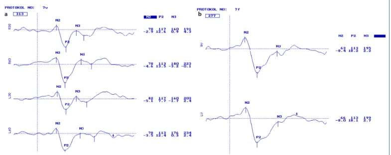
com-Figure 1. Pattern (a) and Flash (b) VEP of a healthy child of seven years old. Latency and amplitudes are seen in the first and second lines con-secutively at the right side of the curves. R for right and L for left eye responses. 30 and 60 attribute to check sizes of 30 and 60 mins.
parison could not be make since the majority of the pa-tients were males.
In previous studies, the neurotoxic effects of the anesthesia were investigated in children who had undergone only a single seance of anesthesia.[20, 21] In this study, our patients had at least 15 repetitive anesthesia applications. One of the limitations of this study was that we had no study group consisting of children, who received anesthesia once. Another limitation was that we could not measure VEP responses in our study group before their first anes-thetic application because all patients underwent emer-gency interventions and we randomized the study group from a retrospective patient data pool. Further studies with a larger number of patients are required to support our findings.
Conclusion
In conclusion, our study shows that in children who had multiple anesthetic applications, VEP parameters are sig-nificantly altered. We believe that VEP responses may be a reliable objective criterion for the evaluation of anesthesia neurotoxicity.
Disclosures
Ethics Committee Approval: Sisli Hamidiye Etfal Education and
Research Hospital; 352/02.09.2014.
Peer-review: Externally peer-reviewed. Conflict of Interest: None declared.
Authorship Contributions: Concept – S.O., S.K.; Design – S.O.,
S.K.; Supervision – A.İ.D.; Data collection &/or processing – C.T.I.,
H.Ş.T., S.A., M.K.; Analysis and/or interpretation – L.K., C.T.I., H.Ş.T.; Literature search – S.O., S.K., C.T.I., H.Ş.T.; Writing – S.O., L.K., H.Ş.T.; Critical review – A.İ.D.
References
1. Nemergut ME, Aganga D, Flick RP. Anesthetic neurotoxicity: what to tell the parents? Paediatr Anaesth 2014;24:120–6.
2. Hudson AE, Hemmings HC Jr. Are anaesthetics toxic to the brain? Br J Anaesth 2011;107:30–7.
3. Sun L. Early childhood general anaesthesia exposure and neuro-cognitive development. Br J Anaesth 2010;105 Suppl 1:i61–8. 4. Brambrink AM, Back SA, Riddle A, Gong X, Moravec MD, Dissen
GA, et al. Isoflurane-induced apoptosis of oligodendrocytes in the neonatal primate brain. Ann Neurol 2012;72:525–35. 5. Zou X, Patterson TA, Sadovova N, Twaddle NC, Doerge DR, Zhang
X, et al. Potential neurotoxicity of ketamine in the developing rat brain. Toxicol Sci 2009;108:149–58.
6. Paule MG, Li M, Allen RR, Liu F, Zou X, Hotchkiss C, et al. Ket-amine anesthesia during the first week of life can cause long-lasting cognitive deficits in rhesus monkeys. Neurotoxicol Teratol 2011;33:220–30.
7. Jevtovic-Todorovic V, Absalom AR, Blomgren K, Brambrink A, Crosby G, Culley DJ, et al. Anaesthetic neurotoxicity and neuro-plasticity: an expert group report and statement based on the BJA Salzburg Seminar. Br J Anaesth 2013;111:143–51.
8. Lenassi E, Likar K, Stirn-Kranjc B, Brecelj J. VEP maturation and visual acuity in infants and preschool children. Doc Ophthalmol 2008;117:111–20.
9. Sprung J, Flick RP, Wilder RT, Katusic SK, Pike TL, Dingli M, et al. An-esthesia for cesarean delivery and learning disabilities in a popu-Figure 2. Pattern (a) and Flash (b) VEP of a child of six years old who had his first anesthesia before three years old. Latency and amplitudes are seen in the first and second lines consecutively at the right side of the curves. R for right and L for left eye responses. 30 and 60 attribute to check sizes of 30 and 60 mins. Note that the latency of the N2 and P2 components are longer than normal, and there are even later com-ponents.
lation-based birth cohort. Anesthesiology 2009;111:302–10. 10. Sokol S. Visually evoked potentials: theory, techniques and
clini-cal applications. Surv Ophthalmol 1976;21:18–44.
11. Sun LS, Li G, Miller TL, Salorio C, Byrne MW, Bellinger DC et al. As-sociation Between a Single General Anesthesia Exposure Before Age 36 Months and Neurocognitive Outcomes in Later Child-hood. JAMA 2016 7;315:2312–20.
12. Davidson AJ, Disma N, de Graaff JC, Withington DE, Dorris L, Bell G, et al. Neurodevelopmental outcome at 2 years of age after general anaesthesia and awake-regional anaesthesia in infancy (GAS): an international multicentre, randomised controlled trial. Lancet 2016 16;387:239–50.
13. Wilder RT, Flick RP, Sprung J, Katusic SK, Barbaresi WJ, Mickelson C, et al. Early exposure to anesthesia and learning disabilities in a population-based birth cohort. Anesthesiology 2009;110:796– 804.
14. Millar K, Bowman AW, Burns D, McLaughlin P, Moores T, Morton NS, et al. Children's cognitive recovery after day-case general an-esthesia: a randomized trial of propofol or isoflurane for dental procedures. Paediatr Anaesth 2014;24:201–7.
15. Kayaalp L, Bozkurt P, Odabasi G, Dogangun B, Cavusoglu P, Bolat N, et al. Psychological effects of repeated general anesthesia in
children. Paediatr Anaesth 2006;16:822–7.
16. Jevtovic-Todorovic V, Hartman RE, Izumi Y, Benshoff ND, Dikra-nian K, Zorumski CF, et al. Early exposure to common anesthetic agents causes widespread neurodegeneration in the developing rat brain and persistent learning deficits. J Neurosci 2003;23:876– 82.
17. DiMaggio C, Sun LS, Li G. Early childhood exposure to anesthesia and risk of developmental and behavioral disorders in a sibling birth cohort. Anesth Analg 2011;113:1143–51.
18. Lee BH, Chan JT, Kraeva E, Peterson K, Sall JW. Isoflurane expo-sure in newborn rats induces long-term cognitive dysfunction in males but not females. Neuropharmacology 2014;83:9–17. 19. Stratmann G. Review article: Neurotoxicity of anesthetic drugs in
the developing brain. Anesth Analg 2011;113:1170–9.
20. Lunardi N, Ori C, Erisir A, Jevtovic-Todorovic V. General anesthesia causes long-lasting disturbances in the ultrastructural properties of developing synapses in young rats. Neurotox Res 2010;17:179– 88.
21. DiMaggio C, Sun LS, Kakavouli A, Byrne MW, Li G. A retrospective cohort study of the association of anesthesia and hernia repair surgery with behavioral and developmental disorders in young children. J Neurosurg Anesthesiol 2009;21:286–91.