Delay of cognitive gamma responses in Alzheimer's disease
Erol Ba
şar
a,⁎
, Derya Durusu Emek-Sava
ş
a,b,c, Bahar Güntekin
a, Görsev G. Yener
a,c,d,e aBrain Dynamics, Cognition and Complex Systems Research Center, Istanbul Kultur University, Istanbul 34156, Turkey
b
Department of Psychology, Dokuz Eylül University, Izmir 35160, Turkey
cDepartment of Neurosciences, Dokuz Eylül University, Izmir 35340, Turkey d
Brain Dynamics Multidisciplinary Research Center, Dokuz Eylül University, Izmir 35340, Turkey
e
Department of Neurology, Dokuz Eylül University Medical School, Izmir 35340, Turkey
a b s t r a c t
a r t i c l e i n f o
Article history: Received 3 August 2015
Received in revised form 5 January 2016 Accepted 18 January 2016
Available online 20 January 2016
Event-related oscillations (EROs) reflect cognitive brain dynamics, while sensory-evoked oscillations (SEOs) re-flect sensory activities. Previous reports from our lab have shown that those with Alzheimer's disease (AD) or mild cognitive impairment (MCI) have decreased activity and/or coherence in delta, theta, alpha and beta cogni-tive responses. In the current study, we investigated gamma responses in visual SEO and ERO in 15 patients with AD and in 15 age-, gender- and education-matched healthy controls. The following parameters were analyzed over the parietal-occipital regions in both groups: (i) latency of the maximum gamma response over a 0– 800 ms time window; (ii) the maximum peak-to-peak amplitudes for each participant's averaged SEO and ERO gamma responses in 3 frequency ranges (25–30, 30–35, 40–48 Hz); and (iii) the maximum peak-to-peak amplitudes for each participant's averaged SEO and ERO gamma responses over a 0–800 ms time block contain-ing four divided time windows (0–200, 200–400, 400–600, and 600–800 ms). There were main group effects in terms of both latency and peak-to-peak amplitudes of gamma ERO. However, peak-to-peak gamma ERO tude differences became noticeable only when the time block was divided into four time windows. SEO ampli-tudes in the 25–30 Hz frequency range of the 0–200 ms time window over the left hemisphere were greater in the healthy controls than in those with AD. Gamma target ERO latency was delayed up to 138 ms in AD patients when compared to healthy controls. Thisfinding may be an effect of lagged neural signaling in cognitive circuits, which is reflected by the delayed gamma responses in those with AD. Based on the results of this study, we pro-pose that gamma responses should be examined in a more detailed fashion using multiple frequency and time windows.
© 2016 The Authors. Published by Elsevier Inc. This is an open access article under the CC BY license (http://creativecommons.org/licenses/by/4.0/). Keywords: Event-related Sensory-evoked Oscillation Gamma Alzheimer's disease EEG P300 1. Introduction
Alzheimer's disease (AD) is the most common dementing illness. In the majority of cases, mild cognitive impairment (MCI) is considered to be prodromal AD (Petersen et al., 2001; Rasquin et al., 2005; Alexopoulos et al., 2006). Current diagnostic methods are heavily weighted by the amyloid and tau levels in the cerebrospinalfluid (CSF) and by volumetric magnetic resonance imaging (MRI) measure-ments. The full potential of electrophysiological methods for use in predicting (Cichocki et al., 2005; Babiloni et al., 2006a; Rossini et al., 2006), diagnosing (Yener et al., 1996; Polich and Herbst, 2000; Jeong, 2004; Babiloni et al., 2006b; Karrasch et al., 2006), and monitoring treat-ment or progress (Jelic et al., 2000; Dauwels et al., 2010) in AD/MCI pa-tients has not been fully examined in routine clinical practice.
Brain oscillatory responses can be used for the non-invasive analysis of local neuronal synchronization, corticortical connectivity, and co-herence of oscillations (Rossini et al., 2007). Cognitive stimuli can elicit event-related oscillations (EROs), which is a powerful technique with high temporal resolution. ERO has been described as a useful tool for de-tecting subtle abnormalities of cognitive processes (Basar, 1980, 2004). In our previous work, we explored ERO, sensory-evoked oscillations (SEOs), and the evoked or event-related coherence of AD/MCI patients using visual and auditory sensory modalities (Yener et al., 2008, 2009, 2012; Güntekin et al., 2008; Basar et al., 2010; Yener and Başar, 2010). The term“event-related” is used for a “potential” that is elicited after a cognitive task, while the term“sensory-evoked” is used for a “potential” that is elicited after a sensory stimulus (Başar et al., 1997).
The history of gamma activity began in the 1940s (Adrian, 1942). In subsequent years,Freeman (1975)andBaşar et al. (1975a, 1975b, 1975c)indicated that gamma oscillatory responses reflect a wide varie-ty of functions. In 1972,Başar and Özesmiintroduced the terminology “gamma response” to describe hippocampal gamma band activity elicit-ed by external stimuli in cats. In human studies,Galambos (1981)later
⁎ Corresponding author at: Istanbul Kültür University, Brain Dynamics, Cognition and Complex Systems Research Center, Faculty of Science and Letters, Ataköy Campus, Bakırköy, 34156 Istanbul, Turkey.
E-mail address:[email protected](E. Başar).
http://dx.doi.org/10.1016/j.nicl.2016.01.015
2213-1582/© 2016 The Authors. Published by Elsevier Inc. This is an open access article under the CC BY license (http://creativecommons.org/licenses/by/4.0/).
Contents lists available atScienceDirect
NeuroImage: Clinical
indicated that there are sensory and cognitive correlates of gamma re-sponses. Gamma oscillatory responses are selectively distributed in the brain, but they do not appear to reflect a specific function in the ner-vous system. Gamma activity has been related to both sensory and cog-nitive responses from the cortex, hippocampus, thalamus, and reticular formations in both animal and human brains (Başar, 2013). Thus, it can be hypothesized that gamma-band synchronization is most likely a fun-damental process in all brain functions [seeBaşar et al. (1999, 2013)and
Başar-Eroglu et al. (1996a)].
In the last decade, there have been several studies published on gamma activity in cognitive impairment, especially in schizophrenia. Most of the results related to these functions indicate a decrease in gamma responses. It is important to note that studies on healthy partic-ipants and particpartic-ipants with cognitive impairment have contradictory results and interpretations. Gamma oscillatory responses have been found to play role in perception, attention and memory processes, object recognition, face recognition and emotional paradigms (Güntekin and Başar, 2014; Keil et al., 1999; Busch et al., 2004, 2006; Tallon-Baudry et al., 1998; Gruber et al., 2004; Herrmann et al., 2004a; Müller and Keil, 2004; Senkowski and Herrmann, 2002; for further information on gamma responses please see reviewsBaşar, 2013; Başar-Eroglu et al., 1996b; Herrmann et al., 2004b; Jensen et al., 2007; Singer, 1999; Tallon-Baudry and Bertrand, 1999). In the literature, gamma responses have mostly been analyzed in single frequency and single time windows. There are few studies analyzing gamma responses in multiple frequency and time windows. However, our recent study (Başar et al., 2015) showed that analyzing the gamma responses in multiple frequency and time windows is extremely important.Başar et al. (2015)showed that, especially during cognitive paradigm, there are at least 3–4 phase/ time-locked gamma responses in the 25–45 Hz frequency windows that occur in multiple time windows (between 0 and 800 ms). In most cases cognitive responses are late (200–400 ms, 400–600 ms), and they depict higher frequencies. Since there were many differences in the gamma responses in multiple frequency and time domains, this manuscript aims to analyze gamma responses in multiple time and fre-quency windows.
The literature regarding gamma responses in AD or MCI indicates that auditory steady state gamma responses with amplitudes of 40 Hz are increased in AD (Osipova et al., 2006) and MCI (van Deursen et al., 2011) patients when compared to controls. Another study comparing gamma activity during the N-back paradigm in stable and progressive MCI patients indicates that the progressive MCI group has lower aver-age changes in gamma values (Missonnier et al., 2010). In the present study, we aimed to analyze gamma responses elicited by sensory or cog-nitive stimulation in AD patients using multiple frequency bands and many time windows. A new strategy was used that involved the analy-sis of three gamma frequency bands within four time windows. We hy-pothesized that cognitive gamma responses would be delayed in AD due to lagged neural signals in cognitive circuits.
2. Materials and methods 2.1. Participants
A total of 15 probable mild AD patients who were diagnosed accord-ing to DSM-IV and NINCDS-ADRDA criteria and 15 age-, gender-, and education-matched healthy controls were consented to participate in the study. All AD patients were within thefirst year of their diagnosis, and six of these patients were taking a cholinesterase inhibitor (donepezil, rivastigmine). The mean age of the healthy controls was 67.47 years (SD 4.14), while the mean age of the AD patients was 67.53 years (SD 6.48). The mean educational years was 8.73 (SD 6.03) for the healthy controls and 8.67 (SD 4.75) for the AD patients. There were 7 females and 8 males in each group. The mini-mental state exam-ination (MMSE) scores ranged between 28 and 30 for the healthy con-trols and 16–27 for the AD patients, out of a possible 30 points. The
general demographic and clinical features of both groups are shown in
Table 1. All participants and/or their relatives provided informed con-sent for the study, which was approved by the local ethical committee. 2.2. Acquisition of visual sensory-evoked oscillations (SEOs) and visual event-related oscillations (EROs)
2.2.1. Sensory-evoked oscillations (SEOs)
A visual sensory paradigm was administered to each participant. A white screen with 40 cd/cm2luminance was used as the stimulus. The
duration of the stimulation was 1000 ms. Sixty stimulation signals were applied, and the inter-stimulus intervals varied randomly be-tween 3 and 7 s.
2.2.2. Event-related oscillations (EROs)
A classical visual oddball paradigm was administered to all partici-pants. There were 40 target and 80 standard stimulations. The probabil-ity of the target stimuli was 0.33. A white screen with a 10 cd/cm2
luminance was used for standard signal stimulation and 40 cd/cm2
was used for the target signals. The duration of the stimulation was 1000 ms. The light appeared at full size on a 22-inch computer monitor with a refresh rate of 75 Hz. The target stimuli were embedded random-ly within a series of standard stimuli in all of the paradigms. The task re-quired the target stimuli to be counted, and the inter-stimulus interval varied randomly between 3 and 7 s.
Ten of the healthy controls counted 40 target stimulations; three of the healthy controls made one mistake while counting the target stim-ulation; and two made more than one mistake. Eight of the AD patients counted 40 target stimulations; two of the AD patients made one mis-take; andfive of them made more than one mistake. There was no sig-nificant difference between groups in terms of counting the target stimulation (p = 0.389).
2.3. Electrophysiological recording
EEGs were recorded according to the International 10–20 system using 30 Ag-AgCl electrodes mounted in an elastic cap (Easy-cap). Two additional linked Ag-AgCl earlobe electrodes (A1 + A2) were used as ref-erences. The electrooculogram (EOG) was registered from both the me-dial upper and the lateral orbital rim of the right eye. All electrode impedances were less than 10 kΩ. The EEG was amplified with a BrainAmp 32-channel DC system with band limits of 0.01–250 Hz, and a sampling rate of 500 Hz was used.
Prior to averaging the data, epochs containing artifacts were rejected by a manual off-line technique (i.e., single sweep EOG recordings were visually studied, and trials with eye movement or blink artifacts were rejected). Sweep numbers were randomly equalized between the target and simple visual stimulation.
2.4. Measurements
Gamma SEO and ERO responses were digitallyfiltered in three gamma ranges measured from P3, Pz, P4, O1, Ozand O2locations and
usingfilter limits of 25–30, 30–35, and 40–48 Hz. The slope of the
Table 1
General demographic and clinical features of participants.
Healthy controls (N = 15) AD patients (N = 15) p Age (SD) 67.47 (4.14) 67.53 (6.48) 0.973a
Education (SD) 8.73 (6.03) 8.67 (4.75) 0.973a
Gender (M/F) 8/7 8/7 1.000b
MMSE (SD) 28.73 (2.02) 21.85 (3.46) 0.000a
SD: standard deviation, M: male, F: female, AD: Alzheimer's disease, MMSE: mini-mental state examination
a
Independent sample t-test.
b
band-passfilter was 48 dB/octave. Parietal and occipital locations were chosen as the regions of interest since gamma activity has been reported to produce significant results upon visual stimulation, especially in the posterior parts of the brain (Başar et al., 2015; Busch et al., 2006; Castelhano et al., 2013; Sakowitz et al., 2001). In our previous study (Başar et al., 2015), we investigated multiple gamma oscillatory re-sponses in healthy young subjects upon presentation of simple visual stimuli and visual oddball paradigm. Gamma responses in multiple time and frequency windows were analyzed from the frontal, central, parietal and occipital electrodes. This manuscript showed that the most significant results were found in the parietal and occipital regions. Accordingly, in the present manuscript, parietal and occipital locations were chosen as the regions of interest.
First, the peak-to-peak amplitude and the latency of the maximum gamma oscillatory activity over a 0–800 ms time window were mea-sured in 3 gamma ranges (25–30, 30–35, 40–48 Hz), which were chosen from the power spectra evaluation. Then, the peak-to-peak amplitude values of the gamma responses were measured in 3 frequency ranges (25–30 Hz, 30–35 Hz, 40–48 Hz) over 4 time windows (0–200 ms, 200–400 ms, 400–600 ms, 600–800 ms). These time periods were cho-sen based on the evaluation of the grand average pictures. In our previ-ous study, we analyzed the multiple gamma responses of healthy young subjects upon presentation of visual sensory stimulation and visual odd-ball paradigm (Başar et al., 2015). In that study, we showed that there were multiple gamma responses in different time domains. Late gamma responses mostly appeared during cognitive load. The differ-ence between cognitive stimulation and sensory stimulation in the pa-rietal locations was seen mostly in the 600–800 ms time window. Furthermore, we showed that 40–48 Hz gamma response oscillations were significantly greater in the third time window (400–600 ms) upon application of target stimulation in comparison to simple light stimulation. The present study therefore takes our previousfindings into consideration. The observations of subject averages as well as the statistical results of the present study show that AD patients have signif-icantly later responses than healthy controls. It would not be possible to see this difference without analyzing the gamma responses in different time windows, including the late responses (400–600 ms, 600–800 ms). Three different measurements were performed in order to deter-mine the entire dynamic properties of the gamma responses. An earlier report from our lab (Başar et al., 2015) showed that there are multiple gamma responses over different time and frequency windows during cognitive stimulation. Therefore, it is more appropriate to analyze gamma responses over multiple time and frequency domains. In addi-tion, we (Başar et al., 2015) previously showed that cognitive stimula-tion elicited more gamma responses over different time and frequency windows than did simple sensory stimulation.
2.5. Statistical analysis
Statistical analyses were performed with Statistica Software. Repeat-ed measures ANOVA was usRepeat-ed for statistical analysis. RepeatRepeat-ed mea-sures ANOVAs were run separately for three different measurements as follows: (1) maximum peak-to-peak gamma amplitudes for three different gamma frequency ranges (25–30 Hz, 30–35 Hz, 40–48 Hz) over a 0–800 ms time window; (2) maximum peak-to-peak gamma amplitudes for three different frequency ranges (25–30 Hz, 30–35 Hz, 40–48 Hz) and over four different time windows (0–200 ms, 200– 400 ms, 400–600 ms, 600–800 ms); and (3) latency of maximum gamma amplitude for three different gamma frequency ranges (25– 30 Hz, 30–35 Hz, 40–48 Hz) over a 0–800 ms time window.
The repeated measures ANOVA analysis of“maximum peak-to-peak gamma amplitudes for three different gamma frequency ranges over a 0–800 ms time window” included 2-level GROUP (AD patients and healthy controls) as between-subject factors, and frequency range (FR [3 levels] = 25–30 Hz, 30–35, 40–48 Hz), anterior–posterior
distribution (AP [2 levels] = parietal, occipital), and lateral distribution (LAT [3 levels] = left, midline, right) as within-subject factors.
The repeated measures ANOVA analysis for“maximum peak-to-peak gamma amplitudes for three different frequency ranges and over four dif-ferent time windows” included 2-level GROUP (AD patients and healthy controls) as between-subject factors, and time window (TW [4 levels] = 0–200 ms, 200–400 ms, 400–600 ms, 600–800 ms), frequency range (FR [3 levels] = 25–30 Hz, 30–35, 40–48 Hz), anterior–posterior distribution (AP [2 levels] = parietal, occipital), and lateral distribution (LAT [3 levels] = left, midline, right) as within-subject factors.
The repeated measures ANOVA analysis for“latency of maximum gamma amplitude for three different gamma frequency ranges over a 0–800 ms time window” included 2-level GROUP (AD patients and healthy controls) as between-subject factors, and frequency range (FR [3 levels] = 25–30 Hz, 30–35, 40–48 Hz), anterior–posterior distribu-tion (AP [2 levels] = parietal, occipital), and lateral distribudistribu-tion (LAT [3 levels] = left, midline, right) as within-subject factors.
These analyses were carried out for both SEO and ERO responses separately. In summary, six different repeated measures ANOVAs were run as described above and listed below:
1) SEO:“maximum peak-to-peak gamma amplitudes for three differ-ent gamma frequency ranges (25–30 Hz, 30–35 Hz, 40–48 Hz) over a 0–800 ms time window”
2) SEO:“maximum peak-to-peak gamma amplitudes for three differ-ent frequency ranges (25–30 Hz, 30–35 Hz, 40–48 Hz) and over four different time windows (0–200 ms, 200–400 ms, 400–600 ms, 600–800 ms)”
3) SEO:“latency of maximum gamma amplitude for three different gamma frequency ranges (25–30, 30–35, 40–48 Hz) over a 0– 800 ms time window”
4) ERO in response to target stimulation:“maximum peak-to-peak gamma amplitudes for three different gamma frequency ranges (25–30 Hz, 30–35 Hz, 40–48 Hz) over a 0–800 ms time window” 5) ERO in response to target stimulation:“maximum peak-to-peak
gamma amplitudes for three different frequency ranges (25–30 Hz, 30–35 Hz, 40–48 Hz) and over four different time windows (0– 200 ms, 200–400 ms, 400–600 ms, 600–800 ms)”
6) ERO in response to target stimulation:“latency of maximum gamma amplitude for three different gamma frequency ranges (25–30, 30– 35, 40–48 Hz) over a 0–800 ms time window”
Greenhouse–Geisser corrected p-values are reported. Post-hoc com-parisons were analyzed with t-tests used with Bonferroni correction. Levels of pb 0.05 were considered significant for all comparisons. 3. Results
3.1. Visual sensory-evoked oscillation (SEO) amplitudes
3.1.1. Maximum peak-to-peak gamma SEO amplitudes at three frequency ranges over a 0–800 ms time window
There was no main GROUP effect on visual SEO amplitudes for the three frequency ranges over a 0–800 ms time window. However, there was a main FR effect [F2.56= 14.112; p = 0.000], with lower
am-plitudes in the 40–48 Hz frequency range than in both the 25–30 Hz (p = 0.000) and the 30–35 Hz (p = 0.024) frequency ranges. It is also important to keep in mind that higher frequency brain oscillations almost always show lower amplitudes.
3.1.2. Maximum peak-to-peak gamma SEO amplitudes at three frequency ranges over four time windows
Repeated measures ANOVA revealed main effects for FR [F2.56=
17.244; p = 0.000], TW [F3.84= 10.454; p = 0.000] and AP [F1.28=
14.203; p = 0.001]. Interaction-effects for FR × TW [F6.168= 2.956;
p = 0.037], FR × AP [F2.56 = 13.685; p = 0.000], and
Post-hoc analysis of FR revealed that the 25–30 Hz frequency range had a significantly higher amplitude than the 30–35 Hz (p = 0.000) and the 40–48 Hz (p = 0.001) frequency ranges. Moreover, the 30–35 Hz frequency range had a significantly higher amplitude than the 40– 48 Hz (p = 0.01).
Post-hoc analysis for TW revealed that the 0–200 ms time window had the highest amplitude when compared to other three time win-dows (p = 0.001, p = 0.003, p = 0.045, respectively). The 600– 800 ms time window had the second highest amplitude, while both the 200–400 ms and the 400–600 ms time windows had the lowest amplitudes.
Post-hoc analysis for AP indicated that the parietal locations had sig-nificantly higher gamma SEO amplitudes than the occipital electrodes (p = 0.001).
Post-hoc analysis of FR × AP revealed that the parietal 25–30 Hz gamma SEO amplitude was significantly higher than all of the others (pb 0.001 for all comparisons), while 40–48 Hz gamma SEO amplitude in both the parietal and the occipital leads was the lowest.
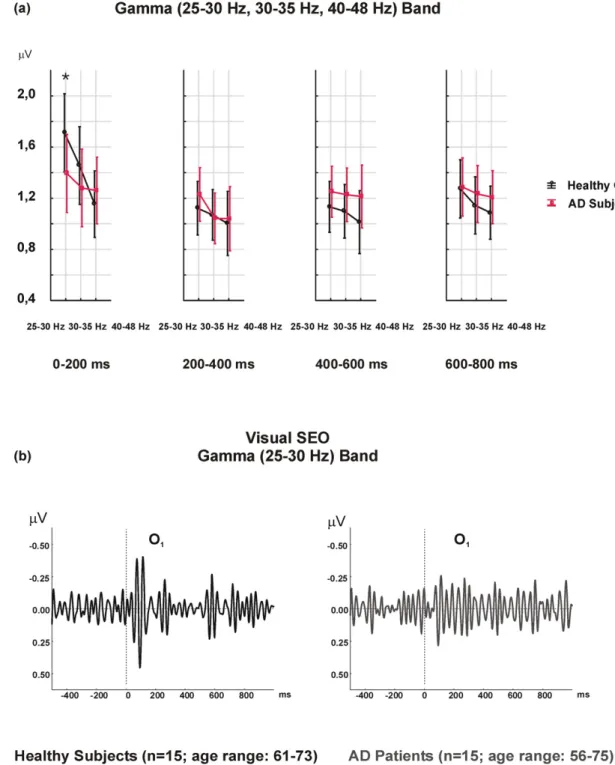
The post-hoc analysis of FR × TW × LAT × GROUP indicated non-significant results. However, the differences between the healthy con-trols and AD patients were greatest in the 0–200 ms time window. The healthy controls had higher gamma amplitudes than AD patients, especially in the 25–30 Hz frequency range, over the 0–200 ms time window, and in the left hemisphere (mean value = 1.71μV; SD = 0.72μV for healthy controls) (mean value = 1.39 μV, SD = 0.57 μV for AD patients) (Fig. 1).
3.2. Gamma target ERO amplitudes
3.2.1. Maximum peak-to-peak gamma target ERO amplitudes in three fre-quency ranges over a 0–800 ms time window
There was no main GROUP effect on maximum peak-to-peak gamma target ERO amplitudes in three frequency ranges over a 0– 800 ms time window. However, there was a main FR effect [F2.56=
14.569; p = 0.000], with higher amplitudes in the 25–30 Hz frequency range than in the 30–35 Hz (p = 0.003) and the 40–48 Hz (p = 0.001) frequency ranges. Moreover, the 30–35 Hz had higher amplitudes than did the 40–48 Hz (p = 0.009).
A significant interaction-effect for the FR × LAT × GROUP [F4.112=
3.379; p = 0.022] was also observed, and post-hoc analysis showed that the difference between healthy controls and AD patients was greatest in the 40–48 Hz frequency range over the right hemisphere (p = 0.04). AD patients had a mean value of 1.708μV (SD 0.646 μV), while the healthy controls had a mean value of 1.314 μV (SD 0.408μV) (Fig. 2).
3.2.2. Maximum peak-to-peak gamma target ERO amplitudes in three fre-quency ranges over four time windows
There was a main GROUP effect in maximum peak-to-peak gamma target ERO amplitudes in three frequency ranges over four time win-dows [F1.28= 4.259; p = 0.048], with higher values in AD compared
to healthy controls [mean 1.423μV (SD 0.67) and 1.145 μV (SD 0.54), respectively].
A main FR effect [F2.56= 8.790; p = 0.004] was also observed, with
higher amplitudes in the 25–30 Hz frequency range than in both the 30– 35 Hz and 40–48 Hz frequency ranges (p = 0.006, p = 0.013, respec-tively). The 30–35 Hz and 40–48 Hz frequency ranges did not differ. In addition, there was a main TW effect [F3.84= 11.146; p = 0.000],
with the 0–200 ms time window showing higher amplitudes than the 200–400 ms, the 400–600 ms, and the 600–800 ms time windows (p = 0.000, p = 0.001, p = 0.006, respectively). The data from the three later time blocks did not differ.
There was an interaction-effect for TW × GROUP [F3.84= 4.744; p =
0.009]. Post-hoc comparisons indicated that for healthy controls, the 0– 200 ms time window elicited a higher gamma response than each of the other time windows, namely the 200–400 ms (p = 0.0003), the 400–
600 ms (p = 0.000007), and the 600–800 ms (p = 0.00002) windows. However, no significant differences between time windows were ob-served in AD patients.
An interaction-effect for FR × TW [F6.168= 6.921; p = 0.000] was
also observed. Post-hoc comparisons indicated that the 25–30 Hz fre-quency range in the 0–200 ms time window was higher than all of the other time window × frequency range combinations (pb 0.001 for all comparisons). Furthermore, the 30–35 Hz frequency range in the 0– 200 ms window was the second highest time window × frequency range combination, and was higher than any of the other combinations. 3.3. Visual gamma SEO latency in three frequency ranges over a 0–800 ms time window
There was no main GROUP effect on latency of maximum gamma SEO responses in three frequency ranges over a 0–800 ms time window. The mean latency of the maximum gamma response in the overall fre-quency ranges was 312.79 ms (SD 235.29 ms) in healthy controls and 363.61 ms (SD 251.49 ms) in AD patients.
There was an interaction-effect on AP × LAT [F2.56= 3.411; p =
0.042]. Post-hoc comparisons revealed that the right parietal location had earlier gamma responses [317.98 (SD 228.16) ms] than the left pa-rietal location [385.96 (SD 249.17) ms] (p = 0.037).
3.4. Visual gamma target ERO latency at three frequency ranges over a 0–800 ms time window
There was a main GROUP effect on latency of maximum gamma ERO responses in 3 frequency ranges over the 0–800 ms time window [F1.28= 6.132; p = 0.02]. The mean value of latency for the maximum
gamma response in the overall frequency ranges was 237.38 ms (SD 212.04 ms) in the healthy controls and 333.87 ms (SD 241.12 ms) in the AD patients, which was significantly different (p b 0.05). As shown inFig. 3, there are significant delays in the cognitive gamma responses of the AD patients.
Moreover, there was a main FR effect [F2.56= 3.645; p = 0.038],
with earlier gamma latency responses in the 25–30 Hz frequency range than in the 40–48 Hz frequency range (p = 0.027).
Table 2presents the ANOVA results of the maximum peak-to-peak amplitudes and latency values of event-related and evoked gamma oscillations.
Across all frequency ranges, the mean value of latency of gamma ERO in healthy controls appeared 39–138 ms before those in the AD group.
The mean latency values of maximum cognitive gamma responses in parietal and occipital locations for 25–30 Hz were 213.1 (SD 187.6) ms and 215.4 (SD 207.3) ms in healthy controls and 296.6 (SD 208.7) ms and 211.3 (SD 197.2) ms in AD patients. These values were 230.4 (SD 194.95) ms and 199.4 (SD 178.98) ms in healthy controls and 339.02 (SD 236.3) ms and 365.8 (SD 256.7) ms in AD patients in the 30– 35 Hz frequency range; and 292.1 (SD 248.8) ms and 273.9 (SD 240.2) ms in healthy controls and 461.4 (SD 242.7) ms and 329.1 (SD 240.5) ms in AD patients in the 40–48 Hz frequency range (Fig. 4).
Fig. 4suggests that there was a delay in the cognitive gamma re-sponses of AD patients within the divided frequency ranges. The gamma ERO responses of the AD group were delayed 84 ms in parietal locations in the 25–30 Hz frequency range, 108 ms in parietal and 166 ms in occipital locations in the 30–35 Hz frequency range, and 169 ms in parietal and 55 ms in occipital locations in the 40–48 Hz fre-quency range when compared to healthy controls (Fig. 4a).
4. Discussion
In this paper, we investigated both sensory and cognitive gamma re-sponses in three frequency ranges over four time windows. Our results indicated significant and diverse differences between AD patients and
healthy controls, implying a separation of sensory and cognitive gamma response-related circuits in AD. Our earlier work on the delta band range in AD (Yener and Başar, 2010) and MCI (Yener et al., 2014) also demonstrated the involvement and separation of sensory and cognitive circuits in AD/MCI.
Earlier results from our laboratory showed differences in gamma re-sponses from healthy controls in three frequency bands and over four time windows; these data imply that diverse sensory/cognitive circuits exist (Başar et al., 2015). Cognitive impairment can differentially alter these circuits.
4.1. Sensory gamma responses
Sensory gamma responses yielded an interaction-effect on fre-quency ranges × time window × laterality × group, indicating great-er amplitudes in healthy controls than in AD patients.Fig. 1b might provoke the idea that AD patients produced larger temporal inter-individuality across time windows compared to more burst-like temporal signal in healthy controls. This sustained character of sen-sory gamma responses in AD patients may potentially differentiate these two groups.
Fig. 1. a) Visual gamma SEO responses reveal higher amplitude values in healthy controls with a FR × TW × LAT × GROUP interaction-effect over the left hemisphere, in a 25–30 Hz frequency range, and over a 0–200 ms time window (*p b 0.05). b) Grand averages of visual gamma SEO in the 25–30 Hz frequency range in the left occipital location indicate higher amplitudes over the 0–200 ms time window in healthy controls.
4.2. Cognitive gamma responses
In the current study, cognitive gamma responses showed higher am-plitudes and prolonged latencies in AD patients compared to healthy controls. Overall, cognitive gamma latency responses were delayed over 100 ms in AD patients when compared to healthy controls. The la-tency gap of cognitive gamma responses between groups was most prominent in the 30–35 Hz frequency range. This delay in cognitive gamma activity may be related to lagged connections between limbic and association areas during memory and other cognition related pro-cesses, which are caused by neurodegeneration in AD. Additional expla-nation for thesefindings could be that there is a relationship between memory and gamma oscillatory responses. Another study reported that increased delta and gamma band responses along with increased gamma band connectivity in parieto-occipital regions was related to a memory task in healthy participants (Imperatori et al., 2014).
When the cognitive gamma responses were analyzed in the 0– 800 ms time block, only an interaction effect was found, indicating higher responses in the 40–48 Hz frequency range in the right hemi-sphere in AD patients. However, when the time windows were divided and analyzed, a main group effect was found, showing higher amplitudes in AD patients. The earliest time window (0–200 ms) appeared to be im-portant for cognitive gamma responses, as healthy controls exhibited the highest amplitude values over this particular time window, whereas AD patients continuously discharge gamma responses across all time win-dows. A possible explanation for higher amplitudes in AD could be relat-ed to this continuous gamma discharge. Analyzing multiple time windows made it possible to monitor gamma changes across time.
Previously reported data suggest that early components of the gamma response are more likely to relate to sensory functions and cog-nitive functions (Sakowitz et al., 2001; Senkowski et al., 2008). One study showed that phase-locked gamma activity was a part of the human auditory visual and auditory function (Başar et al., 1987), while another reported that a 40 Hz response in thefirst 100 ms had a sensory origin independent of cognitive tasks (Karakaş and Başar, 1998). In addition, an early response in thefirst 150 ms following stim-ulation was observed in the cortex, thalamus, hippocampus, and reticu-lar formation of a cat brain (Demiralp et al., 1996). It has been shown that interwoven oscillatory activity, such as increased gamma and theta activity, allows for differential maintenance of temporal or spatial information during working memory tasks (Roberts et al., 2013). In an-other study, frontal theta phase and posterior gamma power demon-strated enhanced cross-frequency coupling during the encoding of visual stimuli; participants later remembered these stimuli while they forgot others (Friese et al., 2012). Moreover, emotional pictures or cog-nitive reappraisal tasks induce greater parietal gamma activity, suggest-ing that parietal gamma activity may be involved in the process of multiple cognitive reappraisals (Kang et al., 2012). Both experimental (Sakowitz et al., 2001) and human data (Senkowski et al., 2008) imply that there is an intersensory facilitation of gamma responses. Subcorti-cal structures (e.g., superior colliculus) and higher cognitive areas (Başar et al., 2015) may be involved in the processes of multimodal con-vergence in the early time domain.
According toFries (2009), it appears as though many different gamma band synchronization phenomena subserve many different functions. Fries also argues that gamma band synchronization is a fundamental
Fig. 2. Visual gamma target ERO responses over a 0–800 ms time window show a significant FR × LAT × GROUP interaction-effect, indicating higher amplitude values in AD patients in the right hemisphere in the 40–48 Hz frequency range (*p b 0.05).
Fig. 3. The mean latency values of the maximum gamma ERO in the overall gamma frequency ranges in healthy controls and AD patients indicate that AD patients display later gamma responses. The mean values include the latency values of all electrodes.
process that subserves an elementary operation of cortical computation, which is in accordance with thefindings ofBasar (1980, 1998, 1999). However,Başar (2006)indicated that gamma band synchronization should be measured in many subcortical areas because there are many cognitive processing strategies during whole-brain operation. Therefore, gamma responses should be analyzed in a more detailed fashion, taking into consideration multiple time windows and frequency ranges. 4.3. Neuroanatomic basis of gamma responses
Target stimulation in the oddball paradigm elicits a compound re-sponse, including a sensory response and a cognitive rere-sponse, in rela-tion to a working memory task. The stimulating target induces an “attend” order. A light signal, including a cognitive load, can be tracked by the transmission of an electrical signal from the retina over the tha-lamic system to the occipital cortex. Cognitive responses use not only the simple visual pathway; the target signal is processed by bottom-up connections. It is expected that cognitive signal processing requires more time and occurs with a delay.
Earlier experimental studies reported that gamma oscillations exist in different parts of the cat brain (Başar-Eroglu and Basar, 1991; Başar, 1998, 1999, 2011). Several studies have estimated a post-stimulus hippocampal-cortical loop time of gamma activity in the range of 120–300 ms in experimental (Miller, 1991) and human studies (Dastjerdi et al., 2011). It is possible that there are several reverbera-tions between association areas and the limbic system. These reverber-ations may also cause considerable delays in cognitive gamma responses in AD patients, which we believe may be due to neurodegen-erative processes.
Every sensation in the brain induces cognitive activity, and all of the presented cognitive stimuli also evoke sensations. Globally-related con-nections can be summarized as 1)“Purely sensory connections” to the cortex over the thalamic nuclei; 2)“Secondary connections” to the cor-tex over the reticular formation; 3)“Secondary connections” over the limbic system; and 4)“Connections within the cortex” between associ-ation areas (Başar et al., 2015).
The present study once more indicates that multiple gamma win-dows exist in different time and frequency domains (Başar, 2013; Başar
et al., 2015). However, the functionality of these different gamma win-dows remains unclear.
4.4. Concluding remarks
In this paper, we report that the latency of cognitive gamma re-sponses was delayed about 100 ms in AD patients. This delay was most likely due to delays in propagation, reverberation of signals, or re-current excitation. Since it has been reported that all signals are con-veyed to the hippocampal and heteromodal cortical areas, brain degeneration and related atrophy may cause the delay in the transmis-sion of signals in AD/MCI patients (Yener et al., 2015).
Furthermore, we described globally separated sensory and cognitive gamma responses between AD patients and healthy controls. In the fu-ture, we may be able to make more precise statements regarding specif-ic functions, as these experiments can be performed by modifying the function related to the stimulus. It is almost imperative to use a pure sensory stimulation (with the same luminance or sound level) as a baseline to separate sensory components from more complex cognitive responses (Başar et al., 2015).
An approach that takes into consideration time and frequency win-dows is important for gamma frequency ranges. For gamma band anal-yses, a different approach was needed to investigate the differences between groups. A matrix of time domains and frequency windows may help to understand the underlying time-based differences of brain dynamics in AD patients and healthy controls.
Finally, gamma activity in multiple frequency bands over several time windows may add additional value to our and other re-searchers' previousfindings. The present report aims to add an addi-tional electrophysiological biomarker to the Alzheimer's literature. Altogether, multiple electrophysiological biomarkers specific to AD can be listed:
1) Decrease in delta responses, especially in a cognitive paradigm for both visual and auditory stimulations (Caravaglios et al., 2008; Polikar et al., 2007; Yener et al., 2008, 2012, 2013; Kurt et al., 2014) and its relation to frontal atrophy as an index of neurodegen-eration (Yener et al., 2015).
Table 2
ANOVA results of the maximum peak-to-peak amplitude and latency of event-related and evoked gamma oscillations.
Effects F df effect df error p Greenhouse–Geisser adjusted p-value Greenhouse–Geisser epsilon Maximum peak-to-peak gamma SEO amplitudes at three frequency ranges over a 0–800 ms time window
FR 14.112 2 56 0.000 0.000 0.924
Maximum peak-to-peak gamma SEO amplitudes at three frequency ranges over four time windows
FR 17.244 2 56 0.000 0.000 0.589 TW 10.454 3 84 0.000 0.000 0.803 AP 14.203 1 28 0.001 0.001 1.000 FR × TW 2.956 6 168 0.009 0.037 0.505 FR × AP 13.685 2 56 0.000 0.000 0.588 FR × TW × LAT × GROUP 2.503 12 336 0.004 0.03 Maximum peak-to-peak gamma target ERO amplitudes at three frequency ranges over a 0–800 ms time window
FR 14.569 2 56 0.000 0.000 0.612
FR × LAT × GROUP 3.379 4 112 0.012 0.022 Maximum peak-to-peak gamma target ERO amplitudes at three frequency ranges over four time windows
GROUP 4.259 1 28 0.048
FR 8.790 2 56 0.000 0.004 0.589
TW 11.146 3 84 0.000 0.000 0.750
TW × GROUP 4.744 3 84 0.004 0.009
FR × TW 6.921 6 168 0.000 0.000 0.500
Gamma SEO latency at three frequency ranges over a 0–800 ms time window
AP × LAT 3.411 2 56 0.04 0.042 0.960
Gamma target ERO latency at three frequency ranges over a 0–800 ms time window
GROUP 6.132 1 28 0.02
FR 3.645 2 56 0.033 0.038 0.898
2) Decrease of event-related delta and theta coherence values, especially in a cognitive paradigm, which showed significant con-nectivity deficits in AD (Güntekin et al., 2008; Basar et al., 2010). 3) Decrease of theta event-related phase-locking in AD (Yener et al.,
2007).
4) In addition to the listed results, the present study showed that AD patients had reduced early sensory gamma responses and delayed cognitive gamma responses. Overall, cognitive gamma latency re-sponses were delayed over 100 ms in AD patients when compared to healthy controls.
Acknowledgments
This work (grant number 112S459) was supported by the Turkish National Science and Research Council (TUBITAK). The funding agency
had no involvement in the conduct of the research or preparation of the article.
References
Adrian, E.D., 1942.Olfactory reactions in the brain of the hedgehog. J. Physiol. 31, 459–473.
Alexopoulos, P., Grimmer, T., Perneczky, R., Domes, G., Kurz, A., 2006.Progression to de-mentia in clinical subtypes of mild cognitive impairment. Dement. Geriatr. Cogn. Disord. 22, 27–34.
Babiloni, C., Ferri, R., Binetti, G., Cassarino, A., Dal Forno, G., Ercolani, M., Ferreri, F., Frisoni, G.B., Lanuzza, B., Miniussi, C., Nobili, F., Rodriguez, G., Rundo, F., Stam, C.J., Musha, T., Vecchio, F., Rossini, P.M., 2006a.Fronto-parietal coupling of brain rhythms in mild cognitive impairment: a multicentric EEG study. Brain Res. Bull. 69, 63–73.
Babiloni, C., Benussi, L., Binetti, G., Bosco, P., Busonero, G., Cesaretti, S., Dal Forno, G., Del Percio, C., Ferri, R., Frisoni, G., Ghidoni, R., Rodriguez, G., Squitti, R., Rossini, P.M., 2006b.Genotype (cystatin C) and EEG phenotype in Alzheimer disease and mild cog-nitive impairment. NeuroImage 29, 9948–9964.
Fig. 4. a) The latency of the maximum gamma target ERO responses over parietal and occipital electrodes in the 0–800 ms time domain for the 3 frequency ranges shows a main GROUP effect, with later gamma responses in AD patients compared to healthy controls. b) Grand averages of the visual gamma ERO responses in the 40–48 Hz frequency range over the right occipital location indicate delayed visual gamma ERO responses in the AD group.
Basar, E., 1980.EEG–Brain Dynamics. Relation Between EEG and Brain Evoked Potentials. Elsevier, Amsterdam, pp. 1–411.
Başar, E., 1998.Brain Oscillations I: Principles and Approaches. Springer, Heidelberg.
Başar, E., 1999.Brain function and oscillations (volume II, integrative brain function. Neu-rophysiology and cognitive processes). Springer Series in Synergetics.
Başar, E., 2004.Memory and Brain Dynamics. Oscillations Integrating Attention, Percep-tion, Learning, and Memory. CRC Press, Florida, pp. 1–261.
Başar, E., 2006.The theory of whole-brain-work. Int. J. Psychophysiol. 60, 133–138.
Başar, E., 2011.Brain–Body–Mind in the Nebulous Cartesian System: A Holistic Approach by Oscillations. Springer, Berlin, Heidelberg.
Başar, E., 2013.A review of gamma oscillations in healthy subjects and in cognitive im-pairment. Int. J. Psychophysiol. 90, 99–117.
Başar, E., Özesmi, C., 1972.The hippocampal EEG-activity and a systems analytical in-terpretation of averaged evoked potentials of the brain. Kybernetik 12, 45–54.
Başar, E., Gönder, A., Ozesmi, C., Ungan, P., 1975a.Dynamics of brain rhythmic and evoked potentials. III. Studies in the auditory pathway, reticular formation, and hippocampus during sleep. Biol. Cybern. 20, 161–169.
Başar, E., Gönder, A., Ozesmi, C., Ungan, P., 1975b.Dynamics of brain rhythmic and evoked potentials. II. Studies in the auditory pathway, reticular formation, and hippocampus during the waking stage. Biol. Cybern. 20, 145–160.
Başar, E., Gönder, A., Ozesmi, C., Ungan, P., 1975c.Dynamics of brain rhythmic and evoked potentials. I. Some computational methods for the analysis of electrical signals from the brain. Biol. Cybern. 20, 137–143.
Başar, E., Rosen, B., Başar-Eroglu, C., Greitschus, F., 1987.The associations between 40 Hz-EEG and the middle latency response of the auditory evoked potential. Int. J. Neurosci. 33, 103–117.
Başar, E., Schürmann, M., Başar-Eroglu, C., Karakas, S., 1997.Alpha oscillations in brain functioning: an integrative theory. Int. J. Psychophysiol. 26, 5–29.
Başar, E., Demiralp, T., Schürmann, M., Başar-Eroğlu, C., Ademoğlu, A., 1999.Oscillatory brain dynamics, wavelet analysis and cognition. Brain Lang. 66, 146–183.
Basar, E., Guntekin, B., Tulay, E., Yener, G.G., 2010.Evoked and event related coherence of Alzheimer patients manifest differentiation of sensory-cognitive networks. Brain Res. 1357, 79–90.
Başar, E., Başar-Eroğlu, C., Güntekin, B., Yener, G.G., 2013.Brain's alpha, beta, gamma, delta, and theta oscillations in neuropsychiatric diseases: proposal for biomarker strategies. Suppl. Clin. Neurophysiol. 62, 19–54.
Başar, E., Tülay, E., Güntekin, B., 2015.Multiple gamma oscillations in the brain: a new strat-egy to differentiate functional correlates and P300 dynamics. Int. J. Psychophysiol. in press.
Başar-Eroglu, C., Basar, E., 1991.A compound P300-40 Hz response of the cat hippocam-pus. Int. J. Neurosci. 60, 227–237.
Başar-Eroglu, C., Strüber, D., Schürmann, M., Stadler, M., Başar, E., 1996a.Gamma-band re-sponses in the brain: a short review of psychophysiological correlates and functional significance. Int. J. Psychophysiol. 24, 101–112.
Başar-Eroglu, C., Strüber, D., Kruse, P., Başar, E., Stadler, M., 1996b.Frontal gamma band enhancement during multistable visual perception. Int. J. Psychol. Physiol. 24, 113–125.
Busch, N.A., Debener, S., Kranczioch, C., Engel, A.K., Herrmann, C.S., 2004.Size matters: ef-fects of stimulus size, duration and eccentricity on the visual gamma-band response. Clin. Neurophysiol. 115, 1810–1820.
Busch, N.A., Herrmann, C.S., Müller, M.M., Lenz, D., Gruber, T., 2006.A cross-laboratory study of event-related gamma activity in a standard object recognition paradigm. NeuroImage 33, 1169–1177.
Caravaglios, G., Costanzo, E., Palermo, F., Muscoso, E.G., 2008.Decreased amplitude of au-ditory event-related delta responses in Alzheimer's disease. Int. J. Psychophysiol. 70, 23–32.
Castelhano, J., Rebola, J., Leitão, B., Rodriguez, E., Castelo-Branco, M., 2013.To perceive or not perceive: the role of gamma-band activity in signaling object percepts. PLoS One 8, e66363.
Cichocki, A., Shishkin, S.L., Musha, T., Leonowicz, Z., Asada, T., Kurachi, T., 2005.EEG filter-ing based on blind source separation (BSS) for early detection of Alzheimer's disease. Clin. Neurophysiol. 116, 729–737.
Dastjerdi, M., Foster, B.L., Nasrullah, S., Rauschecker, A.M., Dougherty, R.F., Townsend, J.D., Chang, C., Greicius, M.D., Menon, V., Kennedy, D.P., Parvizi, J., 2011.Differential elec-trophysiological response during rest, selfreferential, and non-self-referential tasks in human posteromedial cortex. Proc. Natl. Acad. Sci. U. S. A. 108, 3023–3028.
Dauwels, J., Vialatte, F., Cichocki, A., 2010.Diagnosis of Alzheimer's disease from EEG sig-nals: where are we standing? Curr. Alzheimer Res. 7, 487–505.
Demiralp, T., Başar-Eroglu, C., Başar, E., 1996.Distributed gamma band responses in the brain studied in cortex, reticular formation, hippocampus and cerebellum. Int. J. Neurosci. 84, 1–13.
Freeman, W.J., 1975.Mass Action in the Nervous System. Academic Press, New York.
Fries, P., 2009.Neuronal gamma-band synchronization as a fundamental process in corti-cal computation. Annu. Rev. Neurosci. 32, 209–224.
Friese, U., Köster, M., Hassler, U., Martens, U., Trujillo-Barreto, N., Gruber, T., 2012. Suc-cessful memory encoding is associated with increased cross-frequency coupling be-tween frontal theta and posterior gamma oscillations in human scalp-recorded EEG. NeuroImage 66, 642–647.
Galambos, L., 1981.A prospectus of knowledge. Science 212, 775–776.
Gruber, T., Tsivilis, D., Montaldi, D., Müller, M.M., 2004.Induced gamma band responses: an early marker of memory encoding and retrieval. Neuroreport 15, 1837–1841.
Güntekin, B., Başar, E., 2014.A review of brain oscillations in perception of faces and emo-tional pictures. Neuropsychologia 58, 33–51.
Güntekin, B., Saatçi, E., Yener, G., 2008.Decrease of evoked delta, theta and alpha coher-ences in Alzheimer patients during a visual oddball paradigm. Brain Res. 1235, 109–116.
Herrmann, C.S., Lenz, D., Junge, S., Busch, N.A., Maess, B., 2004a.Memory-matches evoke human gamma-responses. BMC Neurosci. 5 (13).
Herrmann, C.S., Munk, M.H., Engel, A.K., 2004b.Cognitive functions of gamma-band activ-ity: memory match and utilization. Trends Cogn. Sci. 8, 347–355.
Imperatori, C., Brunetti, R., Farina, B., Speranza, A.M., Losurdo, A., Testani, E., Contardi, A., Della Marca, G., 2014.Modification of EEG power spectra and EEG connectivity in au-tobiographical memory: a sLORETA study. Cogn. Process. 15, 351–361.
Jelic, V., Johansson, S.E., Almkvist, O., Shigeta, M., Julin, P., Nordberg, A., Winblad, B., Wahlund, L.O., 2000.Quantitative electroencephalography in mild cognitive impair-ment: longitudinal changes and possible prediction of Alzheimer's disease. Neurobiol. Aging 21, 533–540.
Jensen, O., Kaiser, J., Lachaux, J.P., 2007.Human gamma-frequency oscillations associated with attention and memory. Trends Neurosci. 30, 317–324.
Jeong, J., 2004.EEG dynamics in patients with Alzheimer's disease. Clin. Neurophysiol. 115, 1490–1505.
Kang, J.H., Ahn, H.M., Jeong, J.W., Hwang, I., Kim, H.T., Kim, S.H., Kim, S.P., 2012.The mod-ulation of parietal gamma oscillations in the human electroencephalogram with cog-nitive reappraisal. Neuroreport 23, 995–999.
Karakaş, S., Başar, E., 1998.Early gamma response is sensory in origin: a conclusion based on cross-comparison of results from multiple experimental paradigms. Int. J. Psychophysiol. 31, 13–31.
Karrasch, M., Laine, M., Rinne, J.O., Rapinoja, P., Sinervä, E., CM, K., 2006.Brain oscillatory responses to an auditory verbal working memory task in mild cognitive impairment and Alzheimer's disease. Int. J. Psychophysiol. 59, 168–178.
Keil, A., Müller, M.M., William, J.R., Gruber, T., Elbert, T., 1999.Human gamma band activ-ity and perception of a gestalt. J. Neurosci. 19, 7152–7161.
Kurt, P., Emek-Savaş, D.D., Batum, K., Turp, B., Güntekin, B., Karşıdağ, S., Yener, G.G., 2014.
Patients with mild cognitive impairment display reduced auditory event-related delta oscillatory responses. Behav. Neurol. 2014, 268967.
Miller, R., 1991.Cortico-hippocampal Interplay and the Representation of Contexts in the Brain. Springer, Berlin.
Missonnier, P., Herrmann, F.R., Michon, A., Fazio-Costa, L., Gold, G., Giannakopoulos, P., 2010.Early disturbances of gamma band dynamics in mild cognitive impairment. J. Neural Transm. 117, 489–498.
Müller, M.M., Keil, A., 2004.Neuronal synchronization and selective color processing in the human brain. J. Cogn. Neurosci. 16, 503–522.
Osipova, D., Pekkonen, E., Ahveninen, J., 2006.Enhanced magnetic auditory steady-state response in early Alzheimer's disease. Clin. Neurophysiol. 117, 1990–1995.
Petersen, R.C., Stevens, J.C., Ganguli, M., Tangalos, E.G., Cummings, J.L., DeKosky, S.T., 2001.
Practice parameter, early detection of dementia, mild cognitive impairment (an evidence-based review). Report of the Quality Standards Subcommittee of the American Academy of Neurology. Neurology 56, 1133–1142.
Polich, J., Herbst, K.L., 2000.P300 as a clinical assay: rationale, evaluation, andfindings. Int. J. Psychophysiol. 38, 3–19.
Polikar, R., Topalis, A., Green, D., Kounios, J., Clark, C.M., 2007.Comparative multiresolution wavelet analysis of ERP spectral bands using an ensemble of classifiers approach for early diagnosis of Alzheimer's disease. Comput. Biol. Med. 37, 542–558.
Rasquin, S.M., Lodder, J., Visser, P.J., Lousberg, R., Verhey, F.R., 2005.Predictive accuracy of MCI subtypes for Alzheimer's disease and vascular dementia in subjects with mild cognitive impairment, a 2-year follow-up study. Dement. Geriatr. Cogn. Disord. 19, 113–119.
Roberts, B.M., Hsieh, L.T., Ranganath, C., 2013.Oscillatory activity during maintenance of spatial and temporal information in working memory. Neuropsychologia 51, 349–357.
Rossini, P.M., Del Percio, C., Pasqualetti, P., Cassetta, E., Binetti, G., Dal Forno, G., Ferreri, F., Frisoni, G., Chiovenda, P., Miniussi, C., Parisi, L., Tombini, M., Vecchio, F., Babiloni, C., 2006.Conversion from mild cognitive impairment to Alzheimer's disease is predicted by sources and coherence of brain electroencephalography rhythms. Neuroscience 143, 793–803.
Rossini, P.M., Rossi, S., Babiloni, C., Polich, J., 2007.Clinical neurophysiology of aging brain: from normal aging to neurodegeneration. Prog. Neurobiol. 83, 375–400.
Sakowitz, O.W., Quiroga, R.Q., Schürmann, M., Başar, E., 2001.Bisensory stimulation in-creases gamma-responses over multiple cortical regions. Brain Res. Cogn. Brain Res. 11, 267–279.
Senkowski, D., Herrmann, C.S., 2002.Effects of task difficulty on evoked gamma activity and ERPs in a visual discrimination task. Clin. Neurophysiol. 113, 1742–1753.
Senkowski, D., Schneider, T.R., Foxe, J.J., Engel, A.K., 2008.Crossmodal binding through neu-ral coherence: implications for multisensory processing. Trends Neurosci. 31, 401–409.
Singer, W., 1999.Neuronal synchrony: a versatile code for the definition of relations? Neuron 24, 49–65.
Tallon-Baudry, C., Bertrand, O., 1999.Oscillatory gamma activity in humans and its role in object representation. Trends Cogn. Sci. 3, 151–162.
Tallon-Baudry, C., Bertrand, O., Peronnet, F., Pernier, J., 1998.Inducedγ-band activity dur-ing the delay of a visual short-term memory task in humans. J. Neurosci. 18, 4244–4254.
van Deursen, J.A., Vuurman, E.F., van Kranen-Mastenbroek, V.H., Verhey, F.R., Riedel, W.J., 2011.40-Hz steady state response in Alzheimer's disease and mild cognitive impair-ment. Neurobiol. Aging 32, 24–30.
Yener, G.G., Başar, E., 2010.Sensory evoked and event related oscillations in Alzheimer's disease: a short review. Cogn. Neurodyn. 4, 263–274.
Yener, G.G., Leuchter, A.F., Jenden, D., Read, S.L., Cummings, J.L., Miller, B.L., 1996. Quanti-tative EEG in frontotemporal dementia. Clin. Electroencephalogr. 27, 61–68.
Yener, G.G., Güntekin, B., Oniz, A., Başar, E., 2007.Increased frontal phase-locking of event-related theta oscillations in Alzheimer patients treated with cholinesterase in-hibitors. Int. J. Psychophysiol. 64, 46–52.
Yener, G., Güntekin, B., Başar, E., 2008.Event-related delta oscillatory responses of Alzheimer patients. Eur. J. Neurol. 15, 540–547.
Yener, G.G., Güntekin, B., Tülay, E., Başar, E., 2009.A comparative analysis of sensory visual evoked oscillations with visual cognitive event related oscillations in Alzheimer's dis-ease. Neurosci. Lett. 462, 193–197.
Yener, G.G., Güntekin, B., Örken, D.N., Tülay, E., Forta, H., Başar, E., 2012.Auditory delta event-related oscillatory responses are decreased in Alzheimer's disease. Behav. Neurol. 25, 3–11.
Yener, G.G., Kurt, P., Emek-Savaş, D.D., Güntekin, B., Başar, E., 2013.Reduced visual event-relatedδ oscillatory responses in amnestic mild cognitive impairment. J. Alzheimers Dis. 37 (4), 759–767.
Yener, G.G., Emek-Savaş, D.D., Güntekin, B., Başar, E., 2014.The visual cognitive network is affected in amnestic mild cognitive impairment, but not visual sensory network: a study of brain oscillatory responses. Brain Res. 1585, 141–149.
Yener, G.G., Emek-Savaş, D.D., Lizio, R., Çavuşoğlu, B., Carducci, P., Ada, E., Güntekin, B., Babiloni, C., Başar, E., 2015. Delta event-related oscillations relate to frontal volume and gray matter density in mild cognitive impairment.“Brain oscillations in a new take-off state”. Special issue. Int. J Psychophysiol.http://dx.doi.org/10.1016/j. ijpsycho.2015.02.005.