ORIGINAL PAPER
Preparation, characterization and evaluation of a novel
CMC/Chitosan‑α‑Fe
2O
3nanoparticles‑coated 17–4 PH
stainless‑steel foam
Selcan Karakuş1 · İnci Albayrak2 · Nuray Beköz Üllen3 · Mert Akin Insel4 · Ayben Kilislioğlu5
Received: 19 December 2020 / Revised: 25 March 2021 / Accepted: 12 April 2021
© The Author(s), under exclusive licence to Springer-Verlag GmbH Germany, part of Springer Nature 2021
Abstract
In this study, we fabricated a novel CMC/Chitosan-α-Fe2O3 nanoparticles
(NPs)-coated 17–4 PH stainless-steel foam. The CMC/Chitosan matrix was preferred as a stabilizing role in the uniform dispersion of α-Fe2O3 NPs in the colloidal solution.
The BET, SEM/EDX, XRD, and FT-IR techniques were used to determine the func-tional groups and surface of the nanostructure. The α-Fe2O3 NPs were uniformly
dispersed and had a spherical shape with an average particle size of 10–20 nm with a surface area of 180.37 m2/g. Additionally, we examined to identify the
physico-chemical properties of the α-Fe2O3 NPs under ultrasonic irradiation. According to
SEM results, we found that the average particle size of CMC/Chitosan-α-Fe2O3 NPs
was between 10 and 20 nm with a spherical shape. We observed the effects of the operating parameters such as the concentration (0–1 ppm), mass fraction (10–20%) of silica, mass fraction (1–5%) of CTAB, temperature (25–45 °C), pH (3–10), soni-cation time (5–20 min), and the amplitude of sonisoni-cation (10–40%). A mathematical modeling was performed, which related surface tension via the operating variables. The critical micelle concentrations were found 0.399, 0.428, and 0.573 ppm for mass fractions 10, 15, and 20% of silica, respectively. An error analysis was con-ducted in order to evaluate the physicochemical properties of the constructed mod-els. Results showed that the effect of ultrasonic irradiation on the surface tension of biopolymer blend based α-Fe2O3 NPs and the design of uniform stainless-steel foam
with α-Fe2O3 NPs coatings.
Keywords Α-Fe2O3 nanoparticles · Surface tension · Chitosan · Biopolymer
* Nuray Beköz Üllen [email protected]
Introduction
Iron oxide nanoparticles have been commonly preferred due to their unique bio-compatibility, tunable surface modifications, magnetic, chemical, biological, and electrical properties in various fields of industrial applications and approved by the US Food and Drug Administration (FDA) for use on the clinical studies [1–5]. Surface modification with nanomaterials such as iron oxide nanoparticles provide an advantage in the field of drug delivery, magnetic resonance imaging (MRI), and theragnostic for biomedical applications [6].
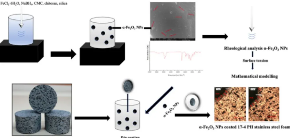
Recently, the various studies have reported on the NPs have received atten-tion due to their large surface area, excellent catalytic activity and morphological features (shape, size and dimension, surface activity, and low reduction potential [7–12]. Especially, the preparation of iron oxide NPs was reported using different methods such as chemical precipitation [13], sol–gel [14], ultrasonic irradiation [15], and emulsion methods [16] in previous studies. In this study, we prepared the CMC/Chitosan-α-Fe2O3 NPs using the green and low-cost ultrasonic irradia-tion method and we coated onto the surface of 17–4 PH stainless-steel foam with polymer blend-α-Fe2O3 NPs to fabricate the novel bio-nanomaterial. We noticed that no studies had reported in the literature regarding the coating of 17–4 PH stainless-steel foam with polymer blend-α-Fe2O3 NPs. Therefore, we focused on this surface modification of 17–4 PH stainless-steel foam in this study.
17–4 PH stainless-steel foam is suitable for use as a biomaterial due to its excellent mechanical and biological properties such as easy producibility, high porosity, corrosion resistance, strength, and biocompatibility [17–19]. Surface modification of the stainless-steel foams depends on some critical factors such the homogeneous distribution, particle morphology, sinter density, and sensitiza-tion [20]. Based on our previous experience, we used synthesized CMC/Chitosan-α-Fe2O3 NPs due to their characteristic properties such as their nanoscale size, shape, high surface area, chemical property, and surface chemistry and we inves-tigated the effect of surface modification on the surface characteristics and surface chemistry of the structure [21]. We evaluated the change in surface tension with different sonication inputs such as time, power, and amplitude on nanosystem formation and stabilization based on the surface tension behaviors. Furthermore, we defined the correlation between surface tension and surface modification, which depends on the types of sonication inputs and experimental parameters. This correlation was examined with the characteristic surface factor based on the surface modification of the surface with a mathematical approach. The novelty of this study was to prepare the novel CMC/Chitosan-α-Fe2O3 NPs coated 17–4 PH stainless-steel foam and investigate the surface characteristics of the coating. We investigated surface properties of the nanostructure and CMC/Chitosan-α-Fe2O3 NPs coated 17–4 PH stainless-steel foam using Brunauer–Emmett–Teller (BET), Scanning Electron Microscopy/Energy-Dispersive X-Ray Spectroscopy (SEM/EDX), X-Ray diffraction (XRD), and Fourier transform infrared spectros-copy (FT-IR) techniques and to compare the surface tensions of CMC/Chitosan-α-Fe2O3 NPs. The surface characterization results showed that the α-Fe2O3 NPs
had a spherical shape, nanosize (10–20 nm), large surface area (180.37 m2/g) and
homogeneous distribution in colloidal solutions. The surface tensions of CMC/ Chitosan-α-Fe2O3 NPs were compared to determine the surface properties of the
nanostructure as affected by the operating parameters such as the concentration (0–1 ppm), mass fraction (10–20%) of silica, mass fraction (1–5%) of cetyltri-methylammonium bromide (CTAB), temperature (25–45 °C), pH (3–10), sonica-tion time (5–20 min), and the amplitude of sonicasonica-tion (10–40%). Furthermore, experimental results provided a mathematical model to describe the effect of CMC/Chitosan-α-Fe2O3 NPs on the surface tension for colloidal solutions. Error
analysis is also conducted to assess the validity of the obtained models. Accord-ing to previous reported studies, this study is the first report of the rheological properties of the CMC/Chitosan-α-Fe2O3 NPs as a coating biomaterial for foams
under ultrasonic irradiation. As far as we know, the coating of CMC/Chitosan-α-Fe2O3 NPs on the pore channels of 17–4 PH stainless-steel foam has not been
reported. For the first time, a mathematical model was formulated to determine the change in surface tension of α-Fe2O3 NPs for homogeneous coating process.
This novel nanomaterial can be a candidate for to be used as a new implant with excellent surface property in bone tissue engineering applications. Consequently, we have suggested that CMC/Chitosan-α-Fe2O3 NPs has a great potential for
designing a promising nanomaterial for biomedical applications.
Materials and methods Materials
Iron (III) chloride hexahydrate (FeCl3·6H2O) (≥ 99%), chitosan (low molecular weight), chitosan (Mw: 50–190 kDa) and carboxymethyl cellulose (CMC) (Mw: 250 kDa, high viscosity, the degree of substitution: 0.90) were purchased from Sigma-Aldrich Company (Germany). Polyvinyl alcohol (PVA), carbamide particles (CO(NH2)2), sodium borohydride (NaBH4), ethyl alcohol (C2H5OH), and silica gel (SiO2) (pore size 60 Å, 70–230 mesh), acetic acid (glacial, 100%), NaOH (> 99% purity), and cetyltrimethylammonium bromide (CTAB) (Average mass: 364.448 Da) were purchased from Merck Company. 17–4 PH stainless-steel powder was pur-chased from Carpenter Company (Sweden). The chemical composition of 17–4 PH stainless-steel powder was presented in Table 1. All chemicals and reagents were analytical grade and used without further purification.
Characterization part
Characterization of the CMC/Chitosan‑α‑Fe2O3 NPs
The surface morphology of all samples was investigated using a scanning electron microscope (SEM, Quanta FEG 450). An X-ray diffractometer (Bruker D8 Advance X-ray Diffractometer) was used (Cu Kα, λ = 0.15418 nm, 40 kV, 40 mA, 2θ angles
from 2 to 75 with a step size of 0.02°/s). FTIR spectroscopy was recorded using the Perkin Elmer FTIR emission spectrometer (Spectrum Two) with KBr powder (4000–600 cm−1 frequency range with a resolution of 4 cm−1 and 8 scans).
Meas-urements of surface tension of all colloidal solutions were performed with a force tensiometer (K11, KRUSS, Germany) at 25 ± 0.5 °C.
Characterization of the uncoated and coated 17–4 PH stainless‑steel foams with CMC/Chitosan‑α‑Fe2O3 NPs
The density and porosity content of sintered 17–4 PH stainless-steel foam were determined using Archimedes’ principle in a Sartorius precision balance equipped with a density determination kit. Mechanical properties of the specimens were stud-ied by the compression test performed on a Zwick–Roell Z050 materials testing machine. The surface roughness of the uncoated and coated 17–4 PH stainless-steel foams with CMC/Chitosan-α-Fe2O3 NPs was measured using a Surftest 210 type Mitutoyo surface profilometer (Mitutoyo, Japan) (5 μm tip radius). The arithmetic average of the surface roughness (Ra) was achieved by significantly bigger than the pores with a chisel point pen to eliminate the effects of pore size change on the measurements. The thickness of the coatings was examined using optical micro-scope (Nikon ME600). The pores of the steel foam were filled with a cold-hardening epoxy resin and then etched in 2% Nital solution for optical analysis.
Preparation of the CMC/Chitosan‑α‑Fe2O3 NPs
The CMC/Chitosan-α-Fe2O3 NPs were prepared following the procedure described
in our previous paper [21]. A solution of FeCl3·6H2O (0.54 g/30 mL) in a mixture
of 24 mL ethanol/6 mL of distilled water was dissolved at 25 °C. 0.1 M NaBH4 and
FeCl3·6H2O solutions were mixed then it was stirred vigorously under nitrogen gas
atmosphere for 15 min. 0.1 g of CMC was dissolved in 100 mL of distilled water for 24 h, and 0.05 g of chitosan was dissolved in 50 mL 2% (v/v) glacial acetic solution
Table 1 Chemical composition of 17–4 PH stainless-steel powder Element % Cr 15.18 Ni 4.470 Cu 3.470 Mn 0.650 Si 0.380 Nb + Ti 0.200 Mo 0.150 S 0.030 C 0.020 P 0.016 Fe Balance
for 30 min. Then, the CMC and chitosan solutions were mixed, and 0.02 g of silica was added in 100 mL water. All solutions were mixed and sonicated for 10 min at room temperature under nitrogen gas atmosphere. Finally, the synthesized CMC/ Chitosan-α-Fe2O3 NPs were stored at 25 °C until further use.
Preparation of 17–4 PH stainless‑steel foam
17–4 PH stainless-steel foam as the substrate material was produced by the space holder-water leaching technique in powder metallurgy. This technique involved four main stages: powder mixing, compaction, removal of space holder by water leaching, and sintering.Initially, 2.5 g of PVA was dissolved in 97.5 mL of distilled water. 250 g of steel powder was added to PVA solution. The blend was mixed for 30 min. Then, 20 g of blend was mixed with 80 g of irregular shaped carbamide particles (710–1000 μm) in a Turbula type mixer for 60 min. The mixture was com-pacted uniaxially at 200 MPa in a steel die using a hydraulic press into cylindrical specimens with a diameter of 10 mm and heights of 15 mm. The green steel foams were immersed in distilled water at room temperature to leach the space holder. The PVA in the green specimens was thermally removed as part of sintering cycle, which consisted of heating at a ramp rate of 5 °C/min to 410 °C (debinding) with a dwell time of 40 min, followed by heating at rate of 10 °C/min to sintering tempera-tures. The steel foams were sintered at 1260 °C for 40 min under high purity hydro-gen gas atmosphere in a horizontal tube furnace.
Coating process of CMC/Chitosan‑α‑Fe2O3 NPs/17–4 PH stainless‑steel foam
For the coating process, the cylindrical foam samples were cut into slices with a small diameter (〜0.5 cm) by EDM (Electrical Discharge Machining) with process parameters such as peak current: 3 A, pulse-on time: 50 µs, pulse-of time: 30 μs, open circuit voltage: 60 V, and wire feed speed: 5 m/s. CuZn37 master brass wire with 0.15 mm diameter (900 N/mm2 tensile strength). After the cutting step, the dust
was removed from the sample surface with compressed air. The uncoated 17–4 PH stainless-steel foam was immersed in the coating α-Fe2O3 NPs solution in a
direc-tion perpendicular, kept in the soludirec-tion for 5 min at room temperature. The sample was dried at 50 °C with a 30% relative humidity for 3 h. Based on weight difference results of the uncoated foam before and after coating, 3.3 mg of solution was loaded on the foam.
Surface tension measurement of CMC/Chitosan‑α‑Fe2O3 NPs
In this study, applying a force tensiometer, the surface tensions of all colloidal solu-tions were measured at 25 ± 0.5 °C. The measuring error and measurement range were 0.1 mN/m and 0.000015–0.0001 mN/m, respectively. To calibrate the system, the surface tension of distilled water was measured at 25 °C. We focused on the operating parameters such as the mass fraction (1–10%) of silica, the concentration (0–1 ppm) of solutions, sonication time (1–15 min), the amplitude of sonication
(10–40%), and temperature (25–45 °C) on the surface tensions of synthesized nano-particles and results were compared to design a promising nanomaterial for biomed-ical applications.
Construction of mathematical model
Critical micelle concentrations are obtained for each mass fraction (10%, 15%, and 20%) of silica by intersecting the lines, which are obtained by linear regressions applied to the decreasing and stable part of the concentration data [22]. A mathe-matical model for surface tension is also obtained as a function of the operating var-iables, namely, concentration, mass fraction of silica, mass fraction of CTAB, tem-perature, pH, sonication time, and amplitude of sonication. In order to obtain such a model, first an exponential regression is applied to the concentration surface tension data corresponding to concentration, for each mass fraction (10, 15, and 20%) of silica. The R2 values of these regressions are computed as 0.958, 0.988, and 0.985,
respectively. Then, another exponential regression is applied to the surface tension data corresponding to temperature (R2 = 0.997). Linear regressions are conducted
for pH, sonication time and surface tension data corresponding to mass fraction of CTAB, and linear relations are obtained for the stated variables with R2 values of
0.955, 0.971, 0.915, respectively. For modeling of amplitude of sonication, a logistic regression is performed (R2 = 0.985). Then, the equations obtained using regressions
are normalized with respect to the fixed parameters in the surface tension data cor-responding to concentration. The values of these fixed parameters are 25 °C (tem-perature), 5 (pH), 20 min (sonication time), 5% (mass fraction of CTAB), and 40% (amplitude). After the normalization of equations for each operating parameter, all functions reconstructed have been multiplied by the exponential function of surface tension data corresponding to concentration for each mass fraction of silica.
Error analysis methods
In this study, we used four different error analysis methods such as the sum of squares of errors (SSE), the hybrid fractional error function (HYBRID), Mar-quart’s percentage standard deviation (MPSD), and the average relative error (ARE) all expressions were given in Table 2 [23–26]. Where, 𝛾e: experimental surface Table 2 The error analysis
methods Expressions Error analysis methods References
∑n i=1(𝛾cal− 𝛾e) 2 i SSE [23] 100 n−p ∑n i=1 � (𝛾e−𝛾cal)2 i 𝛾e � HYBRID [24] 100 � 1 n−p ∑n i=1 � 𝛾e−𝛾cal 𝛾e �2 i MPSD [25] 100 n−p ∑n i=1��� 𝛾e−𝛾cal 𝛾e � � �i ARE [26]
tension, 𝛾cal: calculated surface tension, n: number of data points and p: number of
parameters.
Results and discussion
Characterization of 17–4 PH stainless‑steel foam
The photograph of the sintered steel foam and SEM image of stainless-steel foam were shown in Fig. 1a and b, respectively. The specimens had a relatively uniform distribution of pores. Cell walls separating each pore from its neighbors were clearly seen. No cracks were observed in the highly porous structures, but large numbers of micropores were formed in the cell walls. Although the macropores in foams appear to be isolated from each other, they were in fact connected owing to the micropores in the cell walls. In spite of sintering between steel particles, the macroporous structure remained with a small shrinkage. This suggested that the pore structures could be designed by using morphological properties such as proper size, shape and content of the carbamide particles.
Total porosity and pore size had a critical role in tissue ingrowth into the implant. The minimum pore size was considered to be 100 mm and larger pore size results in greater tissue ingrowth. In the case of pores with size less than 100 mm, cells did not grow into the pores because of spanning of pores by the cells. The morphology of the final pores of the sintered 17–4 PH stainless-steel foams replicated the initial shape of the carbamide particles that were used as space holder. In this study, the total porosity of the specimen consisted of about 58% open and 14% closed porosity.
The mean size and the mean sphericity of the pores were determined to be 620 μm and 0.58, respectively. These values for carbamide particles were determined to be 780 μm and 0.64, respectively. The decrease in the size and sphericity was attrib-uted to crushing of the carbamide particles during pressing and moistening before mixing. Compression tests were conducted on the foam to evaluate their elastic and
plastic deformation behavior. The compressive yield strength and Young’s modulus of 17–4 PH stainless-steel foam were 48 MPa and 0.51 GPa, respectively. Compres-sive yield strength and Young’s modulus of cancellous bone are 40–150 MPa and 0.09–1.5 GPa, respectively [27]. As a result, mechanical properties of the 17–4 PH stainless-steel foams were close to cancellous bones. The average surface rough-ness value of uncoated steel foam was measured in the range of 3.98–4.25 μm. As known, these optimum parameters (surface roughness, mean size and sphericity val-ues of the pores) are suitable for biomedical applications [27].
Characterization of α‑Fe2O3 NPs and α‑Fe2O3 NPs coated 17–4 PH stainless‑steel foam
In this study, the synthesized CMC/Chitosan-α-Fe2O3 NPs were used in the first
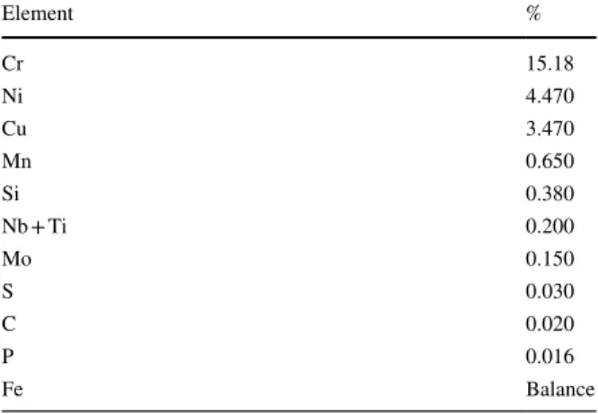
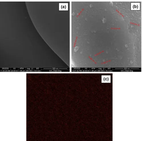
time as a green coating material for 17–4 PH stainless-steel foam. There have been several reported studies on the enhanced surface properties of stainless-steel foam and improving the biocompatibility of the stainless-steel foams, but limited studies have been reported on surface properties of NPs coated stainless-steel foam [28–32]. The surface morphologies of the 17–4 PH stainless-steel foam and CMC/Chitosan-α-Fe2O3 NPs coated 17–4 PH stainless-steel foam in Fig. 2a and b. EDX image of
CMC/Chitosan-α-Fe2O3 NPs was given in Fig. 2c. In Fig. 2, the surface
morphol-ogy results showed the treated surface of 17–4 PH stainless-steel foam was com-pletely covered with CMC/Chitosan-α-Fe2O3 NPs and CMC/Chitosan-α-Fe2O3 NPs
were uniformly dispersed in the CMC/Chitosan@silica [33]. The average particle size of CMC/Chitosan-α-Fe2O3 NPs was 10–20 nm with a spherical shape.
Accord-ing to BET results, the surface area of CMC/Chitosan-α-Fe2O3 NPs was measured
180.37 m2/g.
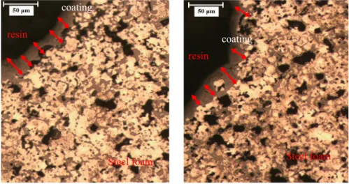
The average surface roughness value of the CMC/Chitosan-α-Fe2O3 NPs coated
steel foam was measured in the range of 4.59–5.91 μm. The coating thickness val-ues were measured using an optical microscope in the range of 15 and 30 μm closer view of pore walls of coated steel foams were observed in Fig. 3 and it was clear that there were micropores obvious in the cell walls. The coating thickness values were measured using an optical microscope in the range of 10 and 20 μm. In this study, we observed that the coating process of stainless-steel foam was effective and there were no cracks or delamination was observed along the interface. According to our experimental results, we demonstrated that the coating process had a major role in the surface and structural properties of the foam due to the changes in the morphol-ogy and surface roughness parameters.
In our previous study, X-ray powder diffraction (XRD) was used to identify the structural properties of α-Fe2O3 NPs and to determine the crystallinity of α-Fe2O3
NPs.
The XRD patterns of CMC/Chitosan-α-Fe2O3 NPs were given in Fig. 4.
Accord-ing to XRD results, the characteristic peaks of CMC/Chitosan-α-Fe2O3 NPs (2 theta:
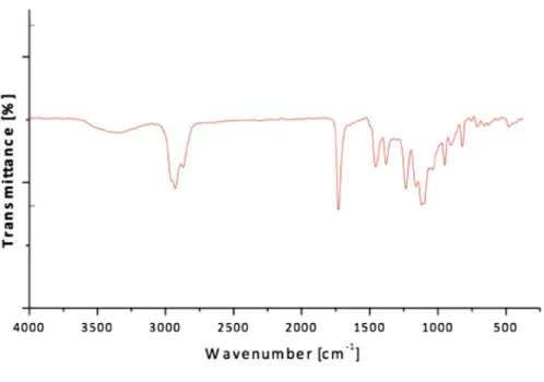
31° and 45°) were observed. The FTIR spectra of the α-Fe2O3 NPs were given in
Fig. 5. In our previous FTIR results, the characteristic peaks of CMC/Chitosan-α-Fe2O3 NPs were observed at 3479 cm−1 (–OH), 2924 cm−1 (–OH), 1750 cm−1 (C=O
stretching), 1514 cm−1 (–NH), 1318 cm−1 (–CH
2OH) and at 530 cm−1 (α-Fe2O3)
[21].
The measurement of surface tension of the CMC/Chitosan‑α‑Fe2O3 NPs
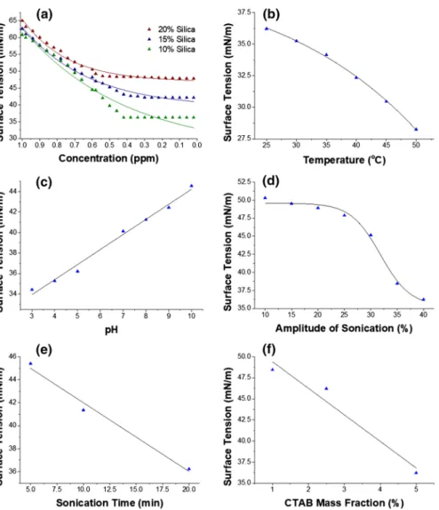
There have been limited studies on the surface tension of the synthesized nano-structures [34–36]. In this study, we focused on the effect of the mass fraction (1–10%) of silica, the concentration (0–1 ppm) of solutions, sonication time (1–15 min), the amplitude of sonication (10–40%), and temperature (25–45 °C) on the surface tension of CMC/Chitosan-α-Fe2O3 NPs solutions were shown in
Figs. 6 and 7. The surface tension of NPs /water from 47.83 to 36.22 mN/m by changing the concentration of colloidal solutions in a range of 0–0.05 ppm (pressure: 1 atm, temperature: 25 °C) was measured (Fig. 4). We investigated
Fig. 2 SEM images of a 17–4 PH stainless-steel foam with × 200.000 magnification and b CMC/ Chitosan-α-Fe2O3 NPs coated 17–4 PH stainless-steel foam with × 200.000 magnification c EDX images
the effect of the mass fraction on the NPS/water nanofluid behavior based on the surface tension and found that there was a relationship between the additive functions of the systems and the surface tension using a mathematical model (Figs. 6 and 7). According to results, the surface tension of α-Fe2O3 NPs/water
was increased with the increase in the concentration of α-Fe2O3 NPs [37]. There
have been few studies reported on a mathematical approach to evaluate the
resin
coating
coating
resin
Steel foam Steel foam
Fig. 3 Cross-sectional surface microstructure of pore wall of the CMC/Chitosan-α-Fe2O3 NPs coating on
steel foams
effects of the sonication inputs such as time, power, and amplitude on the nano-system formation and stabilization based on surface tension behaviors [38]. We showed that the sonication method reduced the surface tension and improved the stability of colloidal solutions. Also, we observed that surface tension results, and surface analysis results were compatible. The effect of temperature (25–45 °C) on the surface tension of α-Fe2O3 NPs/water system (the interface
between NPs and water phase) was proven. The results showed that the tem-perature was increased, and the surface tension decreased as the attraction forces between the particles decreased [38]. The novelty of this study was that it had a large amount of data on the surface tension of NPs/water system, a mathemati-cal relationship was established between the values of the inputs (mass fraction, concentration, sonication time and amplitude) and the output (surface tension) and all parameters was used to fit the equation for the surface tension of nano-particles [39–41].
Determination of critical micelle concentrations
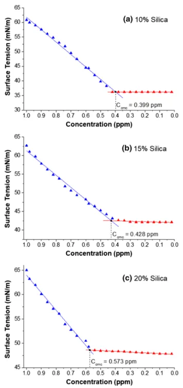
Critical micelle concentrations are determined by intersecting the lines, which are obtained by linear regressions applied to the decreasing and stable part of the data. The critical micelle concentrations were found to be 0.399, 0.428, and 0.573 ppm for mass fractions of 10, 15 and 20% silica, respectively (Fig. 6). The experimental data are available in the Supplementary Materials.
Fig. 6 Determination of critical micelle concentrations for 10% (a), 15% (b), 20% (c) mass fractions of silica
Modeling of surface tension
In this study, we have obtained surface tension as a function of operating variables con-centration (C), temperature (T), pH, amplitude of sonication (AMP), sonication time (ts), and mass fraction of CTAB (mCTAB) for each mass fraction (10, 15, and 20%) of sil-ica. Firstly, an exponential regression is applied to the surface tension data correspond-ing to the concentration, while the remaincorrespond-ing variables are kept constant at T = 25 °C, pH = 5, AMP = 40%, ts = 20 min, and mCTAB = 5%. As a result of these regressions, the functions of 𝛾C10(C) Eq. 3.1, 𝛾C15(C) Eq. 3.2, and 𝛾C20(C) Eq. 3.3 are provided for mass
Fig. 7 The effects of concentration for each mass fraction of silica (a), temperature (b) pH (c), amplitude of sonication (d), sonication time (e) and mass fraction of CTAB (f) on surface tension of α-Fe2O3 NPs/
fraction of 10%, 15%, and 20% silica, respectively. These functions are illustrated with the experimental data (see Supplementary Materials) in Fig. 7a.
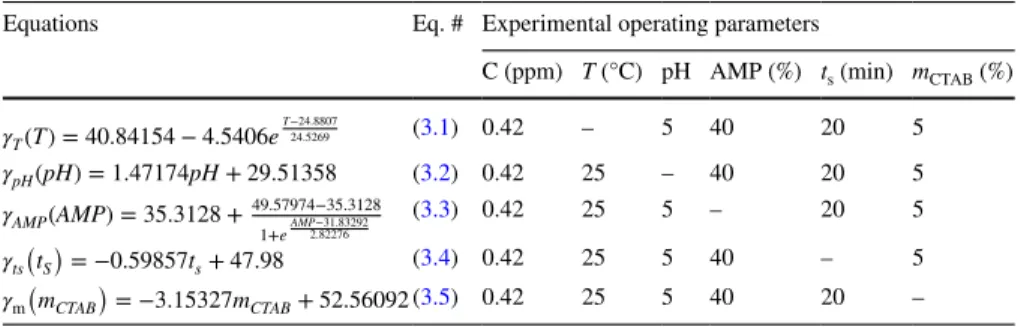
Then, regressions are applied to the surface tension data corresponding to tempera-ture, pH, amplitude of sonication, sonication time, and mass fraction of CTAB respect to fixed parameters given in Table 3 (Eqs. 3.3–3.8). Thus, equations in Table 3 are obtained and all functions and the experimental data (see Supplementary Materials) are illustrated in Fig. 7.
After obtaining Eqs. 3.4–3.8, the functions 𝛾T(T) , 𝛾pH(pH), 𝛾AMP(AMP) , 𝛾ts(tS) , and 𝛾m
CTAB
(
mCTAB) are normalized as in Eqs. 3.9–3.14, respectively. The normalization is
performed in a way that the normalized functions result 1 at fixed parameters T = 25 °C, pH = 5, AMP = 40% min, ts = 20 min, and mCTAB = 5%.
(3.1) 𝛾C10(C) = 26.31286 + 6.64406e C 0.5880114 (3.2) 𝛾C15(C) = 39.08639 + 1.80472e C 0.3861944 (3.3) 𝛾C20(C) = 46.86883 + 0.3793e C 0.2566275 (3.9) 𝛾TN(T) = 𝛾T(T) 𝛾T(25) = 1.12477 − 0.045384e0.040772T (3.10) 𝛾pHN(pH) = 𝛾pH(pH) 𝛾pH(5) = 0.039915pH + 0.80043 (3.11) 𝛾AMPN(AMP) = 𝛾AMP(AMP) 𝛾AMP(40) = 0.97924 + 0.39563 1 + e0.35426AMP−11.27723 (3.12) 𝛾tsN(tS)= 𝛾ts(tS) 𝛾ts(20) = −0.016623ts+ 1.33246
Table 3 Equations via experimental fixed operating parameters of α-Fe2O3 NPs
Equations Eq. # Experimental operating parameters
C (ppm) T (°C) pH AMP (%) ts (min) mCTAB (%)
𝛾T(T) = 40.84154 − 4.5406e T−24.8807 24.5269 (3.1) 0.42 – 5 40 20 5 𝛾pH(pH) = 1.47174pH + 29.51358 (3.2) 0.42 25 – 40 20 5 𝛾AMP(AMP) = 35.3128 + 49.57974−35.3128 1+e AMP−31.83292 2.82276 (3.3) 0.42 25 5 – 20 5 𝛾ts ( tS ) = −0.59857ts+ 47.98 (3.4) 0.42 25 5 40 – 5 𝛾m ( mCTAB ) = −3.15327mCTAB+ 52.56092(3.5) 0.42 25 5 40 20 –
Finally, multiplying all normalized functions by 𝛾C10(C) , 𝛾C15(C) , and 𝛾C20(C) ;
we obtain the surface tension as a function of the operating variables for mass fractions of 10%, 15%, and 20% silica, respectively Eqs. 3.14–3.16.
Error analysis
In this study, we investigated the effects of ultrasonic cavitation on surface mor-phology evolution of NPs. Also, the successful optimization strategy of NPs was experimentally designed with different operating parameters (ultrasonic irradia-tion inputs) using the mathematical model with to the surface tension data. Addi-tionally, we highlighted that the interaction of the water with NPs under ultra-sonic irradiation plays a major role in modulating particle size and distribution due to performance of physicochemical properties.
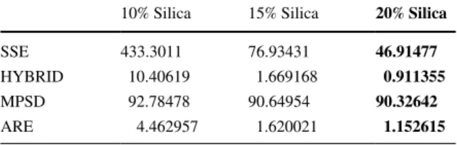
Also, we conducted an error analysis for functions obtained in Eqs. 3.14–3.16
in order to evaluate the performance of the obtained models. SSE, HYBRID, MPSD and ARE values for each function are determined by comparing the results of each equation with 104 experimental data points. The results were given in Table 4 and the schematic diagram of the experimental procedure was given in Fig. 8. By applying Eqs. 3.14–3.16, we compared in terms of accuracy, it is seen that Eq. 3.16 (20% silica) represents the experimental data the best. While the absolute error is relatively higher in Eq. 3.14 (10% silica), the similarity in MPSD value suggests that the standard deviation percentage is similar for each relation. These error values provide a reference for the modeling of surface ten-sion with respect to similar operating variables.
(3.13) 𝛾mN(mCTAB)= − 𝛾m(mCTAB) 𝛾m(5) = −0.085312mCTAB+ 1.42656 (3.14)
𝛾10%(T, pH, AMP, ts, mCTAB, C)= 𝛾TN(T)𝛾pHN(pH)𝛾AMPN(AMP)𝛾tsN(tS)𝛾mN(mCTAB)𝛾C10(C)
(3.15) 𝛾15% ( T, pH, AMP, ts, mCTAB, C ) = 𝛾TN(T)𝛾pHN(pH)𝛾AMPN(AMP)𝛾tsN ( tS ) 𝛾mN ( mCTAB ) 𝛾C15(C) (3.16)
𝛾20%(T, pH, AMP, ts, mCTAB, C)= 𝛾TN(T)𝛾pHN(pH)𝛾AMPN(AMP)𝛾tsN(tS)𝛾mN(mCTAB)𝛾C20(C)
Table 4 Error analysis results of mathematical models for CMC/ Chitosan-α-Fe2O3 NPs
10% Silica 15% Silica 20% Silica
SSE 433.3011 76.93431 46.91477
HYBRID 10.40619 1.669168 0.911355
MPSD 92.78478 90.64954 90.32642
Conclusıon
In this study, preparation, characterization and surface properties of the synthe-sized CMC/Chitosan-α-Fe2O3 NPs and CMC/Chitosan-α-Fe2O3 NPs coated 17–4 PH stainless-steel foam were investigated. Surface tension behaviors of CMC/ Chitosan-α-Fe2O3 NPs were experimentally determined as a function of the oper-ating parameters. The influence of CMC/Chitosan-α-Fe2O3 NPs coating on mor-phology properties of 17–4 PH stainless-steel foam was performed. This study proposed a mathematical model to determine the change in surface tension of NPs for homogeneous coating process. Our research model presented as a meth-odological prediction, which explains the relation between surface tension and operating parameters using different error analysis methods with minimum error values. The novel CMC/Chitosan-α-Fe2O3 NPs coated 17–4 PH stainless-steel foam is a candidate nanomaterial for biomedical applications.
Supplementary Information The online version contains supplementary material available at https:// doi. org/ 10. 1007/ s00289- 021- 03700-2.
Declarations
Conflict of interest The authors declare no conflict of interests.
References
1. Israel LL, Galstyan A, Holler E, Ljubimova JY (2020) Magnetic iron oxide nanoparticles for imaging, targeting and treatment of primary and metastatic tumors of the brain. J Control Release 320:45–62. https:// doi. org/ 10. 1016/j. jconr el. 2020. 01. 009
2. Phul R, Shrivastava V, Farooq U, Sardar M, Kalam A, Al-Sehemi AG, Ahmad T (2019) One pot synthesis and surface modification of mesoporous iron oxide nanoparticles. Struct Nano-Objects 19:100343. https:// doi. org/ 10. 1016/j. nanoso. 2019. 100343
3. Gupta AK, Gupta M (2005) Synthesis and surface engineering of iron oxide nanoparticles for biomedical applications. Biomaterials 26(18):3995–4021. https:// doi. org/ 10. 1016/j. bioma teria ls. 2004. 10. 012
4. Alphandéry E (2020) Iron oxide nanoparticles for therapeutic applications. Drug Discovery Today 25(1):141–149. https:// doi. org/ 10. 1016/j. drudis. 2019. 09. 020
5. Guo L, Chen H, He N, Deng Y (2018) Effects of surface modifications on the physicochemical properties of iron oxide nanoparticles and their performance as anticancer drug carriers. Chin Chem Lett 29(12):1829–1833. https:// doi. org/ 10. 1016/j. cclet. 2018. 10. 038
6. Navarro-Palomares E, González-Saiz P, Renero-Lecuna C, Martín-Rodríguez R, Aguado F, González-Alonso D, Barquín LF, González J, López MB, Valiente R (2020) Dye-doped biode-gradable nanoparticle SiO 2 coating on zinc-and iron-oxide nanoparticles to improve biocom-patibility and for in vivo imaging studies. Nanoscale 12(10):6164–6175. https:// doi. org/ 10. 1039/ c9nr0 8743e
7. Ali I (2018) Microwave assisted economic synthesis of multi walled carbon nanotubes for arse-nic species removal in water: batch and column operations. J Mol Liq 71:677–685. https:// doi. org/ 10. 1016/j. molliq. 2018. 09. 021
8. Burakova EA, Dyachkova TP, Rukhov AV, Tugolukov EN, Galunin EV, Tkachev AG, Ali I (2018) Novel and economic method of carbon nanotubes synthesis on a nickel magnesium oxide catalyst using microwave radiation. J Mol Liq 253:340–346. https:// doi. org/ 10. 1016/j. molliq. 2018. 01. 062
9. Ali I, Suhail M, Alothman ZA, Alwarthan A (2018) Recent advances in syntheses, properties and applications of TiO2 nanostructures. RSC Adv 8(53):30125–30147. https:// doi. org/ 10. 1039/
c8ra0 6517a
10. Ali I, Alharbi OM, Alothman ZA, Alwarthan A (2018) Facile and eco-friendly synthesis of func-tionalized iron nanoparticles for cyanazine removal in water. Coll Surf B Biointerfaces 171:606– 613. https:// doi. org/ 10. 1016/j. colsu rfb. 2018. 07. 071
11. Ali I, Kucherova A, Memetov N, Pasko T, Ovchinnikov K, Pershin V, Kuznetsov D, Galunin E, Grachev V, Tkachev A (2019) Advances in carbon nanomaterials as lubricants modifiers. J Mol Liq 279:251–266. https:// doi. org/ 10. 1016/j. molliq. 2019. 01. 113
12. Ali I, Alharbi OM, Alothman ZA, Badjah AY (2018) Kinetics, thermodynamics, and modeling of amido black dye photodegradation in water using Co/TiO2 nanoparticles. Photochem
Photo-biol 94(5):935–941. https:// doi. org/ 10. 1111/ php. 12937
13. Lassoued A, Dkhil B, Gadri A, Ammar S (2017) Control of the shape and size of iron oxide (α-Fe2O3) nanoparticles synthesized through the chemical precipitation method. Results Phys
7:3007–3015. https:// doi. org/ 10. 1016/j. rinp. 2017. 07. 066
14. Meyerstein D, Adhikary J, Burg A, Shamir D, Albo Y (2020) Zero-valent iron nanoparticles entrapped in SiO2 sol-gel matrices: a catalyst for the reduction of several pollutants. Catal
Com-mun 133:105819. https:// doi. org/ 10. 1016/j. catcom. 2019. 105819
15. Dolores R, Raquel S, Adianez GL (2015) Sonochemical synthesis of iron oxide nanoparticles loaded with folate and cisplatin: effect of ultrasonic frequency. Ultrason Sonochem 23:391–398. https:// doi. org/ 10. 1016/j. ultso nch. 2014. 08. 005
16. Ahmed N, Ahmad NM, Fessi H, Elaissari A (2015) In vitro MRI of biodegradable hybrid (iron oxide/polycaprolactone) magnetic nanoparticles prepared via modified double emulsion evapora-tion mechanism. Colloids Surf B 130:264–271. https:// doi. org/ 10. 1016/j. colsu rfb. 2015. 04. 022 17. Szewczyk-Nykiel A (2014) The effect of the addition of boron on the densification,
micro-structure and properties of sintered 17–4 PH stainless steel. Czasopismo Techniczne 13:85–96. https:// doi. org/ 10. 4467/ 23537 37XCT. 14. 293. 3381
18. Zhang Q, Hu Z, Su W, Zhou H, Liu C, Yang Y, Qi X (2017) Microstructure and surface prop-erties of 17–4PH stainless-steel by ultrasonic surface rolling technology. Surf Coat Technol 321:64–73. https:// doi. org/ 10. 1016/j. surfc oat. 2017. 04. 052
19. Gu H, Gong H, Pal D, Rafi K, Starr T, Stucker B (2013) Influences of energy density on poros-ity and microstructure of selective laser melted 17–4PH stainless steel. In 2013 Solid Freeform Fabrication Symposium.
20. Vieira MT, Martins AG, Barreiros FM, Matos M, Castanho JM (2008) Surface modification of stainless-steel powders for microfabrication. J Mater Process Technol 201:651–656. https:// doi. org/ 10. 1016/j. jmatp rotec. 2007. 11. 162
21. Karakus S, Ilgar M, Tan E, Kilislioglu A (2018) Sonochemical preparation of α-Fe2 O3 nano-particles in a dual biopolymer matrix. Curr Trends Chem Eng Process Technol. DOI: 10.29011. CTCEPT-107/100007
22. Le TTY, Hussain S, Lin SY (2019) A study on the determination of the critical micelle concen-tration of surfactant solutions using contact angle data. J Mol Liq 294:111582. https:// doi. org/ 10. 1016/j. molliq. 2019. 111582
23. Demirbas E, Kobya M, Konukman AES (2008) Error analysis of equilibrium studies for the almond shell activated carbon adsorption of Cr (VI) from aqueous solutions. J Hazard Mater 154(1–3):787–794. https:// doi. org/ 10. 1016/j. jhazm at. 2007. 10. 094
24. Kapoor A, Yang RT (1989) Correlation of equilibrium adsorption data of condensible vapours on porous adsorbents. Gas Sep Purif 3(4):187–192. https:// doi. org/ 10. 1016/ 0950- 4214(89) 80004-0
25. Paluri P, Ahmad KA, Durbha KS (2020) Importance of estimation of optimum isotherm model parameters for adsorption of methylene blue onto biomass derived activated carbons: compari-son between linear and non-linear methods. Biomass Conv Biorefinery. https:// doi. org/ 10. 1007/ s13399- 020- 00867-y
26. Karakuş S (2019) A novel ZnO nanoparticle as drug nanocarrier in therapeutic applications: kinetic models and error analysis. J Turk Chem Soc Sect A Chem 6(2):119–132. https:// doi. org/ 10. 18596/ jotcsa. 405505
27. Matsushita T, Fujibayashi S, Kokubo T (2017) Titanium foam for bone tissue engineering. In: Metallic Foam Bone, 1st edn. Woodhead Publishing, pp 111–130. https:// doi. org/ 10. 1016/ B978-0- 08- 101289- 5. 00004-4
28. Halim FSA, Chandren S, Nur H (2020) Carbon-containing-titania coated stainless-steel prepared by high voltage powder spray coating and its adhesion phenomena. Prog Org Coat 147:105782. https:// doi. org/ 10. 1016/j. porgc oat. 2020. 105782
29. Tang S, Chang X, Li M, Ge T, Niu S, Wang D, Jiang Y, Sun S (2021) Fabrication of calcium carbonate coated-stainless-steel mesh for efficient oil-water separation via bacterially induced biomineralization technique. Chem Eng J 405:126597. https:// doi. org/ 10. 1016/j. cej. 2020. 126597 30. Rezaei A, Golenji RB, Alipour F, Hadavi MM, Mobasherpour I (2020) Hydroxyapatite/ hydroxyapatite-magnesium double-layer coatings as potential candidates for surface modification of 316 LVM stainless-steel implants. Ceram Int 46(16):25374–25381. https:// doi. org/ 10. 1016/j. ceram int. 2020. 07. 005
31. Tran NG, Chun DM (2020) Simple and fast surface modification of nanosecond-pulse laser-tex-tured stainless-steel for robust superhydrophobic surfaces. CIRP Ann 69(1):525–528. https:// doi. org/ 10. 1016/j. cirp. 2020. 04. 012
32. Asl SM, Ganjali M, Karimi M (2019) Surface modification of 316L stainless-steel by laser-treated HA-PLA nanocomposite films toward enhanced biocompatibility and corrosion-resist-ance in vitro. Surf Coat Technol 363:236–243. https:// doi. org/ 10. 1016/j. surfc oat. 2019. 02. 052 33. Gholipour-Ranjbar H, Ganjali MR, Norouzi P, Naderi HR (2016) Synthesis of cross-linked
gra-phene aerogel/Fe2O3 nanocomposite with enhanced supercapacitive performance. Ceram Int 42(10):12097–12104. https:// doi. org/ 10. 1016/j. ceram int. 2016. 04. 140
34. Gibson LJ, Ashby MF (1999) Cellular solids: structure and properties. Cambridge University Press, Cambridge
35. Gibson LJ (2005) Biomechanics of cellular solids. J Biomech 38(3):377–399. https:// doi. org/ 10. 1016/j. jbiom ech. 2004. 09. 027
36. Balla VK, Bodhak S, Bose S, Bandyopadhyay A (2010) Porous tantalum structures for bone implants: fabrication, mechanical and in vitro biological properties. Acta Biomater 6(8):3349– 3359. https:// doi. org/ 10. 1016/j. actbio. 2010. 01. 046
37. Huminic A, Huminic G, Fleaca C, Dumitrache F, Morjan I (2015) Thermal conductivity, viscos-ity and surface tension of nanofluids based on FeC nanoparticles. Powder Technol 284:78–84. https:// doi. org/ 10. 1016/j. powtec. 2015. 06. 040
38. Cui Z, Chen J, Xue Y, Gan J, Chen X, Duan H, Zhang R, Liu J, Hao J (2020) Determination of surface tension and surface thermodynamic properties of nano-ceria by low temperature heat capacity. Fluid Phase Equilib 518:112627. https:// doi. org/ 10. 1016/j. fluid. 2020. 112627
39. Zhang S, Han X, Tan Y, Liang K (2018) Effects of hydrophilicity/lipophilicity of nano-TiO2
on surface tension of TiO2-water nanofluids. Chem Phys Lett 691:135–140. https:// doi. org/ 10.
1016/j. cplett. 2017. 11. 005
40. Yan SR, Kalbasi R, Nguyen Q, Karimipour A (2020) Sensitivity of adhesive and cohesive inter-molecular forces to the incorporation of MWCNTs into liquid paraffin: experimental study and modeling of surface tension. J Mol Liq 310:113235. https:// doi. org/ 10. 1016/j. molliq. 2020. 113235
41. Silva KCG, Sato ACK (2019) Sonication technique to produce emulsions: the impact of ultrasonic power and gelatin concentration. Ultrason Sonochem 52:286–293. https:// doi. org/ 10. 1016/j. ultso nch. 2018. 12. 001
Publisher’s Note Springer Nature remains neutral with regard to jurisdictional claims in published maps and institutional affiliations.
Authors and Affiliations
Selcan Karakuş1 · İnci Albayrak2 · Nuray Beköz Üllen3 · Mert Akin Insel4 · Ayben Kilislioğlu5
1 Department of Chemistry, Faculty of Engineering, Istanbul University-Cerrahpasa,
34320 Istanbul, Turkey
2 Department of Mathematical Engineering, Yıldız Technical University, 34210 Avcilar, Istanbul,
Turkey
3 Department of Metallurgical and Materials Engineering, Istanbul University-Cerrahpasa,
34320 Istanbul, Turkey
4 Department of Chemical Engineering, Yıldız Technical University, 34210 Istanbul, Turkey 5 Department of Electrical – Electronics, Faculty of Engineering and Natural Sciences, Kadir Has