http://journals.tubitak.gov.tr/veterinary/ © TÜBİTAK
doi:10.3906/vet-1311-51
In vivo embryo production in some native goat breeds in Turkey
Ali Reha AĞAOĞLU1,*, Kübra KARAKAŞ2, Mustafa KAYMAZ21Department of Obstetrics and Gynecology, Faculty of Veterinary Medicine, Mehmet Akif University, Burdur, Turkey 2Department of Obstetrics and Gynecology, Faculty of Veterinary Medicine, Ankara University, Ankara, Turkey
1. Introduction
Turkey has rich domestic animal genetic resources and genetic diversity as it is located at the crossroads of Europe, Asia, and Africa (1). Native goat breeds are important genetic resources in livestock due to their socioeconomic value. There are numerous native goat breeds in Turkey, such as Angora (AG), Kilis (KL), Honamli (HO), Hair (HA), and Norduz (NO) goat breeds (2). While in 1961 there were 24 million heads of goat in Turkey, this number has decreased to 5 million heads starting from the 1990s (3). Therefore, there is a certain need for developing strategies and methodologies that aim to conserve and support the sustainability of native goat breeds. In vivo embryo production and storage allow the conservation of endangered species from domestic and wild animals to maintain biodiversity (4,5).
Superovulation is the most important step in the conservation of animal genetic resources via production of in vivo embryos. The most effective way of obtaining embryos from females is the induction of superovulation with various gonadotropins. In recent years, several follicle-stimulating hormones have been used to induce superovulation in livestock (6). In order to ensure the
continuity of programs for multiple ovulation and embryo transfer (MOET) and conservation of genetic resources, a continuous supply of good quality embryos throughout the year is crucial. These programs in small ruminants (sheep and goats) are limited to the natural breeding season, since small ruminants have seasonal cyclic activity (7). The normal breeding season for goats is August to March, and especially October to December in temperate northern latitudes. During this season they are polyestrous animals. Similarly, the highest ovulation rate and embryo yields have been recorded during the natural breeding season while the lowest embryo yields were recorded during the anestrus season in goat breeds (8,9).
A considerable amount of research on this topic has led to development of various superovulation protocols. Differences among the protocols have a significant effect on the embryo yield (10). For instance, the use of porcine follicle-stimulating hormone (pFSH) in repeated superovulation procedures in goats has been reported to decrease the number of ovulations, embryos recovered, and the number of transferable embryos (11,12). Furthermore, several other factors cause the reduction in superovulation response, such as the refractoriness of the ovaries and
Abstract: The aim of this study was to assess the efficacy of the repeated administration of superovulatory protocol and surgical
procedure in in vivo embryo production, as well as to determine the differences in the responses to superovulatory treatment among several Turkish native goat breeds (Angora, Kilis, Honamli, Hair, and Norduz). A superovulatory treatment procedure was repeated 3 times in this study. The estrus cycles were synchronized and superovulatory response was induced with porcine follicle-stimulating hormone during the natural breeding season. Natural mating was performed following CIDR removal. Embryo recovery was performed surgically on day 6, after the first mating. According to the recovery results, the fertilization rates showed variability in Angora goats (50, 100, and 24.49, respectively, for cycles 1, 2, and 3) and Honamli goats (100, 42.48, and 56.25) (P < 0.05), while recovery rates showed no difference among the other breeds. The number of transferable embryos (TEs) showed no significant differences after 3 operations for Angora, Kilis, Honamli, and Hair goats, while the number of TEs decreased in in the third cycle in Norduz goats. The results indicate that Turkish native goat breeds can be utilized as donors in multiple ovulation and embryo transfer programs for at least 3 times with suitable operation techniques, superovulation treatments, and waiting periods among treatments.
Key words: In vivo embryo, reproduction, goat
Received: 12.11.2013 Accepted: 17.03.2014 Published Online: 21.04.2014 Printed: 20.05.2014
the formation of gonadotropin antibodies following consecutive superovulation procedure (11,13,14). In addition to these factors, surgery leads to the formation of postoperative adhesions and trauma, which may cause a decrease in embryo recovery rates after repeated surgeries (10). Therefore, the number of times that surgical flushing can be performed on the same animal is limited (15–18). Despite the possible negative effects listed above, the superovulation and embryo recovery process with surgery is still the most optimal and suitable tool for the in vivo embryo production of endangered small ruminant breeds. The objective of this study was to assess the efficacy of the repeated administration of a superovulatory protocol and surgical procedure in in vivo embryo production and to determine the differences in the responses to superovulation treatment among Turkish native goat breeds.
2. Materials and methods
2.1. Animals and superovulatory treatments
This study was conducted with 14 AG, 15 KL, 10 HO, 9 NO, and 10 HA does and with the same number of bucks. The does and bucks were housed in straw bedded semiopen folds. They were fed with concentrate daily together with alfalfa hay and barley straw; water was provided ad libitum. All goats were between 2 and 3 years of age and the mean body weights of AG, KL, HO, NO, and HA goats were 40.5 ± 1.4, 41.2 ± 2.5, 67.4 ± 3.1, 43.4 ± 2.8, and 43.7 ± 4.3 kg, respectively. The does had not been used in any MOET program prior to this study. The experiments were conducted during the breeding season (September to December) in Central Anatolia (40°06′08.50″N, 32°37′18.65″E) at 850 m above sea level.
Estruses were detected by bucks, which were fitted with aprons. The superovulatory treatment was performed in each breed 5 days after the onset of estrus. The estrus cycles were synchronized with controlled integral drug release dispensers containing 0.3 g of progesterone (CIDR; Eazi-Breed; Pharmacia & Upjohn, Australia) for 11 days. Starting on day 9 of CIDR treatment (48 h prior to CIDR removal) and for 3 consecutive days groups of does were treated with intramuscular pFSH (Folltropin, 10 mL, 200 mg NIH-FSH-P1, Bioniche Animal Health, Ireland) in 6 doses of 2.5, 2.5, 1.5, 1.5, 1, and 1 mL twice a day (0600 and 1800 hours) for induction of superovulation. At the time of first pFSH injection in all groups, D-cloprostenol (Dalmazin, Fatro, Turkey) was administrated intramuscularly at a dose of 150 µg to induce luteolysis. The bucks were used to detect estrus in all does starting from 24 h after CIDR removal. All does in estrus were naturally mated twice a day over 3 days. Superovulatory treatment procedure was repeated 3 times once a year between 2009 and 2011.
2.2. Assessment of superovulatory responses and embryos
Ovarian examination and embryo recovery were performed by laparotomy on day 6 after the first mating. All does were fasted for 24 h before surgery. Animals were sedated with 0.1–1 mg/kg atropine subcutaneously (Atrol-F, Sanovel, Turkey) and 0.5 mg/kg of diazepam intravenously (Diazem, Deva, Turkey) and anesthetized intravenously with 2 mg/kg ketamine HCl (Alfamine, EgeVet, Turkey). The ventral abdominal area, to the anterior of the udder, was shaved and cleaned. A midlateral incision was performed after local anesthesia by infiltration of 2% lidocaine HCl (Adokain, Sanovel, Turkey) in the incision area. The number of corpora lutea (CL) was recorded. Each uterine horn was flushed with a flushing medium (20 mL of mD-PBS + 3 mg/mL bovine serum albumin) using a catheter (1.3 × 130 mm) inserted near the utero-tubal junction. The embryos were recovered into a 90-mm petri dish using a 2-way Foley catheter (No. 10, Rüsch, USA) inserted in the base of the uterine horns. To avoid intraabdominal adhesions, 1 L of 2.5% heparin solution was used during flushing. Embryos were morphologically evaluated under a stereomicroscope (Leica, M205 C) and were classified according to the criteria recommended by the International Embryo Transfer Society (IETS) (19). The embryos were classified as unfertilized ova (UFO), degenerate blastocysts (DBl), and transferable/freezable embryos [compact morulae (CM), early blastocysts (EBl), blastocysts (Bl), expanded blastocysts (ExBl)]. The total number of recovered embryos (TREs), degenerated embryos, and transferable embryos (TEs) per doe surgically flushed was recorded. Fertilization rate (FR) and recovery rate (RR) were calculated.
2.3. Statistical analysis
The data obtained from groups were analyzed by the Kolmogorov–Smirnov Z statistic test for assessing normality. Data from groups that were not normally distributed were the FR and RR. The CL, UFO, DBl, CM, EBl, Bl, ExBl, and TE values were normally distributed. The Friedman test was performed for nonnormally distributed values and the chi-square test was performed for normally distributed values. Differences were considered significant at P < 0.05. All data were analyzed using SPSS 15.0. 3. Results
Superovulatory responses and embryo yield values by repeated surgical embryo flushing operations are presented in Tables 1–5 for each breed. Percentage of does in estrus and time interval from CIDR removal to estrus onset did not vary among the does. All does responded to estrus synchronization and, on average, estrus was demonstrated 24.0 ± 6.1 h following CIDR removal.
Table 1. The effect of repeated superovulation treatment and surgical embryo recovery
procedure on the ovarian activity and embryo yields of Angora goat (AG). AG group
Op.1 Op.2 Op.3
(mean ± SD) (mean ± SD) (mean ± SD) No. of CL 8.21 ± 5.2a 8.85 ± 5.30a 8.00 ±6.96a No. of UFO 0.00a 0.21 ± 0.8a 1.42 ± 2.87b No. of DBl 0.14 ± 0.53a 0.00a 0.35 ± 0.84a No. of CM 0.00a 0.00a 0.85 ±1.46b No. of EBl 3.92 ± 4.76a 4.92 ± 4.37a 0.64± 1.33b No. of Bl 0.00a 0.71 ± 1.85a 0.50 ± 0.75a No. of ExBl 0.00a 0.21 ± 0.8a 1.42 ± 2.87b No. of TEs 3.92±4.76a 5.85±5.18a 3.42±4.92a Median
(min–max) Median(min–max) Median(min–max)
FR 50.00a (0–100) 100 a (0–100) 24.49 b (0–100) RR 35.38a (0–100) 82.57 a (0–100) 72.50 a (0–100)
Op.: Surgical embryo flushing operation, TE: transferable embryo, FR: fertilization rate, RR: recovery rate, UFO: unfertilized ova, DBl: degenerate blastocyst, CM: compact morulae, EBl: early blastocyst, Bl: blastocyst, ExBl: expanded blastocyst, SD: standard deviation; within the groups different superscripts in the same row indicate significant differences (P < 0.05).
Table 2. The effect of repeated superovulation treatment and surgical embryo recovery
procedure on the ovarian activity and embryo yields of Kilis goat (KL). KL group
Op.1 Op.2 Op.3
(mean ± SD) (mean ± SD) (mean ± SD) No. of CL 6.06 ± 4.11a 9.80 ± 6.99a 11.53 ± 9.022a No. of UFO 0.00a 1.33 ± 3.86b 3.06 ± 5.24c No. of DBl 0.00a 0.20 ± 0.56a 0.73 ± 2.15a No. of CM 0.00a 0.60 ± 1.29b 0.00a No. of EBl 1.66 ± 2.43a 0.60 ± 1.80b 0.13 ± 0.51b No. of Bl 1.06 ± 1.93a 0.66 ± 1.29a 0.80 ± 1.85a No. of ExBl 0.20 ± 0.41a 0.33 ± 1.29a 4.20 ± 5.0b No. of TEs 2.93 ± 3.61a 2.20 ± 4.17a 5.26 ± 5.83a Median
(min–max) Median(min–max) Median(min–max)
FR 100a (0–100) 25.00 a (0–100) 37.50 a (0–100) RR 37.50a (0–100) 21.73 a (0–100) 70.00 a (0–100)
Op.: Surgical embryo flushing operation, TE: transferable embryo, FR: fertilization rate, RR: recovery rate, UFO: unfertilized ova, DBl: degenerate blastocyst, CM: compact morulae, EBl: early blastocyst, Bl: blastocyst, ExBl: expanded blastocyst, SD: standard deviation; within the groups different superscripts in the same row indicate significant differences (P < 0.05).
Table 3. The effect of repeated superovulation treatment and surgical embryo recovery
procedure on the ovarian activity and embryo yields of Norduz goat (NO). NO group
Op.1 Op.2 Op.3
(mean ± SD) (mean ± SD) (mean ± SD) No. of CL 9.88 ± 7.16a 11.88 ± 2.93a 6.00 ± 4.74a No. of UFO 0.66 ± 1.65a 3.11 ± 5.66a 2.00 ± 3.90a No. of DBl 1.11 ± 1.69a 0.88 ± 1.61a 0.77 ± 1.64a No. of CM 0.00a 0.00a 0.22 ± 0.66a No. of EBl 1.55 ± 2.12a 0.00b 0.22 ± 0.66b No. of Bl 1.55 ± 3.00a 0.66 ± 1.11a 0.11 ± 0.33a No. of ExBl 2.44 ± 4.95a 1.77 ± 1.98a 0.11 ± 0.33b No. of TEs 5.55 ± 6.44a 2.44 ± 2.50a 0.66 ± 1.65b Median
(min–max) Median(min–max) Median(min–max)
FR 33.33a (0–100) 33.33 a (0–85.71) 0.00 b (0–100) RR 77.77a (0–100) 53.84 a (0–100) 71.42 a (0–100)
Op.: Surgical embryo flushing operation, TE: transferable embryo, FR: fertilization rate, RR: recovery rate, UFO: unfertilized ova, DBl: degenerate blastocyst, CM: compact morulae, EBl: early blastocyst, Bl: blastocyst, ExBl: expanded blastocyst, SD: standard deviation; within the groups different superscripts in the same row indicate significant differences (P <0.05).
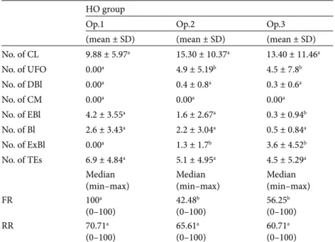
Table 4. The effect of repeated superovulation treatment and surgical embryo recovery
procedure on the ovarian activity and embryo yields of Honamli goat (HO). HO group
Op.1 Op.2 Op.3
(mean ± SD) (mean ± SD) (mean ± SD) No. of CL 9.88 ± 5.97a 15.30 ± 10.37a 13.40 ± 11.46a No. of UFO 0.00a 4.9 ± 5.19b 4.5 ± 7.8b No. of DBl 0.00a 0.4 ± 0.8a 0.3 ± 0.6a No. of CM 0.00a 0.00a 0.00a No. of EBl 4.2 ± 3.55a 1.6 ± 2.67a 0.3 ± 0.94b No. of Bl 2.6 ± 3.43a 2.2 ± 3.04a 0.5 ± 0.84a No. of ExBl 0.00a 1.3 ± 1.7b 3.6 ± 4.52b No. of TEs 6.9 ± 4.84a 5.1 ± 4.95a 4.5 ± 5.29a Median
(min–max) Median(min–max) Median(min–max)
FR 100a (0–100) 42.48 b (0–100) 56.25 b (0–100) RR 70.71a (0–100) 65.61 a (0–100) 60.71 a (0–100)
Op.: Surgical embryo flushing operation, TE: transferable embryo, FR: fertilization rate, RR: recovery rate, UFO: unfertilized ova, DBl: degenerate blastocyst, CM: compact morulae, EBl: early blastocyst, Bl: blastocyst, ExBl: expanded blastocyst, SD: standard deviation; within the groups different superscripts in the same row indicate significant differences (P < 0.05).
In HA goats, the number of CL decreased significantly (P < 0.05) during the third superovulation treatment (12.7 ± 6.2, 14.0 ± 9.06, and 6.8 ± 5.59, respectively, for cycles 1, 2, and 3), while no effects of the cycle were observed in the remainder of the breeds. The number of TEs showed no differences among 3 cycles in AG (3.92 ± 4.76, 5.85 ± 5.18, 3.42 ± 4.92, respectively), in KL (2.93 ± 3.61, 2.20 ± 4.17, 5.26 ± 5.83, respectively), in HO (6.96 ± 4.84, 5.1 ± 4.95, 4.5 ± 5.29, respectively), and in HA (5.7 ± 5.45, 6.6 ± 6.13, 4.7 ± 5.55, respectively) does (P > 0.05). On the other hand, the number of TEs was found significantly decreased in the third cycle in NO does (5.55 ± 6.44, 2.44 ± 2.50, 0.66 ± 1.65, respectively) (P < 0.05). The number of ExBl increased considerably during the third cycle in AG (0, 0.21 ± 0.8, and 1.42 ± 2.87, respectively, for cycles 1, 2, and 3), KL (0.2 ± 0.41, 0.33 ± 1.29, and 4.2 ± 5.0), and HO (0, 1.3 ± 1.7, and 3.6 ± 4.52) does, while a significant decrease was observed in NO does (2.44 ± 4.95, 1.77 ± 1.98, and 0.11 ± 0.33; P < 0.05). The mean numbers of UFO were found significantly increased in AG (0, 0.21 ± 0.8, and 1.42 ± 2.87, respectively, for cycles 1, 2, and 3), KL (0, 1.33 ± 3.86, and 3.06 ± 5.24), and HO (0, 4.9 ± 5.19, and 4.5 ± 7.8) goats (P < 0.05). As a result, fertilization rates (%) showed a decrease in AG (50, 100, 24.49, respectively,
for cycles 1, 2, and 3) and HO (100, 42.48, 56.25) goats (P < 0.05). On the other hand, recovery rates showed no differences among the different breeds.
4. Discussion
The time interval from CIDR removal to the onset of estrus was not affected by repeated superovulatory treatment in all groups and cycles. The time response from CIDR removal (24.0 ± 6.1) was shorter in comparison with previous studies, which found intervals of 32.0 ± 3.5 and 27.6 ± 3.5, respectively (20,21). The shorter time interval from CIDR removal to the onset of estrus may be due to an age factor and good nutritional and housing conditions. The percentage of estrus response (100%) obtained in this study indicates that Turkish native goats seem to respond well to induced estrus following hormonal stimulation.
Repeated superovulation treatment had a significant effect on the number of CL. Especially following the fourth treatment and after superovulation treatment, the numbers of CL have been found to be reduced (11,22). This study’s findings contradict these previous data as the number of CL recorded was not affected by the repeated superovulation treatment. In all goats except HA, repeated induction of superovulation and surgery did not have an
Table 5. The effect of repeated superovulation treatment and surgical embryo recovery
procedure on the ovarian activity and embryo yields of Hair goat (HA). HA group
Op.1 Op.2 Op.3
(mean ± SD) (mean ± SD) (mean ± SD) No. of CL 12.70 ± 6.20a 14.0 ± 9.06a 6.8 ± 5.59b No. of UFO 2.1 ± 3.51a 3.5 ± 4.76a 0.4 ± 0.96a No. of DBl 0.6 ± 1.34a 0.3 ± 0.94a 0.7 ± 1.05a No. of CM 0.4 ± 1.26a 0.00a 0.00a No. of EBl 0.00a 0.1 ± 0.31a 0.7 ± 0.94a No. of Bl 2.5 ± 4.0a 3.3 ± 3.52a 1.2 ± 2.14a No. of ExBl 2.8 ± 3.22a 2.9 ± 2.64a 2.6 ± 4.35a No of TEs 5.7 ± 5.45a 6.6 ± 6.13a 4.7 ± 5.55a Median
(min–max) Median(min–max) Median(min–max)
FR 51.01a (0–100) 70.83 a (0–100) 50.00 a (0–100) RR 73.21a (0–100) 86.11 a (26.09–100) 79.46 a (0–100)
Op.: Surgical embryo flushing operation, TE: transferable embryo, FR: fertilization rate, RR: recovery rate, UFO: unfertilized ova, DBl: degenerate blastocyst, CM: compact morulae, EBl: early blastocyst, Bl: blastocyst, ExBl: expanded blastocyst, SD: standard deviation; within the groups different superscripts in the same row indicate significant differences (P < 0.05).
effect on the number of CL recorded. This result may have occurred as a result of the 1-year rest period among the treatments, which allowed enough time for the uterus and ovaries to regenerate. The mean CL numbers of AG, KL, NO, HO, and HA goats were recorded as 8.21 ± 5.2, 6.06 ± 4.11, 9.88 ± 7.16, 9.88 ± 5.97, and 12.7 ± 6.20, respectively, in the first cycle. These values were lower compared to the mean CL numbers in Nubian (23) and Angora (24) goats (28.7 ± 2.3 and 15.00, respectively) and were higher compared to mean CL numbers in Jamunapari goats (25). The increased number of UFO is associated with the repeated treatment of FSH since the formation of FSH antibodies, which might affect the process of oocyte development, could occur after repeated superovulatory treatments (15,26).
The numbers of TEs showed no significant difference in AG, KL, HO, and HA groups among the cycles. In the other hand, the number of TEs was found significantly decreased in the third cycle in NO goats. The use of intraabdominal heparinized washing solutions is thought to have a positive effect on RR and TEs. In the NO group, the formation of postoperative adhesions may have decreased the number of TEs recovered, especially adhesions in the uterine lumen (27,28).
The FR and RR following repeated superovulation treatment may be falling in goats (24). Likewise, in the present study, in groups AG, NO, and HO, FR decreased in the third cycle. On the contrary, in groups KL and HA, FR did not change among the cycles. The lower fertilization rates in the third cycle may be attributed to a reduced
activity of the exogenous gonadotrophin applied, due to it being neutralized by antibodies generated from previous gonadotrophin treatments (13,29).
The reduction of RR can also be ascribed to the formation of postoperative adhesions following repeated surgical collection of the embryos. This factor has also been shown to reduce RR (30). In this study, the RR showed no significant difference due to the use of intraabdominal heparinized washing solutions, which are thought to have a positive effect on the RR.
In conclusion, the use of intravaginal CIDR in combination with pFSH is suitable for estrous synchronization and superovulatory in vivo embryo production in indigenous goats during the breeding season. In addition, the results of this study suggest that use of heparin-based intraabdominal washing solutions be strongly recommended to prevent adhesions and trauma, consequently increasing the efficiency of MOET programs in goats and especially helping in programs for conservation of genetic resources. Furthermore, rest periods among treatments could help in achieving more productive results in repeated superovulatory treatments. Acknowledgments
This study was financed by the Scientific and Technologi-cal Research Council of Turkey [TÜBİTAK, Project No. 106G005(117), TURKHAYGEN-1]. Preliminary findings of this study were presented as a poster at the 39th IETS Annual Conference, 19–22 January 2013, Hannover, Ger-many.
References
1. Korkmaz-Agaoglu O, Ertugrul O. Assessment of genetic diversity, genetic relationship and bottleneck using microsatellites in some native Turkish goat breeds. Small Rum Res 2012; 105: 53–60.
2. Yilmaz O, Kor A, Ertugrul M, Wilson RT. The domestic live-stock resources of Turkey: goat breeds and types and their con-servation status. Anim Genet Res 2012; 51: 105–116. 3. TurkStat. Livestock Statistics. Ankara, Turkey: Turkish
Statisti-cal Institute; 2011.
4. Arat S. In vitro conservation and preliminary molecular iden-tification of some Turkish domestic animal genetic resources (TURKHAYGEN-1). In: Proceedings of the Eighth Global Conference on the Conservation of Animal Genetic Resources. Tekirdağ, Turkey; 2011. pp. 51–58.
5. Andrabi SMH, Maxwell WMC. A review on reproductive biotechnologies for conservation of endangered mammalian species. Anim Reprod Sci 2007; 99: 223–243.
6. Paramio MT. In vivo and in vitro embryo production in goats. Small Rum Res 2010; 89: 144–148.
7. Chagas e Silva J, Lopes da Costa L, Cidadão R, Robalo Silva J. Plasma progesterone profiles, ovulation rate, donor embryo yield and recipient embryo survival in native Saloia sheep in the fall and spring breeding seasons. Theriogenology 2003; 60: 521–532.
8. Chang Z, Fan X, Luo M, Wu Z, Tan J. Factors affecting super-ovulation and embryo transfer in Boer goats. Asian-Aust J Anim Sci 2006; 19: 341–346.
9. Tasdemir U, Agaoglu AR, Kaymaz M, Karakas K. Ovarian re-sponse and embryo yield of Angora and Kilis goats given the day 0 protocol for superovulation in the non-breeding season. Trop Anim Health Prod 2011; 43: 1035–1038.
10. Forcada F, Ait Amer-Meziane M, Abecia JA, Maurel MC, Ce-brian-Perez JA, Muino-Blanco T, Asenjo B, Vazquez MI, Casao A. Repeated superovulation using a simplified FSH/eCG treat-ment for in vivo embryo production in sheep. Theriogenology 2011; 75: 769–776.
11. Nuti LC, Minhas BS, Baker WC, Capehart JS, Marrack P. Su-perovulation and recovery of zygotes from Nubian and Alpine dairy goats. Theriogenology 1987; 28: 481–488.
12. Beckers JF, Baril G, Vallet JC, Chupin D, Remy B, Saumande J. Are porcine follicle stimulating hormone antibodies associated with decreased superovulatory response in goat? Theriogenol-ogy 1990; 33: 192.
13. Brebion P, Baril G, Cognie Y, Vallet JC. Embryo transfer in sheep and goats. Ann Zootech 1992; 41: 331–339.
14. Holtz W. Recent developments in assisted reproduction in goats. Small Rumin Res 2005; 60: 95–110.
15. McKelvey WAC, Robinson JJ, Aitken RP. A simplified tech-nique for the transfer of ovine embryos by laparoscopy. Vet Rec 1985; 117: 492–494.
16. Ishwar, AK, Memon MA. Embryo transfer in sheep and goats: a review. Small Rumin Res 1996; 19: 35–43.
17. Pereira RJTA, Sohnrey B, Holtz W. Nonsurgical embryo col-lection in goats treated with prostaglandin F2α and oxytocin. J
Anim Sci 1998; 76: 360–363.
18. Suyadi B, Sohnrey W, Holtz W. Transcervical embryo collec-tion in Boer goats. Small Rumin Res 2000; 36: 195–200. 19. Stringfellow DA, Seidel SM, editors. Manual of the
tional Embryo Transfer Society. Champaign, IL, USA: Interna-tional Embryo Transfer Society; 1998.
20. Baril G, Vallet JC. Time of ovulations in dairy goats induced to superovulate with porcine follicle stimulating hormone dur-ing and out of the breeddur-ing season. Theriogenology 1990; 34: 303–311.
21. Pendleton RJ, Youngs CR, Rorie RW, Pool SH, Memon MA, Godke RA. Follicle stimulating hormone versus pregnant mare serum gonadotropin for superovulation of dairy goats. Small Rumin Res 1992; 8: 217–224.
22. Baril B, Casamitjana P, Perrin J, Vallet JC. Embryo production, freezing and transfer in Angora, Alpine and Saanen goats. Re-prod Domest Anim 1989; 24: 101–115.
23. Senn BJ, Richardson, ME. Seasonal effects on caprine response to synchronization of estrus and superovulatory treatment. Theriogenology 1991; 37: 579–585.
24. Kiessling AA, Hughes WH, Blankevoort MR. Superovulation and embryo transfer in the dairy goat. JAVMA 1986; 188: 829– 832.
25. Goal AK, Tyagi S, Agrawal KP. Superovulation and embryo re-covery in Jamunapari goats using FSH of equine origin. Indian J Anim Reprod 1993; 14: 18–19.
26. Kumar J, Osborn JC, Cameron AWN. Luteinizing hormone and follicle stimulating hormone induce premature condensa-tion of chromatin in goat (Capra hircus) oocytes. Reprod Fertil Dev 1991; 3: 585–591.
27. Torres S, Sevellec C. Repeated superovulation and surgical recovery of embryos in the ewe. Reprod Nutr Dev 1987; 27: 859–863.
28. Forcada F, Abecia JA, Cebrian-Perez JA, Muino-Blanco T, Va-lares JA, Palacin I, Casao A. The effect of melatonin implants during the seasonal anestrus on embryo production after su-perovulation in aged high-prolificacy Rasa Aragonesa ewes. Theriogenology 2006; 65: 356–365.
29. Remy B, Baril G, Vallet JC, Dufour R, Chouvet C, Saumande J, Chupin D, Beckers JF. Are antibodies responsible for a de-creased superovulatory response in goats which have been treated repeatedly with porcine follicle-stimulating hormone? Theriogenology 1991; 36: 389–399.
30. Cognie Y. State of art in sheep-goat embryo transfer. Therio-genology 1999; 51: 105–116.