Introduction
While determining the basic data such as age, height and sex in physical and forensic anthropology, the differences in calcaneus in between males and females are impor-tant.[1,2]
In cases with the presence of the majority of
skele-ton’s parts, one can say that the individual’s sex will be correctly determined with 90% of accuracy.[3]
Different parts of skeletons have been analysed in the studies of sex determination. Based on Washburn’s stud-ies of sex determination in different societstud-ies, the most successful results were achieved using the pelvis.[4]
Sex determination from the radiographic
measurements of calcaneus*
Muhammet Bora Uzuner1
, Ferhat Geneci1
, Mert Ocak1
, P›nar Bayram2
, ‹brahim Tanzer Sancak3
, An›l Dolgun4
, Mustafa Fevzi Sargon1
1
Department of Anatomy, School of Medicine, Hacettepe University, Ankara, Turkey 2
Department of Histology, School of Medicine, Ankara University, Ankara, Turkey 3
TOBB University of Economics and Technology Hospital, Department of Radiology, Ankara, Turkey 4
Department of Biostatistics, School of Medicine, Hacettepe University, Ankara, Turkey
Abstract
Objectives:The main reason why the calcaneus is chosen for the sex determination is due to its solid structure and resistance to postmortem changes. The comparison of calcanei in radiographies ensures the determination of the sex of corpses whose sex is unknown. A number of skeletons that have been studied as part of the sex determination studies, as well as the variability extents of the male and female samples in the physical and forensic anthropologies which deal with the analysis of the past and present biodiversity, provide information for the observation of data like age, height and sex that are essential for identification.
Methods: In this study, we used the radiographies of patients in the Radiology Department of TOBB University of Economics and Technology Hospital. A total of 143 individuals (including 66 male and 77 female patients) whose calcanei were anatom-ically normal were involved in the study. The participating individuals were divided into three groups: Group 1 consisted of individuals born in and before 1970, Group 2 consisted of individuals born between 1971 and 1985, and Group 3 consist-ed of individuals born in and after 1986. Sex distribution was similar in each of the three age groups. Metric and non-met-ric methods were used in the process of identification held with the aim of sex distinction. Metnon-met-ric measurements were made for eight parameters of the calcaneus, e.g. maximum width, body width, maximum length, minimum length, height of the facies articularis cuboidea, tuber angle, front angle and the tuber plantar angle.
Results: The maximum, minimum and average values of the conducted measurements were obtained. In each of the age groups, differences were observed between the metric lengths of the female and male parameters. Groups 1 and 2 showed similarities in the angular (alpha, beta, sigma) lengths and Group 3 showed similar values in alpha and sigma angles. A statistically signifi-cant difference was observed in the beta angle of Group 3. When all of the measurements of the three groups were compared, the maximum height, the minimum height and alpha angle showed similarities, whereas in other parameters a statistically sig-nificant difference was observed.
Conclusion:This study reveals the importance of calcaneus in the sex determination and suggests that it can be used as an alter-native method in the forensic anthropology and forensic sciences.
Keywords: anatomy; calcaneus; forensic anthropology; radiography; sex determination
Anatomy 2016;10(3):200–204 ©2016 Turkish Society of Anatomy and Clinical Anatomy (TSACA)
*This study has been presented as an oral presentation at the XVII National Anatomy Congress in Eskiflehir, Turkey in 2016.
www.anatomy.org.tr
Received: October 13, 2016; Accepted: December 13, 2016
Meanwhile, according to fiahiner and Yalç›n’s reference to Scheuer, sex can be determined with 80% confidence using the cranium; with 90% confidence using the head and mandible, and with 80% confidence using the pelvis.[5] Sex determination can be made by the radiographic measurements of the calcaneus.[6]
This method ensures an alternative facility to possible problems arising from the post-mortem changes in the skeleton. The relevance of calcaneus in sex determination was asserted by a number of authors.[6–9]The main reason why the calcaneus is cho-sen for the determination of sex is related to its solid struc-ture and its resistance to post-mortem changes.[6]
Materials and Methods
In this study, radiographs were obtained from the Department of Radiology of TOBB University of Economics and Technology Hospital. The calcanei of 66 male and 77 females (a total of 143 individuals), anatomi-cally normal in every aspect, were used (Ethics committee approval numbered GO16 / 68 with meeting number 2016/08).
The radiographs were divided into three groups. Group 1 contained individuals born in and before 1970 (n=43), Group 2 consisted of patients born in between 1971-1985 (n=40), and Group 3 included patients born in 1986 and afterwards (n=60). Grouping was done because of the foot’s anatomy and changes in bone development in different age groups. The bony mass reaches to its maxi-mum density in the ages of 30-35. After this age interval, the bone turnover results in the loss of bone mass.[10] Because of this, individuals younger than 30 were includ-ed in a separate group, Group 3. Group 1 includinclud-ed people older than 45 years old due to the possibility of osteoporo-sis after the age of 45.[11]
In the identification process used for the sex determination, metric and non-metric methods were used. In this study, the metric methods were chosen.
After determining the following eight parameters from the latero-lateral radiographies of calcaneus, we conduct-ed the metric measurements with the eFlim program which is appropriate to the PACS system that is used in the TOBB University of Economics and Technology Hospital (Figure 1).
Maximum width (a–g): The distance in between the most posterior point of calcaneus and the highest point of the height of facies articularis cuboidea.
Body width (b–g): The distance in between the most pos-terior point of calcaneus and lowest point of the height of facies articularis cuboidea.
Maximum length (e–f): The distance in between the most superior and most inferior points of the calcaneus.
Minimum length (c–d): The distance in between the deepest points of the superior and inferior surfaces calca-neus.
The height of facies articularis cuboidea (a–b): The distance in between the highest and lowest points of facies articularis cuboidea.
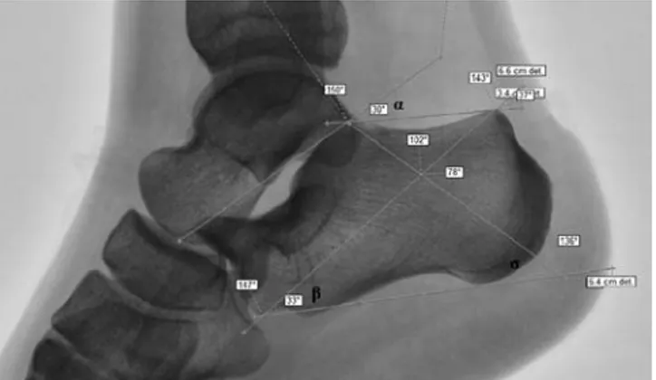
Tuber angle (αα): The angle in between the line drawn from uppermost point of facies articularis cuboidea to most ante-rior point of calcaneus’ supeante-rior surface and the line drawn from the most anterior point of calcaneus’ superior surface to the most posterior point of this surface (Figure 2). Anterior angle (ββ): The angle in between the line drawn from the lowest point of facies articularis cuboidea to most posterior point of calcaneus’s superior surface and the line drawn from the lowest point facies articularis cuboidea to most posterior point of the inferior surface of calcaneus (Figure 2).
Figure 1. Measurements for maximum width (a–g), body width (b–g),
maximum length (e–f), minimum length (c–d), and height of facies artic-ularis cuboidea (a–b).
Figure 2. Tuber angle (αα), anterior angle (ββ) and tuber plantare angle (∑∑).
c e g f d b a
Tuber plantare angle (∑∑): The angle in between the line drawn from the most posterior point of calcaneus to the line of most inferior point of calcaneus and the line drawn from the intersection of the previous parameters to most posterior point of superior surface of calcaneus (Figure 2).
Statistical analysis
Mean±standard deviation (SD) and median values were used to describe the quantitative variables. Also, frequen-cy and percentages were given for the nominal data. Normality assumption was checked by Shapiro Wilk’s test. Since data did not conform to normal distribution, non-parametric statistical tests were used. The distribu-tion of qualitative variables among study groups was ana-lyzed by chi-square test. Within each age group, Mann-Whitney U test was used to compare females and males in terms quantitative variables. Kruskal-Wallis test (with the Conover-Dunn multiple comparison technique) was used to compare quantitative variables among age groups. For all analyses, IBM SPSS Statistics for Windows (Version 21.0, Armonk, NY) was used and statistical significance was set at p<0.05.
Results
Among males 24.2% of (16/66) were in Group 1, 30.3% (20/66) were in Group 2 and 45.5% (30/60) of were in Group 3. Among females, 35.1% of (27/77) were in the Group 1, 26% (20/77) were in Group 2, and 39% (30/77) of were in Group 3. The sexes in the age groups were observed to be similar (p=0.372). Mean, standard deviation and median values for each parameter among age groups are shown in Table 1. In each of the age groups, the defining statistics of each sex were separate-ly defined (Table 2). The statisticalseparate-ly significant
differ-ence according to sex was observed in the parameters a-g, b–a-g, b–a, e–f, c–d for all parameters (p<0.001). The difference between the age groups was determined in a-g (p=0.012), b–a-g (p=0.014), b–a (p=0.025), tuber ana-gle (α) (p=0.002), and anterior angle (β) (p=0.036) measure-ments.
In the first group, significant statistical difference was observed in all of the parameters (p<0.05) except for the tuber angle (α) (p=0.17), the anterior angle (β) (p=0.343) and tuber plantare angle (∑) (p=0.92) (Table 2). In the second group, statistically significant difference was observed in all of the parameters (p<0.05) except for the tuber plantare angle (α) (p=0.184), the anterior angle (β) (p=0.673) and tuber plantare angle (∑) (p=0.585) (Table 2). In the third group, statistically significant difference was observed in all of the parameters (p<0.05) except for the tuber angle (α) (p=0.722), and the tuber plantare angle (∑) (p=0.051) (Table 2).
Discussion
In females and males, Riepert et al.[6]
found the mean value of tuber angle 33.8° and 34.7° mean value of the anterior angle 40.1° and 41.2° and mean value of tuber plantare 73° and 72°, respectively. In our study, the mean value of tuber angle was 33.52° and 35.14°, mean value of anterior angle was 38.96° and 40.32° and mean value of tuber plantare was 46.03° and 46.59°, respec-tively. In the study of Riepert et al.,[6]
there was obvious-ly a greater difference between males and females in the linear measurements than in angles. The authors found the male calcaneus significantly to be larger than the females. However, there was no difference in their pro-portions. In our study, the mean value of tuber plantare was found to be smaller than Riepert’s research.[6]
Variables Group 1 (n=43) Group 2 (n=40) Group 3 (n=60) p Age 55.93±8.19 (54) 37.72±4.36 (38) 22.4±4.69 (22.5) -a-g 7.55±0.96 (7.7) 8.185±0.9 (8.1)* 8.012±0.97 (8.05) 0.012 b-g 6.93± 0.87(7) 7.49±0.93 (7.6)* 7.29±0.91 (7.45) 0.014 b-a 2.3±0.35 (2.3) 2.49±0.33 (2.5)† 2.31±0.35 (2.3) 0.025 e-f 4.36±0.71 (4.5) 4.58± 0.59 (4.65) 4.41±0.68 (4.5) 0.396 c-d 3.62±0.53(3.7) 3.75±0.48 (3.7) 3.77±0.60 (3.8) 0.463 Tuber angle (α) 32.81±5.39 (32)† 33.025±6.35 (33)† 36.13±5.42 (37) 0.002 Anterior angle (β) 41.09±5.39 (41)† 39.35±4.07 (39) 38.67±357 (39) 0.036
Tuber plantare angle (∑) 46.86±3.52 (47) 45.65±3.75 (46.5) 46.3±2.77 (46) 0.339
*Significantly different from Group 3 (p<0.05); †significantly different from the Group 1 (p<0.05)
Table 1
Parameters Groups Males (n=16) Females (n=27) p Mean ± SD (Median) Mean ± SD (Median)
a–g Group 1 8.25; 0.69 (8.3) 7.13; 0.84 (7.3) <0.001 Group 2 8.82; 0.57 (8.8) 7.55; 0.69 (7.65) <0.001 Group 3 8.52; 0.81 (8.7) 7.51; 0.83 (7.65) <0.001 b–g Group 1 7.6; 0.63 (7.5) 6.53; 0.75 (6.8) <0.001 Group 2 8.095; 0.65 (8) 6.88; 0.76 (6.95) <0.001 Group 3 7.75; 0.7 (7.75) 6.85; 0.87 (7) <0.001 b–a Group 1 2.49; 0.26 (2.45) 2.19; 0.34 (2.2) 0.004 Group 2 2.67; 0.26 (2.65) 2.315; 0.28 (2.3) <0.001 Group 3 2.47; 0.3 (2.5) 2.15; 0.31 (2.15) <0.001 e–f Group 1 4.85; 0.48 (4.95) 4.062; 0.65 (4.1) <0.001 Group 2 4.985; 0.38 (4.9) 4.17; 0.46 (4.2) <0.001 Group 3 4.84; 0.52 (4.9) 3.98; 0.52 (4) <0.001 c–d Group 1 3.95; 0.41(4.1) 3.42; 0.50 (3.4) 0.001 Group 2 3.97; 0.43 (4) 3.52; 0.40 (3.55) 0.001 Group 3 4.01;0.51 (4) 3.52; 0.58 (3.45) 0.001 Tuber angle (α) Group 1 33.93; 5.82(35.5) 32.14; 5.11 (31) 0.17
Group 2 34.7; 7.18 (33.5) 31.35; 5.03 (32.5) 0.184 Group 3 36.06; 6.07 (37) 36.2; 4.76 (36.5) 0.722 Anterior angle (β) Group 1 42.56; 7.08 (42) 40.22; 3.99 (41) 0.343 Group 2 39.05; 4.23 (38.5) 39.65; 3.97 (40) 0.673 Group 3 39.96; 2.90 (39.5) 37.36; 3.74 (37) 0.003 Tuber plantare angle (∑) Group 1 3.56; 46.75 (46.5) 46.92; 3.55 (47) 0.92
Group 2 45.8; 2.87 (47) 45.5; 4.53 (46) 0.585 Group 3 47.03; 2.80 (47) 45.56; 2.56 (46) 0.051
Table 2
Descriptive statistics of parameters among gender and within each age group [mean ± SD (median)].
Method Gender (n) a–g e–f c–d a–b
Present study X-ray Males (66) 8.55 4.9 3.9 2.53 Females (77) 7.39 4.05 3.48 2.2 Riepert et al.[6] X-ray Males (436) 8.98 5.18 4.23
Females (364) 8.2 4.67 3.78
Kim et al.[14] Bones Males (50) 8.05 4.91 3.95 2.72
Females (54) 7.37 4.46 3.55 2.49 Zhang et al.[13] X-ray Males (148) 7.19 3.51 2.23
Females (186) 6.52 3.12 1.96 Campobasso et al.[9] Bones Males (40) 7.91 2.45
Females (40) 7.25 2.24
Bidmos et al.[12] Bones Males (58) 7.98 4.31 2.39
Females (58) 7.36 4 2.09 Bidmos et al.[8] Bones Males (53) 8.47 4.77 2.29
Females (60) 7.58 4.33 2.02
Table 3
Morphometric data of the measurements of calcaneus.
In the comparison of the same parameters studied by Riepert et al.,[6]Kim et al.,[14]Zhang et al.,[13]Introna et al.,[9] Bidmos et al.[8]
and our study, morphometric measurements of the males were found to be higher than in females in all of the studies. However, in the study by Zhang et al.,[13]
morphometric values were found to be smaller than our morphometric data both in males and females. This differ-ence was thought to be related with genetical differdiffer-ences (Table 3).[6,8,9,13,14]
Studies found in the literature concluded that the measurements of males were found to be higher
than the females (Table 3). The black community living in Europe had higher calcaneus measurement values than the ones living in Southern African society. Within the frame-work of this conclusion, according to Bidmos,[15]
sex and population cause to a difference in osteometric dimensions of calcaneus.
In the study of Bidmos and Dayal,[16]
the values of males’ parameters were higher than the females’. The authors further maintained that the results gained by the osteometric methods provided an easy and fast way of determining sex.[16]
As already asserted in a number of studies. Kim et al.,[14]also supported Bidmos and Dayal,[16] claim that males possess higher measurement values as compared to females.[14]
On the other hand, while stating that it is difficult to statistically differentiate between white and black Americans by osteometric methods. Steele claimed that these methods could be used to determine sex in the community of Pueblo.[17]
Conclusion
This research shows that the calcaneus plays an important role in the determination of sex. As it was observed that the measurements of length were an easy method for sex determination, the participants’ angular measurements did not reveal any statistical differences. In conclusion, if supported by non-metric methods which are mostly used by the forensic anthropologists, radiographic analysis of the calcaneus can be used as an alternative method in the forensic anthropology and the forensic sciences.
References
1. Cox M, Mays S. Human osteology in archaeology and forensic sci-ence. London (UK): Greenwich Medical Media; 2000. p. 548. 2. France DL. Observational and metric analysis of sex in the skeleton.
In: Reichs KJ, editor. Forensic osteology: advances in the identifica-tion of human remains. 2nd ed. Springfield (IL): Charles C. Thomas; 1998. p. 163–186.
3. Zeyfeo¤lu Y. Hanc› H. ‹nsanlarda kimlik tespiti. Sürekli T›p E¤itimi Dergisi 2001;10:375–7.
4. Washburn SL. Sex differences in the pubic bone. Am J Phys Anthropol 1948;6:199–207.
5. fiahiner Y, Yalç›n H. Erkek ve bayanlarda kafatas› kemi¤inden geometrik morfometri metoduyla cinsiyet tayini ve ramus flexure. Atatürk Üniversitesi Veteriner Bilimleri Dergisi 2007;2:132–40. 6. Riepert T, Drechsler T, Schild H. Nafe B. Mattern R. Estimation of
sex on the basis of radiographs of the calcaneus. Forensic Sci Int 1996;77:133–40.
7. Gualdi-Russo E. Sex determination from the talus and calcaneus measurements. Forensic Sci Int 2007;171:151–6.
8. Bidmos MA, Asala SA. Discriminant function sexing of the calcaneus of the South African whites. J Forensic Sci 2003;48:1213–8. 9. Introna F Jr, Di Vella G, Campobasso CP, Dragone M. Sex
deter-mination by discriminant analysis of calcanei measurements. J Forensic Sci 1997;42:725–8.
10. Demir A, Çivi S. Assessment of 10-year major osteoporotic and
femur fracture risk of postmenopausal women using FRAX®. The
Turkish Journal of Physical Medicine and Rehabilitation 2014;60:11–8.
11. Saka G, Ceylan A, Ertem M, Palanci Y, Toksöz P. Diyarbak›r il merkezinde lise ve üzeri ö¤renim görmüfl 40 yafl üzeri kad›nlar›n menopoz dönemine ait baz› özellikleri ve kalsiyum kayna¤› yiyecek-leri tüketim s›kl›klar›. Dicle T›p Dergisi 2005;32:77–83.
12. Bidmos MA, Asala SA. Sexual dimorphism of the calcaneus of South African blacks. J Forensic Sci 2004;49:446–50.
13. Zhang ZH, Chen XG, Li WK, Yang SQ, Deng ZH, Yu JQ, Yang ZG, Huang L. Sex determination by discriminant analysis of cal-caneal measurements on the lateral digital radiography. Fa Yi Xue Za Zhi 2008;24:122–5.
14. Kim DI, Kim YS, Lee UY, Han SH. Sex determination from calca-neus in Korean using discriminant analysis. Forensic Sci Int 2013;228:177.e1–7.
15. Bidmos M. Adult stature reconstruction from the calcaneus of South Africans of European descent. J Clin Forensic Med 2006;13:247–52. 16. Bidmos MA, Dayal MR. Sex determination from the talus of South african whites by discriminant function analysis. Am J Forensic Med Pathol 2003;24:322–8.
17. Steele DG. The estimation of sex on the basis of the talus and calca-neus. Am J Phys Anthropol 1976;45:581–8.
Correspondence to: Muhammet Bora Uzuner, MD Department of Anatomy, School of Medicine, Hacettepe University, Ankara, Turkey Phone: +90 312 305 21 07 e-mail: bora.uzuner@hacettepe.edu.tr
Conflict of interest statement: No conflicts declared.
This is an open access article distributed under the terms of the Creative Commons Attribution-NonCommercial-NoDerivs 3.0 Unported (CC BY-NC-ND3.0) Licence (http://creativecommons.org/licenses/by-nc-nd/3.0/) which permits unrestricted noncommercial use, distribution, and reproduction in any medium, provided the original work is properly cited. Please cite this article as: Uzuner MB, Geneci F, Ocak M, Bayram P, Sancak ‹T, Dolgun A, Sargon MF. Sex determination from the radiographic measurements of calcaneus. Anatomy 2016;10(3):200–204.
Online available at: www.anatomy.org.tr doi:10.2399/ana.16.039 QR code: