Olgu sunumu/ Case report
An Interesting Cause of Acute Abdomen: Stomach Perforation due to Foreign Body Stomach Perforation due to Foreign Body
Nizamettin KUTLUER 1, Ahmet BOZDAĞ 2, Pınar GÜNDOĞAN BOZDAĞ 3, Ali AKSU1, Barış GÜLTÜRK1
1
Sağlık Bilimleri Üniversitesi, Elazığ Sağlık Uygulama ve Araştırma Merkezi, Genel Cerrahi Kliniği, Elazığ. 2
Fırat Üniversitesi Tıp Fakültesi Genel Cerrahi Anabilim Dalı, Elazığ
3Sağlık Bilimleri Üniversitesi, Elazığ Sağlık Uygulama ve Araştırma Merkezi, Radyoloji Kliniği, Elazığ.
ABSTRACT
Foreign body ingestion is a frequent cause of emergency service admission. Although frequent in children, it can be seen in alcoholics and adults with psychiatric distress or mental retardation. Most of these patients do not require any intervention, however only a few have surgery or endoscopic procedures. Surgery is mostly performed in cases such as obstruction, perforation or hemorrhage. In this article, we present a case along with literature which was evaluated as acute abdomen in the emergency department, The etiology explaining the case was not found in the tests and perforation was determined due to a foreign body after laparotomy.
Keywords: Gastric perforation, Foreign body, Bone.
DOI: 10.30569/adiyamansaglik.402088
Yazışmadan Sorumlu Yazar
Ahmet BOZDAĞ
Fırat Üniversitesi Tıp Fakültesi Genel Cerrahi Anabilim Dalı, Elazığ
Tel : +90 0424 23335 55 Dahili: 2271 Email: [email protected]
Geliş Tarihi: 06.03.2018 Kabul Tarihi: 03.04.2018
708
İlginç Bir Akut Batın Nedeni: Yabancı Cisme Bağlı Mide Perforasyonu Yabancı Cisme Bağlı Mide Perforasyonu
ÖZET
Yabancı cisim yutulması acil servise başvuruların sık sebeplerindendir. Sıklıkla çocuklarda karşımıza çıkmasına rağmen alkolikler, pskiyatrik rahatsızlığı olan ya da mental retarde erişkinlerde de görülebilmektedir. Bu hastaların birçoğu herhangi bir girişim gerektirmemekte ancak az bir kısmına ise cerrahi ya da endoskopik işlem yapılmaktadır. Cerrahi daha çok obstrüksiyon, perforasyon ya da kanama gibi durumlar söz konusu ise yapılmaktadır. Bu makalede acil serviste akut karın nedeniyle değerlendirilip tetkiklerinde tabloyu açıklayacak bir sebep bulunamayan ve laparatomi sonrası yabancı cisme bağlı perforasyon tespit edilen olgu literatür eşliğinde sunulacaktır.
709
Foreign body ingestion is a condition that is seen mostly in children. It is seen less frequently in the adult age; in chronic alcoholics, in those with epileptic or psychiatric disturbances, or in those with mental retardation. Approximately 95% of the bodies that are ingested pass from the stomach to the intestines and are excreted without creating any symptoms through the passage without giving any harm to the body. The remaining cases require surgical or endoscopic treatment (1, 2). Type of treatment differs according to the characteristics of ingested foreign bodies, the place and the time they persist in the gastrointestinal tract, or the clinical characteristics of the patient (3). The aim of this article is; presenting a foreign body ingestion case in the context of the literature that causes gastric perforation in a patient who does not have a history of foreign body swallowing and who does not have any evidence of foreign body in the studies, and reminding this rare situation that causes acute abdominal pain.
710
CASE REPORT
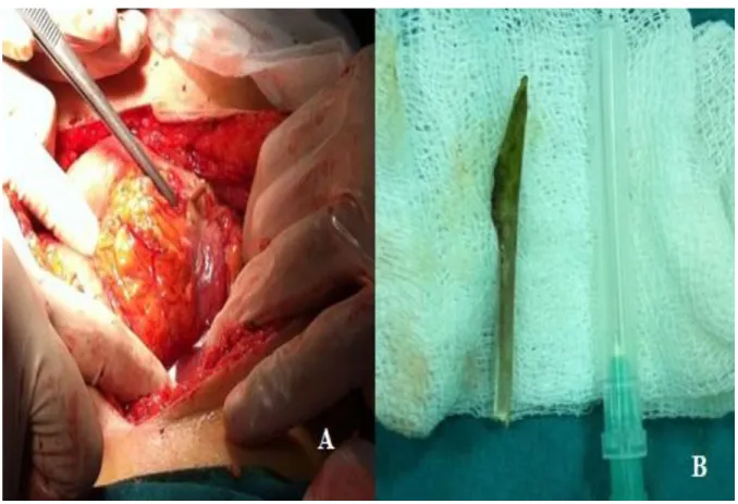
A 60-year-old female patient who was admitted to the emergency room with complaints of diffuse abdominal pain and nausea of two days was evaluated by general surgery with the diagnosis of "Acute Abdomen". There was no story of swallowing foreign bodies in the patient's anamnesis and the patient applied to the emergency room the day before and was given medical treatment and discharged due to normal workup, nonspecific gastrointestinal complaints after the gastroenterological consultation. There was diffuse tenderness, defense and rebound on the physical examination. Biochemical parameters were normal except for high white blood cell count. Radiologic evaluation showed no free air or foreign body opacity on the radiogram. Opacity extending from the anterio rstomach to the lumen on the large curvature side was seen on tomography (Asteion 4, Toshiba, Japan) obtained without contrast, no additional finding was determined (Figure 1). Information was given to the relatives of the patient and operation was decided by receiving their consent. On the evaluation during operation, the foreign body was seen in the anterior gastric side at a distance of 2 cm to pylorus on the side of the large curvature, one side inside the stomach and the other side outside the stomach, it was thought to be a chicken bone also confirmed by the patient after surgery that was perforating the stomach wall. The bone was removed and the area was repaired using graham method (Figure 2). On the third day after the operation, the patient started a regimen and was discharged on the 5th day. Written consent has been obtained for the scientific presentation and use of patient data.
711
Figure1: A)Freeairandforeign body are not seen in the direct radiography. B) Foreign body is seen in tomography in transverse section. C.) Foreign body is seen in tomography in coronal section. D) Foreign body is seen in tomography in sagittal section.
712
Figure 2: A) Intraoperative perforation foreign body in the stomach, one side inside the other side outside the stomach. B) Foreign body removed
DISCUSSION
Foreign body ingestion is among the common causes of emergency service admissions especially in children. It is seen less frequently in adults than children. Most of the patients admit saying that they accidentally ingested the body in the anamnesis however such as our patient, some of them do not know that they ingested. The most important aspects of foreign body ingestion are the delays and difficulties experienced in diagnosis and treatment in cases where the family is not aware of the situation in the case of children, the patient does not express it in the history or the patients who cannot give sufficient anamnesis such as mental retardation (3). Discharge of our patient from the previous admission to after proceeded
713
unique symptom or physical examination finding.
Direct radiographs can be used to obtain information about the number of foreign bodies that are radiopaque, their shape, and their location in the gastrointestinal tract. Of the ingested foreign bodies, the metal products, 86% of the glass products can be seen on direct radiography, only 26% of the bone structures are seen.Iftheforeign body is radiolucent, furtherinvestigationssuch as computedtomography (CT) can be used. Three-dimensional CT has been shown to be successful in cases that are radiologically radiolucent and cannot be detected in CT (1,4). Since the foreign body was not radiopaque in this case, it was not seen in direct abdominal radiography. The foreign body was also detected in the obtained tomography however it caused a clinical delay due to the fact that it was not in the history of the patient. In radiopaque foreign bodies the passage of the body should be monitored by intermittent serial x-rays to see if it is stuck or not in the anatomically narrowing regions or corners of gastrointestinal tract such as C loop of duodenum, ligament of Treitz and ileocecal valve (5,6,7).
Approach to foreign bodies detected in the gastrointestinal tract can vary according to the location, type, shape of the body, the age of the patient, and the determined findings. The vast majority of foreign bodies are excreted without causing any complication. In the literature, 75.6% of foreign bodies are removed spontaneously, 19.5% by endoscopic procedure and 4.8% by surgical intervention (1,2). Passage of bodies that remain for long periods of time in the stomach or small intestine can be accelerated by administering various drugs (magnesium citrate or magnesium sulfate, etc.).
714
methods should be applied and it should be removed if the foreign body persists for more than 4 weeks after it is confirmed that it is in the stomach. We think that four weeks is a long time and it should be evaluated endoscopically earlier and if complicated, surgery should be performed. It has been reported that sharp objects such as wire or bone may be stuck in the gastric mucosa and cause only gastritis without causing perforation. (8,9,10,11) Another criterion for determining the type of the treatment is the size of the foreign body. For adults, this size is 4 cm in the literature. It has been reported that the objects larger than 4 cm, especially piercing sharp ones, are more dangerous and should be intervened.
As a result, foreign bodies can be removed by flexible endoscopy in many cases, especially in adult patients without general anesthesia, and gastroscopy continues to be the most reliable method for foreign body removal today. Laparotomy is indispensable when the ingested material can not be removed or pose a risk. However, it should not be forgotten that the importance of physical examination and the surgical operation are inevitable in these cases, which do not provide any anamnesis for foreign body ingestion, in cases where foreign body presence in the abdomen could not be determined with radiological studies.
715
foreign bodies. AJR Am J Roentgenol. 2014 Jul;203(1):37- 53. doi: 10.2214/AJR.13.12185.
2. Velitchkov NG, Grigorov GI, Losanoff JE, et al. Ingested foreign bodies of the gastrointestinal tract: Retrospective analysis of 542 cases. World J Surg 1996;20:1001-1005.
3. Neves CZ, Maluf-Filho F. Clinical and endoscopic aspects of foreign body ingestion. Gastroenterol Hepatol (NY) 2010;6(9):584-5.
4. Takada M, Kashiwagi R, Sakane M, 3D-CT diagnosis for ingested foreign bodies. Am J Emerg Med 2000;18: 192-193.
5. Ciriza C, Garcia L, Suarez P, Jimenez C, Romero MJ, Urquiza O, Dajil S. What predictive parameters best indicate the need for emergent gastrointestinal endoscopy after foreign body ingestion. Clin Gastroenterol 2000;31:23-28.
6. Gambaracci G, Mecarini E, Franceschini MS, Scialpi M. Gastric Perforation by Ingested Rabbit Bone Fragment. Case Rep Gastroenterol 2016;10:121–126
7. Yang Z, Wu D, Xiong D, Li Y. Gastrointestinal perforation secondary to accidental ingestion of toothpicks A series case report. Medicine (Baltimore). 2017;96(50):9066.
8. American Society for Gastrointestinal Endoscopy. Guideline for the management of ingested foreign bodies. Gastrointest. Endosc. 2002;55:802-806.
9. Ricci G, Campisi N, Capuano G, et al. Liver abscess and pseudotumoral gastric lesion caused by chicken bone perforation: laparoscopic management. Case RepSurg 2012;791-857.
10. Karakaş BR, Bülbüller N, Demirci RK, Arduçoğlu AM. A Rare Cause of Chronic Gastritis: Chicken Bone Ingestion; Okmeydanı MedicalJournal 2014;30(1):51-53.
11. Gültürk B, Bozdağ A, Kanat BH, Girgin M, Gülaçtı U. Foreign Body in the Stomach Surprising the Patient: Case Presentation Göztepe Medical Journal 2013;28(4):219-221.