Journal of Clinical & Analytical Medicine
Original Research
Reşat Dikme1, Mustafa Göz2, Mehmet Salih Aydın2, Hakim Çelik3, Mahmut Padak1, Ömer Göç1, Abdullah Taşkın4 1Perfusion Techniques Program, Harran University Vocational School of Health Services,
2Cardiovascular Surgery, Harran University Faculty of Medicine, 3Physiology, Harran University Faculty of Medicine, 4Nursing Department, Harran University Faculty of Health Sciences, Şanlıurfa, Turkey
DNA damage during cardiopulmonary bypass
Oxidative stress and DNA damage during
cardiopulmonary bypass
DOI: 10.4328/JCAM.5883 Received: 17.04.2018 Accepted: 27.03.2019 Published Online: 27.03.2019
Corresponding Author: Reşat Dikme, Harran University Yenişehir Campus Vocational School of Health Services, 63300, Haliliye, Şanlıurfa, Turkey. GSM: +905354587844 F.: +90 414 3183209 E-Mail: [email protected]
ORCID ID: 0000-0001-9157-7830 Abstract
Aim: The aim of this study is to measure the oxidative stress index (OSI) and DNA damage in patients undergoing cardiopulmonary bypass (CPB) surgery due to coronary artery disease. Material and Method: 25 patients operated for CPB surgery due to coronary artery disease were included in the study. By collect-ing heparin containcollect-ing blood from the patients at 5 different times (Before CPB, durcollect-ing pump inlet, durcollect-ing placcollect-ing of cross clamp, durcollect-ing takcollect-ing out of cross clamp and after CPB) DNA damage was studied by Comet Assay method from mononuclear leucocytes and OSI in plasma was studied. Results: At 5 different times plasma OSI value was found respectively as 4.92 ± 1.73, 5.20 ± 2.69, 5.70 ± 2.06, 9.88 ± 4.88, 9.57 ± 4.54 whereas Arbitrary unit (AU), DNA damage were determined respectively as 28.48 ± 11.30, 29.04 ± 9.43, 31.44 ± 11.74, 33.92 ± 7.96, 38.56 ± 6.20 AU and p<0.001. Discussion: During CPB, OSI and DNA damage gradually increased and a positive meaningful correlation occured between these two parameters (r=0,882 p=0,048). As CPB period extended, oxidative stress and DNA damage increased as well as TOS and OSI increased, or vice versa.
Keywords
Introduction
During CPB, the patient’s blood is in contact with non-physi-ological and non-endothelial surfaces such as cannula, tubing set, reservoir, antifoaming agents, filters, pumps, heat exchang-ers and oxygenators. This contact activates blood proteins and immune system cells [1].As the contact surface increases, the damage in the blood contacting nonendothelial surface increas-es. The immune response starting during CPB may cause organ disfunction, that effects postoperative mortality and morbidity [2]. In continuity of this response, oxidative stress [3] initiated by catecholamines, complement system, oscillated cytokines from activated neutrophiles, free oxygen radicals released during ischemia-reperfusion period, endothelium damage, kallikrein cascade, endotoxine oscillation [4] and stimulated by factors such as systemic heparinization, nonpulsatile current, removal of blood stream by cross clamping, anaesthetic drugs, reper-fusion, acute dilutional anaemia, perfusion pressure, stream changes and lungs being out of circulation, play an important role [5].
Intensive reactive oxygen types (ROS) production or antioxidant defence reduction causes oxidative stress by making structur-al and functionstructur-al modifications in biomolecules [6]. Oxidative processes start a series of events that consequently cause cell death via decrease in mitochondrial energy capacity [7]. Dur-ing CPB, perfusion system, low arterial pressure, embolism and surgical manipulation also cause ischemia [8] resulting in ex-cessive energy phosphate consumption and intracellular Ca ac-cumulation [2]. During ischemia, fatty acid oxidation disorders and creatine phosphate anaerobic metabolism become main energy source of the cell [9]. However creatine phosphate runs short quickly, and rapidly improved acidosis limits glycolysis as well. In addition to the effect of the extracorporeal circulation, the activation of proinflammatory mediators, is also stimulat-ed. As a result, consisted free oxygen radicals damage the cell membrane and myocardium [10]. During the restoration of the heart in CPB, the heart is stopped temporarily by putting cross clamp on aorta and the myocardium is exposed to ischemia. After taking out the cross clamp (after ischemia), reoxygenation of tissues is provided by sudden reperfusion, where the free O2 radicals cause a phenomenon named as ischemia-reperfusion damage [6, 11]. Free oxygen radicals have a property of react-ing fast with proteins, phospholipids and thiols, their attacks on DNA cause mutations, even cell deaths [4, 7]. Various clinical, epidemiologic and experimental studies show that there is a relation between free radicals, lipid peroxidation and its prod-ucts and DNA damage and carcinogenesis [12, 13]. CPB and the pathology of the subjects have importance at least as DNA damage.
DNA damage can be detected in many methods. The recent-ly developed method “Single Cell Gel Electrophoresis” (SCGE) also named as “Comet Assay” or “Microgel Electrophoretic Technique”, is sensitive, fast and cheap in obtaining the spiral fracture [14]. The easily used SCGE method, shows even low level of DNA damages, provides its analysis with only few cells, with different cell and tissue groups, does not require too many equipments and, its reliable results can be obtained economi-cally and evaluated within just few hours [15].
Material and Method
25 patients who were operated (by on pump) in the Cardiovas-cular Surgery Department of Harran University Hospital or The State Hospital in Sanliurfa were included in our study and 5
tubes of blood were collected from each of them consecutively before CPB, during the pump inlet, at cross clamping, at cross clamp removal and after CPB. During CPB, roller pump was used and non-pulsatile perfusion was applied. Commercial crystal-loid cardioplegia (Plegisol®, 4-9 ° C) and tube sets (Bıcakcılar Extracorporeal Tubing Set-Turkey) were used during surgery. Patients were treated with 1200 ml of Ringer’s lactate or isoli-tol-priming solution. Routine CPB procedures were performed during surgery.
Preperation of Samples and Studied Tests
Collected blood samples were transferred immediately to hepa-rin containing vacutainers. DNA oxidation levels and the dam-age was analyzed with Comet Assay method after separating mononuclear leucocytes from fresh blood samples. Then the rest of blood sample was centrifuged at 3000 rpm and the plasma was separated out. The samples were kept frozen and stored at -80 oC until handling. Total oxidative Status (TOS), Total Antioxidant Status (TAS) and Oxidative Stress Index (OSI) were studied as a result of lipid peroxide, lipid hydroperoxide, myeloperoxidase, ceruloplasmin, total peroxide in the separated plasma.
DNA Damage Identification with Alkali Mono Cell Electropho-resis Method (Comet Assay)
Comet assay method is based on agarose gel loads of DNA chain exposed to electrical field in alkali setting and their car-riage according to molecular size. In this measuring method, the processes show differences according to lab conditions. Tice et. al. suggested 8 steps of SCGE method as isolation of cells, preparation of slices, lysis, disintegration of DNA spiral, electrophoresis, neutralization, coloring and evaluation [16]. In Comet assay method mono cells are placed in agar gel. After ly-sis, while the damaged and broken DNA pieces form tail format since they move in electrical field at different degrees due to having different loads and molecular weights, free and undam-aged DNAs do not form tail because they migrate together in electical field.
Since damaged DNA cells were evaluated by visual detection method in our study, in the examination of the undamaged cells, there were circular cells, less dense at sides, luminary outlook with brightness in the middle. This outlook of cells is evaluated as nonmigration. If DNA damage starts to occur, smooth mar-ginated outlook becomes ragged edge outlook due to migra-tion of DNA fractures out of the cell. Depending on the sever-ity of the damage, there occurs a stretch from centre to the edge. This outlook is named as stretch or low migration. As the damage increases, the cells take the shape of a comet, that means high migration [17, 18]. The last stage is apoptosis in which stretching is directly proportional to the damage. Also fluorescence density at the tail is parallel to the degree of the damage. In this method, the damage was classified in 5 catego-ries due to the degree of DNA migration. The length of migra-tion shows difference according to quantity of fragments, DNA chain breakages and alkali-labile fields levels. The evaluation was done in five categories as undamaged DNA is evaluated as category 0, the ones having maximum damage are evaluated as category 4. (Figure 1). Totally 100 cells were evaluated from each slide. The results were calculated as arbitrary unit (AU) by considering maximum damage as 400.
Protein oxidation (PO)
15 µl plasma was mixed with 0,5 ml DNPH (10 milimolar 2,4-di-nitrophenylhydrazine in 2 molar HCl) solution and left to incu-bation for 1 hour at room temperature. After incuincu-bation it was vortexed by adding 0,5 ml TCA (10% trichloroaceticacid in de-ionized water) solution and centrifuged for 3 minutes at 15000 rpm. After expelling supernatant, the pellet was washed 2 times with in the ratio of 1/1 ethanol/ ethyl acetate. 0,6 ml Guanidin HCl solution (6 molar guanidine HCl dissolutes in 20 milimolar pottsium phosphate (pH=2,3) was added and left to incubation for 15 minutes at 37 oC’de for dissolution. The absorbance of obtained colour was measured at 365 nm. After multiplying absorbance coefficient with (emax=22000/M/cm), the results were given as nmol (nmol/mgprot) per mg protein [17].
Plasma Lipide Peroxidation Measurement
25 µl plasma was mixed with 0,5 ml DETBA (10 mmol/L 1,3-di-ethylthiobarbituric acid, dissolved in 75 mol/L phosphate (pH=3)) and left in incubation for an hour at 96 oC. After leaving samples in ice bath for 5 minutes, 2,5 ml n-butanol was added. The mixture was vortexed and centrifuged at 1500 x g at 4 oC for 10 minutes. The supernatant was taken and analyzed in florometer (Extantion= 539 and emission= 553)(Schimatzu, Japan) [19].
Total Antioxidant Status (TAS) Level Measurement
The measurement method, which uses Total Antioxidant Sta-tus Assay Kıt (Rel Assay, Product Code: RL0017), is based on decolorization of all antioxidant molecules in the sample in pro-portion to the total concentration of antioxidant molecules of colourful radicals as a result of reduced colourful ABTS* cat-ionic radical. Trolox, a water-soluable analogue of vitamine E is used as calibrator. The results were given as mmol Trolox Equivalent/L [20].
Total Oxidant Status (TOS) Level Measurement
In total automatic colormetric method, Total Oxidant Status As-say Kıt (Rel AsAs-say, Product Code: RL0024) is used. The colori-metric method is based on oxidization of ferrous ions of oxidant molecules to ferric ion cumulatively.
The results are given as µmol H2O2 Equivalent/ L [21].
Oksidative Stress İndex (OSI)
OSI, as a sign of Oxidative Stress, is defined as percentage of the degree of TOS levels to TAS levels. In calculation of OSI
value of samples, TAS levels are multiplied with 10 and their units are balanced with TOS levels [24]. The results are defined as Arbitrary Units (AU).
TOS, µmol H2O2 Equiv. / L. OSI = ————————————— X 100 TAS, µmol trolox Equiv. / L.
Statistical Analyses
Stastistical analyses were done by using SPSS programme. First of all, normality evaluation was done with normality test ‘’Shapiro-Wilk’’. In normal distribution, ‘’Pearson’’analysis was used for parametric correlations, while ’Spearman’’analysis was used for nonparametric correlations in abnormal distribution. p<0.05 value was considered significant.
Results
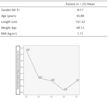
Demographic and characteristic data are given in Table 1. In the table, the gender (8 Male + 17 Female), average age (45.88 years), average height (161.52 cm), average weight (68.12 kg) and average BMI (1.71 kg/m2) values were given.
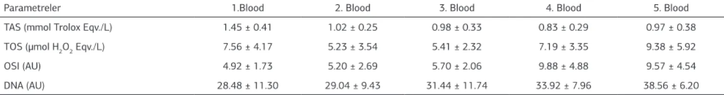
TAS, TOS, OSI and mononuclear leucyte DNA damage of pa-tients are shown in Table 2.
1. Blood before pump, 2. Blood taken at the pump inlet, 3. Blood taken when a cross clamp is placed, 4. Blood taken after cross-clamp removal, 5. Blood taken from the pump
TAS level continuously reduces until the 4th blood sample (after removal cross clamp), (Table 1) and increases a little bit at the 5th blood (pump outlet) (Figure 2). TOS reduced a little as
pass-ing from the 1st blood sample (before pump) to the 2nd blood sample (at the pump inlet), then increased continuously after the 2nd blood (after pump inlet) (Figure 3). Until the 4th blood sample, OSI levels (as cross unclamping) continuously increas-es, at 5th blood sample there is a little reduction (Figure 4). This increase in OSI levels became maximum after unclamping. (4th blood sample). DNA damage on the other hand O. Category 1. Category 2. Category
3. Category 4. Category
Table 1. Comparison of physical values of patients
Patient (n = 25) Mean Gender (M/ F) 8/17 Age (years) 45.88 Length (cm) 161.52 Weight (kg) 68.12 BMI (kg/m2) 1.71
Figure 1. Images of DNA after electrophoresis migration of damage from oxida-tive DNA damage under the microscope.
ously increased from 1st blood sample (before pump) to the 5th blood sample. (pump outlet) (Figure 5). Generally TOS, OSI, DNA damage increase while TAS level decreased.
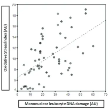
According to the correlation analysis done at the end of study, a significant positive correlation was found (r=0,882 p=0,048) between OSI and DNA damage (Table 3). The distribution of relations between mononuclear leucyte DNA damage and oxi-dative stres index levels were shown in Figure 6.
Discussion
According to the results of our study, TAS continually decreased until unclamping the aorta, myocardium became bloodstained and TAS value increased after this period. This situation shows us that TAS value continuously decreased when there is no bloodstain, but TOS value continuously increased. This
situa-tion shows us that during CPB increases oxidative stress con-tinuously due to either unfamiliar surface material or surgical situations. Besides, the antioxidant defence systems become insufficient due to the decrease of TAS, but the increase of TOS value. In the analysis, TAS and TOS values decreased a little during the pump inlet because of the hemodillution. OSI levels continuously increased at cross clamping and was maximum at unclamping. When the circulation of the heart stopped dur-ing the cross clampdur-ing period, oxygen derivative radicals are generated from ischemi reperfusion as a result of bloodstained heart and increase OSI levels. In other words, oxygen deriva-tive radicals cause oxidaderiva-tive stress. Also due to the decrease of antioxidant level during pump period, oxidant level and oxida-tive stress cannot be eliminated, accordingly OSI continuously increases. Due to the increase of TOS and OSI levels and the decrease in TAS levels, DNA damage continuously increased, because of non endothelial level, non pulsatile current, insuf-ficient perfusion, discontinuation of blood stream with cross clamping, anesthetic drugs, myocardium damage, cells in stress due to reperfusion and immune response and attacks of oxi-dative stress elements to DNA, during CPB. Our study shows that OSI and DNA damage continuously increased during the pump and a positive correlation between these two parameters occurred. As the length of clamping period and pump period extended, oxidative stress increased and more DNA damage oc-cured. Many studies show that ROS levels can severely increase in stress periods and cause serious damage in many molecular molecules such as lipides, proteins and DNA.
The results of our study are parallel to many previous studies. Starkopf et. al. stated that TAS decreases and oxidant damage occurs during ischemia and reperfusion period in CBP. Also, in coronary artery bypass facts during 72 hours after surgery, it was observed that TAS was depressed, lipid peroxidation in-creased and depression in TAS were in reverse relation with
Table 3. Relationships between DNA damage and Oxidant / Antioxidant pa-rameters
TOS TAS OSi
TAS r -0,058 p 0,926 . OSI r -0,330 -0,661 p 0,587 0,224 . DNA r -0,335 -0,576 0,882 p 0,581 0,309 0,048(*) * p < 0.05
lipid peroxidation (23 tubing set, reservoir, antifoaming agents). According to the study of Taşkıran A. et. al. on the damage and relation of plasma TAS levels with ischemia-reperfusion dam-age before CBP coronary artery bypass surgery, they found out that low TAS values before surgery are in relation with isch-emia-reperfusion damage and severity of myocardium damage [24, 25]. Ischemia and reperfusion damage during CPB cause proinflammatory mediators and ROS formation by causing an important myocardial stress, which harms proteins, lipides that affect postoperative cardiac function as well as DNA. The existence of positive correlation between DNA damage and oxidants and negative correlation between DNA damage and antioxidants, force to find better methods that can reduce side effects of CPB to minimum level. As a result, factors such as contact with artificial surfaces, surgical trauma, oxidative stress and ischemia reperfusion injury play an important role in the formation of DNA damage. As a result of these factors, reactions at the molecular level increase DNA damage during CPB. Among the limitations of this study is the absence of a control group from patients operated by non-pump techniques.
Scientific Responsibility Statement
The authors declare that they are responsible for the article’s scientific content including study design, data collection, analy-sis and interpretation, writing, some of the main line, or all of the preparation and scientific review of the contents and ap-proval of the final version of the article.
Animal and human rights statement
All procedures performed in this study were in accordance with the ethical standards of the institutional and/or national re-search committee and with the 1964 Helsinki declaration and its later amendments or comparable ethical standards. No ani-mal or human studies were carried out by the authors for this article.
Funding: None Conflict of interest
None of the authors received any type of financial support that could be considered potential conflict of interest regarding the manuscript or its submission.
References
1. Tavares-Murta BM, Cordeiro AO, Murta EF, Cunha Fde Q, Bisinotto FM. Effect of myocardial protection and perfusion temperature on production of cytokines and nitric oxide during cardiopulmonary bypass. Acta Cir Bras. 2007;22(4):243-50. 2. Clermont G, Vergely C, Jazayeri S, Lahet JJ, Goudeau JJ, Lecour S, et al. Systemic free radical activation is a major event involved in myocardial oxidative stress related to cardiopulmonary bypass. Anesthesiology 2002;96 (1):80-7.
3. Zakkar M, Guida G, Suleiman M.-S., Angelini G. D. Cardiopulmonary bypass and oxidative stress. Oxidative Medicine and Cellular Longevity. 2015;2015:189863. 4. McDonald C. I, Fraser J. F, Coombes J. S, Fung Y. L. Oxidative stress during extra-corporeal circulation. Eur J Cardiothorac Surg. 2014;46(6):937-43.
5. Lefer D. J, Granger D. N. Oxidative stress and cardiac disease. Am J Med. 2000;109(4):315-23.
6. Berg K, Haaverstad R, Astudillo R, Björngaard M, Skarra S,Wiseth R, et al.
Oxi-dative stress during coronary artery bypass operations: Importance of surgical trauma and drug treatment. Scand Cardiovasc J. 2006;40(5):291-7.
7. Ott M, Gogvadze V, Orrenius S, Zhivotovsky B. Mitochondria, oxidative stress and cell death. Apoptosis. vol. 12, no. 5. 2007;12(5):913-22.
8. Warren OJ, Watret AL, de Wit KL, Alexiou C, Vincent C, Darzi AW, Athanasiou T. The inflammatory response to cardiopulmonary bypass: part 2--anti-inflammato-ry therapeutic strategies. J Cardiothorac Vasc Anesth. 2009;23(3):384-93. 9. Suleiman MS, Zacharowski K, Angelini GD. Inflammatory response and cardio-protection during open-heart surgery: the importance of anaesthetics. Br J Phar-macol. 2008;153(1):21-33.
10. Kamata H, Honda S, Maeda S, Chang L, Hirata H, Karin M. Reactive oxygen species promote TNF-induced death and sustained JNK activation by inhibiting MAP kinase phosphatases. Cell. 2005;120(5):649-61.
11. Ulus AT, Aksoyek A, Ozkan M, Katircioglu SF, Basu S. Cardiopulmonary bypass as a cause of free radical-induced oxidative stress and enhanced blood-borne isoprostanes in humans. Free Radic Biol Med. 2003;34(7):911-7.
12. Ames B.N, Shigenara M.K. DNA damage by Endogenous oxsidants and mitho-genesis As Causes of Aging and Cancer. Molecular Biology of free radical scav-enging systems, ed, scandalios. J.G. (Cold Spring Harbor Laboratuary Pres, Plain-viev .p:1-21). 1992.
13. Andican G, Burçak G. Oksidatif DNA Hasarı ve HPLC İle Analizi. II. Ulusal HPLC ve Diğer seperasyon Teknikleri sempozyumu. Özet kitabı. Ankara. 2004. 14. Fairbain D.W, Olive P.L, O’Neill K.L. The Comet Assay: A Comprehensive Review. Mutat Res. 1995;339(1):37-59.
15. Singh NP, Danner DB, Tice RR, Pearson JB, Brant LJ, Schneider EL. DNA dam-age and repair with dam-age in individual human lymphocytes. Mutat Res 237:123–30. 16. Kocyigit A, Keles H, Selek S, Guzel S, Celık H, Erel O. Increased DNA damage and oxidative stress in patients with cutaneous leishmaniasis Mutation. Mutat Res. 2005;585(1-2):71-8.
17. Collins AR. The comet assay for DNA damage and repair: principles, applica-tions, and limitations. Mol Biotechnol 2004;26(3):249-61.
18. Shacter E. Protein oxidative damage. Methods Enzymol. 2000;319:428-36. 19. Yagi K. Lipid peroxides and related radicals in clinical medicine. Adv Exp Med Biol. 1994;366:1-15.
20. Erel O. A novel automated direct measurement method for total antioxidant capacity using a new generation, more stable ABTS radical cation. Clin Biochem. 2004;37(4):277-85.
21. Erel O. A new automated colorimetric method for measuring total oxidant status. Clin Biochem. 2005;38(12):1103-11.
22. Aycicek A, Erel O, Kocyigit A. Increased oxidative stres in infants exposed o passive smoking. Eur J Pediatr. 2005; 164: 775-8.
23. Starkopf J, Zilmer K, Vihalemm T, KulHsaar T, Zilmer M, Samarotel J. Time course of oxidative stres during open-heart surgery. Scandl Thorac Cardiovasc Surg. 1995; 28 (4),181-6.
24. Taşkıran A, Eskiocak S, Ege T, Duran E, Gülen Ş. Investigation of Myocardial Tissue Injury and Oxidant Stress During Coronary Bypass. Turk J Biochem. 2004; 29(2); 193-8.
25. Taşkıran A, Eskiocak S, Çıkırıkçıoğlu M, Ege T, Duran E. The Relationship be-tween Preoperative Plasma Total Antioxidant Capacity and Ischemia-Reperfusion Injury in Patients Undergoing Coronary Artery Bypass Surgery. Balkan Medical Journal 2005;22:16-22.
How to cite this article:
Dikme R, Göz M, Aydın MS, Çelik H, Padak M, Göç Ö, Taşkın A. Oxidative stress and DNA damage during cardiopulmonary bypass. J Clin Anal Med 2019; DOI: 10.4328/JCAM.5883.
Table 2. Comparison of DNA damage and Oxidant / Antioxidant parameters of patients
Parametreler 1.Blood 2. Blood 3. Blood 4. Blood 5. Blood
TAS (mmol Trolox Eqv./L) 1.45 ± 0.41 1.02 ± 0.25 0.98 ± 0.33 0.83 ± 0.29 0.97 ± 0.38 TOS (µmol H2O2 Eqv./L) 7.56 ± 4.17 5.23 ± 3.54 5.41 ± 2.32 7.19 ± 3.35 9.38 ± 5.92
OSI (AU) 4.92 ± 1.73 5.20 ± 2.69 5.70 ± 2.06 9.88 ± 4.88 9.57 ± 4.54