Journal of Clinical and Analytical Medicine | 1
Le
tt
er
s to Editor
Anticoagulant Therapy and Intramural Small Bowel Hematoma and CT
Elif Karadeli, Sermin Tok, Gurcan Erbay Radyoloji Anabilim Dalı, Başkent Üniversitesi Tıp Fakültesi, Adana, Türkiye
CT findings of Intramural Small Bowel
Hematoma Secondary to use of Oral Anticoagulant Therapy
Antikoagulan Kullanımına Bağlı
İncebarsak İntramural Hematom BT Bulguları
DOI: 10.4328/JCAM.3562 Received: 01.05.2015 Accepted: 18.05.2015 Printed: 01.02.2016
Corresponding Author: Elif Karadeli, Radyoloji Anabilim Dalı, Başkent Üniversitesi Tıp Fakültesi, Dadaloğlu Mah. 2591 Sok. No:4/A 01250 Yüreğir, Adana, Türkiye. T.: +90 3223272727 E-Mail: elifkaradeli @gmail.com
To the editor:
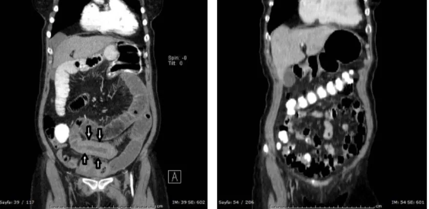
We have read the article with the number DOI: 10.4328/JCAM.176 under the title “Intramural Small Intestinal Hematoma caused by Warfarin Overdose (Case report)” [1] presented by İbrahim Yetim and colleagues in your journal. We wanted to men-tion a case diagnosed by clinical, laboratory and CT findings and treated non-surgically that might contribute to the valuable article. Oral anticoagulants are used for reasons like atrial fibrillation and venous thrombosis. One small intestinal hematoma was reported in 2500 cases using oral anticoagulants [2]. Ultrasonography and CT findings of intramural hematomas might imitate massive formations [3]. The patient history and INR levels are important for the diagnosis. The diagnosis can be con-firmed by endoscopic biopsies [4]. In the treatment, hemostasis is obtained and the regression of the hematoma is monitored. Rarely, surgery is needed. In literature, surgery has been performed in cases where spontaneous regression does not occur or necrosis or peritonitis develops and if the diagnosis is unclear [4]. The 55 year old female case presented with a 3 day history of diffuse abdominal pain and no gas or stool passage. She had been using Coumadin after an ASD operation for atrial fibrillation. Physical examination revealed diffuse abdominal defense. The laboratory test results were high, APTT was 93.70 sec, PT was 111.30 sec and INR was 15.96. In the CT scans with and without contrast, thickening of the intestinal wall was observed in the pelvic region and the mean density was measured 50HU in the scan without contrast and 55HU in the contrasted scan [figure 1]. In the proximal of the intestines defined, diffuse dilatation consistent with ileus was identified. A minimal degree of free fluid was observed. The SMA and SMV were intact in the contrasted images. With consideration of the patient’s INR values the findings were primarily regarded in favor of an
intramural hematoma. Coumadin was stopped and a heparin infusion was started. After the INR levels and abdominal complaints resolved the Coumadin treatment was continued. In the CT scans obtained, regression of the intramu-ral thickness and dilatation of the small intes-tines was noted [figure 2]. As a conclusion, the diagnosis of intramural hemorrhage can be made according to the clinical, laboratory and radiologic findings. In the following, INR levels and ultrasound and CT findings are important. Ultrasonography is noninvasive, so it may be first prefer in the following. The treatment can be stopping coumadin treatment and perform-ing surgery if the situation does not resolve depending on the clinical picture.
References
1. Yetim İ, Semerci E, Özkan OV, Temiz M, Aslan A. Warfarin Dozaşımına Bağlı İntramural İnce Barsak Hematomu(Olgu Sunumu). J Clin Anal Med 2011;2(2):43-5. 2. Lobo L, Koudki R, Prasad K, Shetty B. Colon Obstruction due to an Anticoagulant Induced Intramural Haematoma; A Rare Case Report. Journal of Clinical and Diagnostic Research 2013;7(4):739-41.
3. Ghersin E, Gaitini D, Wills O, Soudack M., Engel A. Intramural Duodenal Hematoma Mimicking an Intestinal Mass on Sonography. J Ultrasound Med 2002;21(6):693-5. 4. Kwon K, Cheung DY, Seo Y, Kim SB, Bae KN, Kim HJ, et al. Supportive management resolved a colonic intramural hematoma in an anticoagulant user. Intern Med 2014;53(14):1505-9.
Figure 1. Thickening of the intestinal wall (ar-rows) was identified in the coronal CT images. The mean HU value was measured as 55. A dif-fuse dilatation consistent with ileus was identi-fied proximally from the thickening of the wall.
Figure 2. In the follow up CT scan, regression of the intramural thickening and dilatation of the small intestines was observed.