Başlık: Macro-anatomical investigation of encephalon in donkeyYazar(lar):OTO, Çağdaş;HAZIROĞLU, R. Merih Cilt: 56 Sayı: 3 Sayfa: 159-164 DOI: 10.1501/Vetfak_0000002219 Yayın Tarihi: 2009 PDF
Tam metin
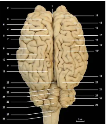
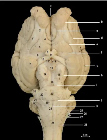
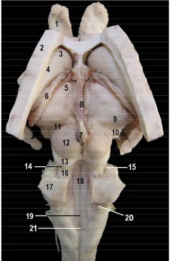
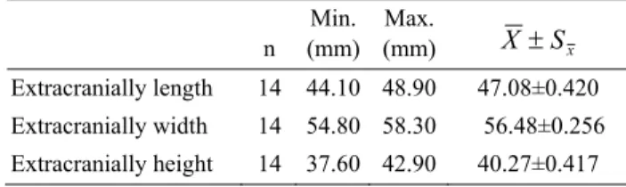
Şekil
Benzer Belgeler
In particular, using the form factors entering the low energy matrix elements both from full QCD as well as HQET, we have investigated the branching ratio, forward-backward
(Re)Making and Undoing of Peace/Conflict (Eds.) Tuğrul İlter, Hanife Aliefendioğlu, Pembe Behçetoğulları, Nurten Kara Famagusta: Eastern Mediterranean University
Cote ve Miners (2006) tarafından yapılan araştırmada duygusal zeka ile örgüte yönelik olarak sergilenen ÖVD davranışları arasında olumlu yönde anlamlı ilişki
25 Physics Department, Brookhaven National Laboratory, Upton NY, United States of America 26 (a) National Institute of Physics and Nuclear Engineering, Bucharest; (b) National
The study revealed that majority of the ELT students experience high or average level of writing anxiety towards writing tasks in general, the participant-related
Son olarak, bir önceki eleştiride de ele alındığı üzere, F&P yaklaşımının berimsel süreç üzerinde yük oluşturduğu bir diğer nokta, dizilim çizelgesine kaydedilen
Bu nedenle sadece kültür, dil içi ve dil dışı bağlam, daha “saygın” olduğu varsayılan dil kullanılması eğilimi, cinsiyet ve yaş, dinleyiciye göre dil tasarımı
Bu türden bir kuramsal taban üzerine kurulan sözkonusu dilbilgisi öğretimi yaklaşı- mında şimdiye değin uygulanmış ve bugün de uygulanmakta olan dilbilgisi öğretiminde
