Research Article
Association of Cytotoxic T Lymphocyte Antigen-4 Gene
Polymorphisms with Psoriasis Vulgaris: A Case-Control
Study in Turkish Population
Hatice Gül Dursun
,
1Hüseyin Osman Y
ılmaz,
1Recep Dursun,
2and Sevsen Kulaks
ızoğlu
31Department of Medical Biology, Meram Faculty of Medicine, Necmettin Erbakan University, 42080 Konya, Turkey 2Department of Dermatology, Meram Faculty of Medicine, Necmettin Erbakan University, 42080 Konya, Turkey 3Department of Clinical Biochemistry, School of Medicine, Baskent University, Konya, Turkey
Correspondence should be addressed to Hatice Gül Dursun; [email protected]
Received 3 January 2018; Revised 6 March 2018; Accepted 14 March 2018; Published 23 April 2018 Academic Editor: Margarete D. Bagatini
Copyright © 2018 Hatice Gül Dursun et al. This is an open access article distributed under the Creative Commons Attribution License, which permits unrestricted use, distribution, and reproduction in any medium, provided the original work is properly cited.
Psoriasis is a common, chronic, and autoimmune skin disease in which dysregulation of immune cells, particularly T cells, is thought to play an important role in the pathogenesis. Cytotoxic T lymphocyte antigen-4 (CTLA-4) expressed only on activated T cells is an immunoregulatory molecule and plays a role in the pathogenesis of autoimmune disorders. We aimed to determine whether CTLA-4 gene polymorphisms are associated with development and/or clinical features of psoriasis vulgaris (Pv). Genotyping of SNPs (−318C>T, +49A>G, and CT60A>G) in CTLA-4 gene was performed using polymerase chain reaction-restriction fragment length polymorphism (PCR-RFLP) in 103 Pv patients and 102 controls. No statistically significant associations were detected in any of the investigated genetic models for the −318C>T polymorphism. The genotype distributions of +49A>G and CT60A>G were associated with Pv development. In haplotype analysis, while frequency of CAA haplotype was significantly higher in the control group, frequencies of CGG and CAG haplotype were significantly higher among the patients. However, all of CTLA-4 polymorphisms and haplotypes do not have an effect on severity and onset age of Pv. In conclusion, the +49A>G and CT60A>G polymorphisms may be risk factors for Pv development. Furthermore, CGG and CAG haplotypes may contribute to Pv development, while CAA haplotype may be protective against Pv.
1. Introduction
Psoriasis is a common in
flammatory skin disease that affects
approximately 125 million people globally [1]. The disease
that exhibits a variable clinical presentation is characterized
by lesions in the form of circular, red papules and plaques
with a grey or silvery-white, dry scale. Psoriatic lesions are
generally distributed symmetrically on the scalp, elbows,
knees, lumbosacral area, and umbilicus [2, 3]. In addition,
nail disease and/or psoriatic arthritis, which can be very
painful and deforming, may develop in many patients with
psoriasis [2
–5]. The incidence of psoriasis in women and
men is almost equal [3]. Psoriasis is associated with several
comorbidities, such as Chron
’s disease [6, 7], cardiovascular
syndrome [8, 9], metabolic syndrome [10–12], depression
[13], and cancer [14, 15]. The disease leads to a serious
reduc-tion in the quality of a patient
’s life, because it is linked with
social stigmatization, pain, discomfort, physical disability, and
psychological distress [2]. Recently, psoriasis has begun to be
defined as a disease spectrum or systemic disease because of
abovementioned concomitant comorbidities. As a result, it
requires lifelong treatment [16]. Although the molecular
path-ogenesis of the disease is still poorly understood, it is generally
agreed that psoriasis is triggered by some environmental
factors such as stress, infections, trauma, and drugs with a
genetic background [17]. The common view about the
molecular pathogenesis of the disease is that alterations in
the complex interactions between T lymphocytes, dendritic
cells, macrophages, mast cells, neutrophils, keratinocytes,
cytokines, and chemokines cause psoriasis, and this wise
Volume 2018, Article ID 1643906, 10 pages https://doi.org/10.1155/2018/1643906
unbalanced immune response contributes to the psoriatic
process [18, 19]. Psoriasis has four major clinical
pheno-types, which are distinguished by the morphological
char-acteristics of their lesions: (i) psoriasis vulgaris, (ii) guttate
psoriasis, (iii) pustular psoriasis, and (iv) erythrodermic
psori-asis [20]. The most common of these clinical phenotypes is
psoriasis vulgaris, responsible for 90% of all cases, and is also
known as plaque psoriasis [3]. In this phenotype, the lesions
are dry, sharply demarcated, oval/circular plaques and can be
localized all over the body, but eventually affecting mostly
the knees, elbows, lumbosacral area, intergluteal cleft, and
scalp[20, 21].
Cytotoxic T lymphocyte antigen-4 (CTLA-4) is an
important immunoregulatory molecule that plays a role in
the maintenance of T cell homeostasis. In T cell-mediated
immunological response, the interaction of MHC on
antigen-presenting cell (APC) with CD28 on T cell is
essen-tial but not su
fficient for T cell activation. However, the
additional costimulatory factors and pathways are required
for T cell activation [22, 23]. One of the costimulatory
path-ways is B7- (CD80/86) CD28 [22]. CD28 expressed on
antigen-presenting cells by naive T cells binds to B7 (CD80/
86) initiates the proliferation, differentiation, and cytokine
production in T cells. Binding of CTLA-4 expressed by T
cells to B7 presented on APC contributes to peripheral
toler-ance leading to the arrest of T cell cycle and termination of T
cell activation [22, 24]. CTLA-4 acts as an inhibitor of
autoimmunity, and the defects in the B7-CD28/CTLA-4
pathway may lower the threshold of autoreactive lymphocyte
activation and which in turn may lead to the development of
an autoimmune disease [25]. CTLA-4 molecule is encoded
by the CTLA-4 gene (gene ID: 1493; OMIM
∗123890) located
on chromosome 2p33 [26]. Several polymorphisms were
identi
fied in the CTLA-4 gene. The polymorphisms reducing
the CTLA-4 expression or function may cause autoimmune
clonal T cell proliferation and thus the development of
autoimmune diseases [27]. In fact, some association studies
indicated that there is an association between several
CTLA-4 gene polymorphisms and various autoimmune
diseases [28–48]. Recently, it has been shown that
polymor-phisms of many genes that are directly or indirectly related
to the immune system and/or inflammation are associated
with psoriasis. These include genes such as ADAM33 (a
disintegrin and metalloprotease33) [49], TLR2 and TLR4
(toll-like receptor 2 and 4) [50], MCP-1 (monocyte
chemoat-tractant protein-1) and RANTES (regulated upon activation
normal T cell expressed and secreted) [51], TNF
α (tumor
necrosis factor alpha) [52], PON1 (paraoxonase) [53], IL-4
and IL-10 (interleukins) [54], HLA [55], VEGF (vascular
endothelial growth factor) [56], and ERAP (endoplazmic
reticulum aminopeptidase) [57]. However, there are a few
studies establishing a possible relationship between CTLA-4
gene polymorphisms and psoriasis.
In the present study, we have conducted a research on
three single-nucleotide polymorphisms (SNPs) in the
CTLA-4 gene, because of its possible e
ffects on expression
level or function of the CTLA-4 molecule:
−318C>T (in
pro-moter), +49A
>G (in exon-1), and CT60A>G (in exon-4).
With this hypothesis, our goal was (i) to investigate whether
the CTLA-4 gene polymorphisms are related to the
develop-ment of Pv (psoriasis vulgaris) and (ii) to detect whether the
CTLA-4 gene polymorphisms have an impact on the clinical
features of P. vulgaris such as onset age and severity. In
the literature, there are few studies which observed the
relationship between CTLA-4 gene polymorphisms and
Pv [58
–61]. Yet, there are no previous studies revealing a
relationship between CTLA-4 genes
−318C>T, +49A>G,
and CT60A
>G SNPs and the development of Pv.
2. Subjects and Methods
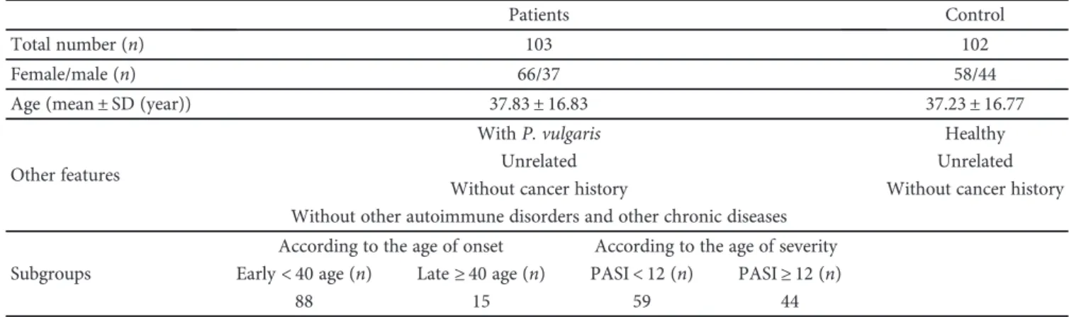
2.1. Research Population. 103 unrelated Turkish Pv patients
were selected for the experimental group, and 102 unrelated
healthy Turkish people were selected for the control group.
Psoriasis vulgaris patients (66 female/37 male; mean age
± SD: 37.83 ± 16.83) were recruited from a dermatologic
clinic. The patients with other chronic and autoimmune
diseases or cancer were excluded from the study. The control
group (58 female/44 male; mean age
± SD: 37.23 ± 16.77) was
formed with healthy individuals who did not have cancer,
psoriasis, and other autoimmune diseases and did not have
a family history of these diseases. The patients and control
subjects were matched according to their gender and age.
Severity of psoriasis was assessed with Psoriasis Area and
Severity Index (PASI), ranging from 0 (no disease) to 72, with
higher scores indicating the severity of disease [62]. To
deter-mine the association of CTLA-4 gene polymorphisms with
the clinical features of Pv, the patients were divided into
two groups according to the severity of disease (PASI
< 12
group and PASI
≥ 12 group) and then assigned into two
groups according to the onset of disease (early-onset group:
<40 age and late-onset group: ≥40 age) (Table 1).
This study was conducted in accordance with the
Decla-ration of Helsinki principles and was approved by the Ethics
Committee of Meram Medical Faculty (number 2010/138).
Informed consent was obtained from all the participants
before the study.
2.2. Genotyping. Peripheral blood sample was taken from
each patient and control subject collected in tubes containing
EDTA and stored at
−20
°C before DNA isolation. Genomic
DNA was extracted from the blood sample using the
QIAamp DNA Blood Mini Kit (Qiagen, Hilden, Germany).
Genotyping of the CTLA-4 gene
−318C>T (rs5742909),
+49A
>G (rs231775), and CT60A>G (rs3087243) was carried
out by the polymerase chain reaction-restriction length
poly-morphism (PCR-RFLP) using MseII, BbvI, and NcoI
enzymes (New England BioLabs, Hitchin, UK). PCR reactions
were performed with mixtures consisting of 0.2
μg genomic
DNA, 5
μl ammonium buffer, 4.5 μl MgCl
2, 20 pmol of each
primer, 5 unit Taq polymerase, and double-distilled H
2O up
to
final volume of 50 μl. The primers were designed according
to the complete CTLA-4 gene sequence derived from NCBI
Sequence Viewer (http://www.ncbi.nlm.nig.gov/). PCR was
carried out with denaturation at 95
°C for 5 minutes, followed
by 35 cycles of 45 seconds at 94
°C, 45 seconds at 55
°C,
and 45 seconds at 72
°C and
finally 10 minutes at 72
°C.
6 U of BbvI, and 10 U of NcoI enzymes and then
electro-phoresed on 2.5% agarose gel, stained with ethidium
bromide, and evaluated. The primers used for PCR,
condi-tions for digestion, products of digestion, and genotypes
determined according to the products of digestion are
listed in Table 2.
2.3. Statistical Analysis. The SPSS 13.0 package programme
was used for data analysis. Comparisons of the
distribu-tions of allele and genotype frequencies were performed
by Pearson
’s chi-squared test. The deviation from the
Hardy-Weinberg equilibrium was tested using chi-square
analysis. To test the association between Pv and CTLA-4
polymorphisms, logistic regression analysis was performed
according to
five inheritance models (codominant 1,
codom-inant 2, domcodom-inant, recessive, and log-additive). Odds ratios
(OR), 95% confidence intervals (CI), and p values were
determined using SNPStats (http://bioinfo.iconcologia.net/
index.php?module=Snpstats) and SPSS 13.0 program. The
linkage disequilibrium (LD) blocks and haplotypes were
esti-mated using Haploview version 4.2 (http://www.broadinstitu
te.org/scienti
fic-community/science/programs/medical-and-population-genetics/haploview).
p values less than 0.05 were
considered signi
ficant.
3. Results
3.1. Genotype Analysis and Association of SNPs with Pv.
Table 3 shows the genotype and allele frequencies of
CTLA-4 polymorphisms (
−318C>T, +49A>G, and CT60A
>G) in Pv patients and the control group. The genotype
distributions of the examined SNPs were consistent with
the Hardy-Weinberg equilibrium (HWE) (Table 3).
In multiple logistic regression analysis,
−318C>T SNP
was not associated with the development of Pv (p > 0 05 for
all genetic models and T allele frequency). However,
+49A>G and CT60A>G SNPs were associated with Pv. The
disease-related risk was observed in the codominant 1 model
(OR = 0.57,
p = 0 04), dominant model (OR = 0.54, p = 0 03),
and log-additive model (OR = 0.62 and
p = 0 03) for +49A>G
and in the codominant 2 model (OR = 0.29,
p = 0 004) and
recessive model (OR = 1.33,
p = 0 001) for CT60. In addition,
G allele (minor allele) frequencies of both +49A>G and
CT60A
>G SNPs were higher in the Pv patient (31% for
+49A
>G and 55% for CT60A>G) than in the control group
(21% for +49A>G and 40% for CT60A>G) (OR = 0.59, p =
0 02 for +49A>G and OR = 0.54, p = 0 002 for CT60A>G).
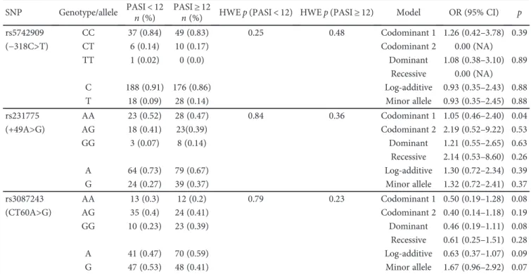
3.2. Genotype Analysis and Association of SNPs with Clinical
Features of Pv. Tables 4 and 5 present the genotype and allele
frequencies of CTLA-4 SNPs in clinical subgroups of Pv
(onset age of disease and severity of disease). None of the
examined SNPs showed no association with onset age and
severity of Pv (for all genetic models). The genotype and
allele frequencies of examined SNPs did not di
ffer between
the early group and late group (Table 4) and PASI
< 12 group
and PASI
≥ 12 group (Table 5). The results indicated that
−318C>T, +49A>G, and CT60A>G SNPs have no effect on
the onset age and severity of Pv.
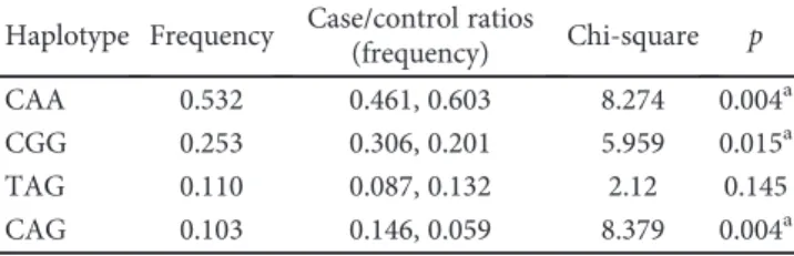
3.3. Linkage Disequilibrium and Haplotype Analysis. We
esti-mated the linkage disequilibrium (LD) block by using
Haplo-view version 4.2. The LD block was strongly made between
−318C>T and +49A>G (D’ = 0.999 and r
2= 0 043),
−318C>T and CT60A>G (D’ = 0.999 and r
2= 0 141), and
+49A>G and CT60A>G (D’ = 1.000 and r
2= 0 383). In
hap-lotype analysis which was performed to investigate the
asso-ciation between the haplotypes of LD block SNPs and Pv,
four major haplotypes were detected which are CAA, CGG,
TAG, and CAG (Table 6). The frequencies of these
haplo-types were 0.532, 0.253, 0.110, and 0.103, respectively. A
sig-ni
ficantly higher frequency of CAA haplotype was found in
controls (0.603) than in Pv patients (0.461,
p = 0 004). In
contrast, signi
ficant increases in the frequencies of CGG
and CAG haplotypes were observed in patients (0.306 and
0.146, resp.) compared to healthy individuals in the control
group (0.201 and 0.059, resp.;
p = 0 015 and p = 0 004). These
results suggest that while the CAA haplotype may have a
pro-tective effect on the development of Pv, the CGG and CAG
haplotypes may be associated with the development of Pv.
In haplotype analysis which was performed to investigate
the association between the haplotypes and the clinical
sub-groups of Pv, three major haplotypes were detected which
are AA, GG, and AG (Table 7). The frequencies of these
hap-lotypes were 0.461, 0.306, and 0.233, respectively.
Consider-ing the onset age of Pv, the frequencies of these haplotypes
Table 1: Characteristics of the study population.
Patients Control
Total number (n) 103 102
Female/male (n) 66/37 58/44
Age (mean± SD (year)) 37.83± 16.83 37.23± 16.77
Other features
With P. vulgaris Healthy
Unrelated Unrelated
Without cancer history Without cancer history
Without other autoimmune disorders and other chronic diseases Subgroups
According to the age of onset According to the age of severity Early< 40 age (n) Late≥ 40 age (n) PASI< 12 (n) PASI≥ 12 (n)
did not di
ffer between the early group and the late
group (
p = 0 467, p = 0 434, and p = 0 243, resp.). There was
no significant difference between the PASI < 12 group
and the PASI
≥ 12 group with respect to the frequencies
of AA, GG, and AG haplotypes (p = 0 069, p = 0 373, and
p = 0 243, resp.).
4. Discussion
Psoriasis is an in
flammatory disease which is characterized
by keratinocyte proliferation and activated T cell
accumula-tion [63]. The incidence of psoriasis in women and men is
almost equal [3]. However, in our study, the number of
female patients (66) was significantly higher than the number
of male patients (37). This situation is entirely coincidental
and only results from the fact that the number of female
patients who applied to the clinic during the study period
was more than the number of male patients. Probably, the
number of female patients and male patients would be close
if the study period was extended a little longer or the number
of patients could be increased.
Although its pathogenesis has not been well understood,
psoriasis bears many features of a T cell-mediated
autoim-mune disease. It reveals a strong HLA association [64]. Since
CTLA-4 regulates T cell activation and the proliferation
through a negative feedback, the CTLA-4 gene is considered
to be a candidate gene for T cell-mediated autoimmune
dis-ease. Hence, in this study, we aimed to investigate the
Table 2: Primers, conditions for digestion, products of digestion, and genotypes according to products of digestion.
SNP Primers Amplicon (bp) RE Temperature and
duration of digestion
Products of digestion (bp) and genotypes −318C>T F: 5′-AAATGAATTGGACTGGATGGT-3′R: 3′-TTACGAGAAAGGAAGCCGTG-5′ 247 MseII 37°C, overnight
CC: 247 CT: 20, 95, 132, 247
TT: 20, 95, 132 +49A>G F: 5′-TTGCTCTACTTCCTGAAGACCTGAA-3′
R: 3′-AAAGTCTCACTCACCTTTGCAGAAG-5′ 166 BbvI 37
°C, overnight AG: 76, 90, 166AA: 166
GG: 76, 90 CT60 A>G F: 5′-CAC CACTATTTGGGATATACC-3′
R: 3′-AGGTCTATATTTCAGGAAGGC-5′ 216 NcoI 37
°C, overnight AG: 20, 196, 216AA: 20, 196
GG: 216
Table 3: Genotype and allele frequencies of CTLA-4 gene polymorphisms in Pv patients and control and the association of these polymorphisms with Pv.
SNP Genotype/allele Casesn (%) Controls n (%) HWE p (cases) HWE p (controls) Model OR (95% CI) p
rs5742909 CC 86 (0.83) 75 (0.74) 0.79 0.44 Codominant 1 1.84 (0.92–3.70) 0.85 (−318C>T) CT 16 (0.16) 26 (0.25) Codominant 2 1.08 (0.07–17.89) 0.22 TT 1 (0.01) 1 (0.01) Dominant 1.80 (0.91–3.56) 0.09 Recessive 0.95 (0.06–15.67) 0.97 C 188 (0.91) 176 (0.86) Log-additive 1.67 (0.88–3.17) 0.11 T 18 (0.09) 28 (0.14) Minor allele 1.66 (0.88–3.11) 0.11 rs231775 AA 51 (0.50) 66 (0.65) 0.53 0.31 Codominant 1 0.57 (0.31–1.04) 0.04a (+49A>G) AG 41 (0.39) 30 (0.29) Codominant 2 0.43 (0.15–1.25) 0.09 GG 11 (0.11) 6 (0.06) Dominant 0.54 (0.31–0.95) 0.03a Recessive 0.53 (0.19–1.50) 0.22 A 143 (0.69) 162 (0.79) Log-additive 0.62 (0.40–0.96) 0.03a G 63 (0.31) 42 (0.21) Minor allele 0.59 (0.38–0.92) 0.02a rs3087243 AA 25 (0.24) 36 (0.35) 0.11 0.65 Codominant 1 0.81 (0.42–1.55) 0.06 (CT60A>G) AG 43 (0.42) 51 (0.50) Codominant 2 0.29 (0.13–0.64) 0.004a GG 35 (0.34) 15 (0.15) Dominant 0.58 (0.17–0.66) 0.07 Recessive 1.33 (0.17–0.66) 0.001a A 93 (0.45) 123 (0.6) Log-additive 1.38 (0.80–2.40) 0.25 G 113 (0.55) 81(0.40) Minor allele 0.54 (0.37–0.80) 0.002a
SNP: single-nucleotide polymorphism; HWE: Hardy-Weinberg equilibrium; OR: odds ratio; CI: confidence interval.aStatistically significant values (p < 0 05).
Codominant 1: major allele homozygotes versus heterozygotes; codominant 2: major allele homozygotes versus minor allele homozygotes; dominant: major allele homozygotes versus heterozygotes + minor allele homozygotes; recessive: major allele homozygotes + heterozygotes versus minor allele homozygotes; log-additive: major allele homozygotes versus heterozygotes versus minor allele homozygotes.
possibility of an association between this candidate gene and
Pv, which is defined as an autoimmune disease. In the
pres-ent study,
−318C>T, +49A>G, and CT60 polymorphisms
were studied to evaluate their contributions to the
pathogen-esis of Pv, focusing on their potential effects on the activity
and function of the CTLA-4 molecule. In fact, it has been
Table 4: Genotype and allele frequencies of CTLA-4 gene polymorphisms in the early-onset subgroup and late-onset subgroup and the association of these polymorphisms with onset age of Pv.
SNP Genotype/allele Early onsetn (%) Late onset n (%) HWE p (early) HWE p (late) Model OR (95% CI) p
rs5742909 CC 74 (0.84) 12 (0.8) 0.62 0.67 Codominant 1 1.42 (0.35–5.75) 0.76 (−318C>T) CT 13 (0.15) 3 (0.2) Codominant 2 0.00 (NA) TT 1 (0.01) 0 (0.0) Dominant 1.32 (0.33–5.30) 0.7 Recessive 0.00 (NA) C 188 (0.91) 176 (0.86) Log-additive 1.19 (0.33–4.30) 0.8 T 18 (0.09) 28 (0.14) Minor allele 1.19 (0.32–4.39) 0.11 rs231775 AA 45 (0.51) 6 (0.4) 0.49 0.98 Codominant 1 1.54 (0.48–5.01) 0.04 (+49A>G) AG 34 (0.39) 7 (0.47) Codominant 2 1.67 (0.29–9.62) 0.72 GG 9 (0.1) 2 (0.13) Dominant 1.57 (0.52–4.78) 0.42 Recessive 1.35 (0.26–6.97) 0.73 A 124 (0.7) 19 (0.63) Log-additive 1.35 (0.62–2.98) 0.45 G 52 (0.3) 11 (0.37) Minor allele 1.38 (0.61–3.10) 0.43 rs3087243 AA 24 (0.27) 1 (0.07) 0.06 0.13 Codominant 1 2.07 (0.59–7.30) 0.45 (CT60A>G) AG 35 (0.4) 10 (0.67) Codominant 2 0.30 (0.03–2.89) 0.08 GG 29 (0.33) 4 (0.27) Dominant 1.35 (0.40–4.61) 0.62 Recessive 0.19 (0.02–1.53) 0.06 A 93 (0.53) 18 (0.6) Log-additive 0.77 (0.36–1.63) 0.49 G 83 (0.47) 12 (0.4) Minor allele 1.34 (0.61–2.94) 0.47
SNP: single-nucleotide polymorphism; HWE: Hardy-Weinberg equilibrium; OR: odds ratio; CI: confidence interval.
Table 5: Genotype and allele frequencies of CTLA-4 gene polymorphisms in PASI < 12 and PASI ≥ 12 and the association of these polymorphisms with the severity of Pv.
SNP Genotype/allele PASI< 12
n (%) PASIn (%)≥ 12 HWEp (PASI < 12) HWE p (PASI ≥ 12) Model OR (95% CI) p
rs5742909 CC 37 (0.84) 49 (0.83) 0.25 0.48 Codominant 1 1.26 (0.42–3.78) 0.39 (−318C>T) CT 6 (0.14) 10 (0.17) Codominant 2 0.00 (NA) TT 1 (0.02) 0 (0.0) Dominant 1.08 (0.38–3.10) 0.89 Recessive 0.00 (NA) C 188 (0.91) 176 (0.86) Log-additive 0.93 (0.35–2.43) 0.88 T 18 (0.09) 28 (0.14) Minor allele 0.93 (0.35–2.45) 0.88 rs231775 AA 23 (0.52) 28 (0.47) 0.84 0.36 Codominant 1 1.05 (0.46–2.40) 0.04 (+49A>G) AG 18 (0.41) 23(0.39) Codominant 2 2.19 (0.52–9.22) 0.53 GG 3 (0.07) 8 (0.14) Dominant 1.21 (0.55–2.65) 0.63 Recessive 2.14 (0.53–8.60) 0.26 A 64 (0.73) 79 (0.67) Log-additive 1.30 (0.72–2.34) 0.39 G 24 (0.27) 39 (0.37) Minor allele 1.32 (0.72–2.41) 0.37 rs3087243 AA 13 (0.3) 12 (0.2) 0.79 0.23 Codominant 1 0.50 (0.19–1.28) 0.08 (CT60A>G) AG 35 (0.4) 24 (0.41) Codominant 2 0.40 (0.14–1.18) 0.19 GG 10 (0.23) 23 (0.39) Dominant 0.46 (0.19–1.11) 0.08 Recessive 0.61 (0.25–1.51) 0.28 A 41 (0.47) 70 (0.59) Log-additive 0.63 (0.37–1.07) 0.09 G 47 (0.53) 48 (0.41) Minor allele 1.67 (0.96–2.92) 0.07
suggested that
−318C>T polymorphism is an effective
pro-moter activity of the CTLA-4 gene and change transcription
of CTLA-4 gene [65]. +49A
>G polymorphism is located in
the leader sequence which is important in the binding of
the CTLA-4 molecule to B7.1 (CD80). CT60A>G
polymor-phism is considered to affect the alternative splicing and
soluble CTLA-4 production [66].
Our data displayed no association between
−318C>T
SNP and the development of Pv. There were no differences
in genotype and allele frequencies between the patient group
and the control group. Likewise,
Łuszczek et al. [60] found an
association between polymorphism and Pv in their study. It
has been also indicated that the association of
−318C>T
polymorphism with other autoimmune disorders supports
our hypothesis. The association of
−318C>T polymorphism
with other autoimmune diseases such as
spondyloarthro-pathy [67], pemphigus foliaceus [30], multiple sclerosis
[38, 68], Behçet’s disease [35], systemic lupus erythematosus
[37, 69], Hashimoto’s thyroiditis [41, 44], ankylosing
spon-dylitis [40], and Graves’ disease [70] supports our findings
in which the researchers did not
find any significant
relation-ship between
−318C>T and other diseases; however, an
association between
−318C>T polymorphism and other
autoimmune disorders was found. The association of
−318C>T polymorphism with childhood Graves’ disease
was reported in a Chinese population [34]. In a study on a
Chinese population, a significant relationship was found
between
−318C>T polymorphism and rheumatoid arthritis
[47, 71]. In the Italian systemic sclerosis patients, an
associa-tion was found between
−318C>T polymorphism and the
susceptibility to develop systemic sclerosis [33].
+49A
>G SNP is a CTLA-4 gene polymorphism which is
probably the most widely studied and most commonly
asso-ciated with autoimmune disorders and cancers. In our study,
+49A
>G polymorphism indicated a strong relationship with
Pv in terms of minor allele frequency (OR = 0.59, 95%
CI = 0.38–0.92, p = 0 02), codominant 1 model (OR = 1.54,
95% CI = 0.48–5.01 p = 0 04), dominant genetic model
(OR = 0.54, 95% CI = 0.40–0.96, p = 0 03), and log-additive
genetic model (OR = 0.62, 95% CI = 0.40
–0.96, p = 0 03). In
addition, +49A
>G SNP might contribute to the risk of Pv
development and G allele might be a risk factor in Pv
devel-opment. This SNP causes substitution of threonine at
position 17 to alanine in the CTLA-4 protein [72]. It has been
postulated that this amino acid substitution may affect T cell
activation by changing the posttranslational modification
and ability of CTLA-4 to bind with B7.1 (CD80) [73].
Vari-ous studies have revealed that the +49G allele leads to
decreased expression of CTLA-4 compared to +49A allele
[27, 74]. Our
findings are probably related to +49A>G SNP
and may be explained by the inability of CTLA-4 to bind to
B7 and/or by decreasing of CTLA-4 expression.
Further-more, decreased expression and/or broken binding with B7
in CTLA-4 may contribute to the pathogenesis of Pv by
changing the T cell response. The
findings of this study are
inconsistent with the results of Tsunemi et al. [58], Kim
et al. [59], and
Łuszczek et al. [61] who evaluated the
associ-ation between +49A>G polymorphism and Pv. Łuszczek
et al. [61] studied on 141 Pv patients recruited from a Polish
population and found that the allele and genotype
distribu-tions of +49A
>G polymorphism are similar for the patients
in the experimental group and healthy individuals in the
control group. In the studies with Japanese [58] and Korean
[59] populations, no association was reported between
poly-morphism and Pv. However, the results of some studies
examining the relationship of +49A
>G polymorphism with
other autoimmune disorder, but not with Pv, are consistent
with the results of our study. These studies revealed the
association of +49A
>G polymorphism with Graves’ disease
[27
–29, 34, 70], rheumatoid arthritis [39], and ankylosing
spondylitis [40]. On the other hand, it has been shown
that there is no relation between +49A
>G polymorphism
and several autoimmune diseases such as rheumatoid
arthritis [43, 71], Behçet
’s disease [35], vitiligo [75],
sys-temic lupus erythematosus [37, 69], syssys-temic sclerosis [33],
spondyloarthropathy [67], ankylosing spondylitis [36, 40],
pemphigus foliaceus [30], multiple sclerosis [38, 68], primary
Sjögren syndrome [31], and ulcerative colitis [48].
In the present study, we observed a strong association
between CT60A>G polymorphism and Pv in terms of
codominant 2 (OR = 0.29, 95% CI = 0.13–0.64, p = 0.004),
recessive (OR = 1.33, 95% CI = 0.17–0.60, p = 0 001), and
minor allele frequency (OR = 0.54, 95% CI = 0.37
–0.80,
p = 0 002). Allele G appears to be a risk factor for the
devel-opment of Pv.
Łuszczek et al. [61] observed no difference in
allele and genotype distributions of CT60A
>G
polymor-phism between Pv patients and control subjects. This SNP
is located in 3′ UTR (untranslated region) of the CTLA-4
gene and is supposed to affect the proportion of soluble
iso-form of CTLA-4 (sCTLA-4) to membrane-bound CTLA-4
(mCTLA-4). sCTLA-4 isoform is generated through
alterna-tive splicing of CTLA-4 mRNA. It has been previously
sug-gested that the G allele on position +6230 (CT60G) may
decrease sCTLA-4 transcript up to 50% [66]. Furthermore,
we also observed higher frequencies of G allele and GG
geno-type in Pv patients than the control group. It is assumed G
allele causes a decrease in CTLA-4 expression and
deteriora-tion of the balance between sCTLA-4/mCTLA-4 by blocking
the alternative splicing of CTLA-4 mRNA. Chong et al. [34]
have suggested that CT60A>G polymorphism plays a role in
susceptibility to childhood Graves’ disease. Kavvoura et al.
[32] have discovered that polymorphism can be an important
marker of genetic risk in Graves
’ disease and Hashimoto
thyroiditis. Furthermore, it has also been suggested that
Table 6: Haplotype distribution belongs to CTLA-4 polymorphisms between Pv patients and control.
Haplotype Frequency Case/control ratios
(frequency) Chi-square p
CAA 0.532 0.461, 0.603 8.274 0.004a
CGG 0.253 0.306, 0.201 5.959 0.015a
TAG 0.110 0.087, 0.132 2.12 0.145
CAG 0.103 0.146, 0.059 8.379 0.004a
Haplotypes were constructed in the following order:−318C>T (rs5742909)/ +49A>G (rs231775)/CT60A>G (rs3087243). aStatistically significant
CT60A>G polymorphism leads to the susceptibility of
vitiligo [75] and ankylosing spondylitis [40].
There are several reasons that could explain these
controversial results among different studies: (i) studied
pop-ulations have di
fferent ethnic features, (ii) studied
popula-tions have di
fferent sizes, and (iii) studied autoimmune
disorders have already a multifactorial nature. In this study,
−318C>T, +49A>G, and CT60A>G polymorphisms were
selected because they can play a role on Pv pathogenesis by
altering the promoter activity and transcription efficiency
(for
−318C>T), by altering T cell activation through
post-translational modification (for +49A>G), and by affecting
the alternative splicing and production of CTLA-4 isoforms
(for CT60A>G). Although our population size was relatively
small, we believe that our results will contribute to
meta-analysis studies which have aimed at understanding the role
of CTLA-4 on the pathogenesis of Pv.
5. Conclusions
To conclude, our data suggest that while there seems to be no
correlation between
−318C>T polymorphism and the
devel-opment of Pv, +49A
>G and CT60A>G polymorphisms may
be associated with the development of Pv. In addition, our
results present that none of the studied polymorphisms were
related with the clinical features of Pv such as severity and
onset age of disease. In performed haplotype analysis, CGG
and CAG haplotypes were found to be the risk factor for
the development of Pv, while CAA haplotype was found to
be a protective haplotype for Pv. The haplotypes showed no
association with severity and onset age of Pv. As a result, all
of these
findings suggest that +49A>G and CT60A>G
polymorphisms of the CTLA-4 gene may play a role in the
pathogenesis of Pv.
Conflicts of Interest
The authors declare that there are no conflicts of interest
regarding the publication of this article.
Acknowledgments
This study was supported by Grant no. 10202049 from the
Scientific Research Project Unit of Selçuk University.
References
[1] International Federation of Psoriasis Associations, “World psoriasis day 2015,” February 2018, https://ifpa-pso.com/our-actions/world-psoriasis-day.
[2] R. B. G. Langley, G. G. Krueger, and C. E. M. Griffiths, “Psori-asis: epidemiology, clinical features, and quality of life,” Annals of the Rheumatic Diseases, vol. 64, Supplement 2, pp. ii18– ii23, 2005.
[3] C. E. M. Griffiths and J. N. W. N. Barker, “Pathogenesis and clinical features of psoriasis,” The Lancet, vol. 370, no. 9583, pp. 263–271, 2007.
[4] J. E. Gudjonsson and J. T. Elder, “Psoriasis: epidemiology,” Clinics in Dermatology, vol. 25, no. 6, pp. 535–546, 2007. [5] A. J. Hueber and I. B. McInnes,“Immune regulation in
psori-asis and psoriatic arthritis—recent developments,” Immunol-ogy Letters, vol. 114, no. 2, pp. 59–65, 2007.
[6] D. J. Najarian and A. B. Gottlieb,“Connections between psori-asis and Crohn’s disease,” Journal of the American Academy of Dermatology, vol. 48, no. 6, pp. 805–824, 2003.
[7] A. B. Kimball, D. Gladman, J. M. Gelfand et al., “National Psoriasis Foundation clinical consensus on psoriasis comor-bidities and recommendations for screening,” Journal of the American Academy of Dermatology, vol. 58, no. 6, pp. 1031– 1042, 2008.
[8] S. C.-H. Hu and C.-C. E. Lan,“Psoriasis and cardiovascular comorbidities: focusing on severe vascular events, cardiovas-cular risk factors and implications for treatment,” Interna-tional Journal of Molecular Sciences, vol. 18, no. 12, p. 2211, 2017.
[9] R. A. Kölliker Frers, R. J. Bisoendial, S. F. Montoya et al., “Psoriasis and cardiovascular risk: immune-mediated cross-talk between metabolic, vascular and autoimmune in flam-mation,” IJC Metabolic & Endocrine, vol. 6, pp. 43–54, 2015. [10] A. Parodi, N. Aste, C. Calvieri et al., “Metabolic syndrome prevalence in psoriasis: a cross-sectional study in the Italian Table 7: Haplotype distribution belongs to CTLA-4 polymorphisms among different subgroups of Pv.
(a)
Haplotype Frequency Late/early ratios (frequency) Chi-square p
AA 0.461 0.472, 0.400 0.529 0.467
GG 0.306 0.295, 0.367 0.612 0.434
AG 0.233 0.233, 0.263 1.364 0.243
(b)
Haplotype Frequency PASI< 12/PASI ≥ 12 ratios(frequency) Chi-square p
AA 0.461 0.534, 0.407 3.288 0.069
GG 0.306 0.273, 0.331 0.793 0.373
AG 0.233 0.193, 0.263 1.364 0.243
population,” American Journal of Clinical Dermatology, vol. 15, no. 4, pp. 371–377, 2014.
[11] I. M. Miller, C. Ellervik, K. Zarchi et al.,“The association of metabolic syndrome and psoriasis: a population- and hospital-based cross-sectional study,” Journal of the European Academy of Dermatology and Venereology, vol. 29, no. 3, pp. 490–497, 2015.
[12] A. S. Lonnberg, L. Skov, A. Skytthe, K. O. Kyvik, O. B. Pedersen, and S. F. Thomsen,“Association of psoriasis with the risk for type 2 diabetes mellitus and obesity,” JAMA Dermatology, vol. 152, no. 7, pp. 761–767, 2016.
[13] E. A. Dowlatshahi, M. Wakkee, L. R. Arends, and T. Nijsten, “The prevalence and odds of depressive symptoms and clinical depression in psoriasis patients: a systematic review and meta-analysis,” Journal of Investigative Dermatology, vol. 134, no. 6, pp. 1542–1551, 2014.
[14] K. Abuabara, R. S. Azfar, D. B. Shin, A. L. Neimann, A. B. Troxel, and J. M. Gelfand,“Cause-specific mortality in patients with severe psoriasis: a population-based cohort study in the U.K,” British Journal of Dermatology, vol. 163, no. 3, pp. 586–592, 2010.
[15] J. M. Gelfand, J. Berlin, A. Van Voorhees, and D. J. Margolis, “Lymphoma rates are low but increased in patients with psori-asis: results from a populationbased study in the United Kingdom,” Archives of Dermatology, vol. 139, no. 11, pp. 1425–1429, 2003.
[16] C. Ryan and B. Kirby,“Psoriasis is a systemic disease with multiple cardiovascular and metabolic comorbidities,” Derma-tologic Clinics, vol. 33, no. 1, pp. 41–55, 2015.
[17] R. Parisi, D. P. Symmons, C. E. Griffiths, D. M. Ashcroft, and Identification and Management of Psoriasis and Associated ComorbidiTy (IMPACT) project team,“Global epidemiology of psoriasis: a systematic review of incidence and prevalence,” Journal of Investigative Dermatology, vol. 133, no. 2, pp. 377–385, 2013.
[18] M. P. Schön and W.-H. Boehncke, “Psoriasis,” The New England Journal of Medicine, vol. 352, no. 18, pp. 1899–1912, 2005.
[19] M. A. Lowes, A. M. Bowcock, and J. G. Krueger, “Pathogen-esis and therapy of psoriasis,” Nature, vol. 445, no. 7130, pp. 866–873, 2007.
[20] L. Naldi and D. Gambini,“The clinical spectrum of psoriasis,” Clinics in Dermatology, vol. 25, no. 6, pp. 510–518, 2007. [21] S. K. Raychaudhuri, E. Maverakis, and S. P. Raychaudhuri,
“Diagnosis and classification of psoriasis,” Autoimmunity Reviews, vol. 13, no. 4-5, pp. 490–495, 2014.
[22] C. B. Thompson and J. P. Allison, “The emerging role of CTLA-4 as an immune attenuator,” Immunity, vol. 7, no. 4, pp. 445–450, 1997.
[23] A. G. Baxter and P. D. Hodgkin,“Activation rules: the two-signal theories of immune activation,” Nature Reviews Immu-nology, vol. 2, no. 6, pp. 439–446, 2002.
[24] M. L. Alegre, K. A. Frauwirth, and C. B. Thompson,“T-cell regulation by CD28 and CTLA-4,” Nature Reviews Immunol-ogy, vol. 1, no. 3, pp. 220–228, 2001.
[25] D. M. Harlan, R. Abe, K. P. Lee, and C. H. June,“Potential roles of the B7 and CD28 receptor families in autoimmunity and immune evasion,” Clinical Immunology and Immunopa-thology, vol. 75, no. 2, pp. 99–111, 1995.
[26] M. Lafage-Pochitaloff, R. Costello, D. Couez et al., “Human CD28 and CTLA-4 Ig superfamily genes are located on
chromosome 2 at bands q33–q34,” Immunogenetics, vol. 31, no. 3, pp. 198–201, 1990.
[27] T. Kouki, Y. Sawai, C. A. Gardine, M.-E. Fisfalen, M.-L. Alegre, and L. J. DeGroot,“CTLA-4 gene polymorphism at position 49 in exon I reduces the inhibitory function of CTLA-4 and contributes to the pathogenesis of Graves’ disease,” The Journal of Immunology, vol. 165, no. 11, pp. 6606–6611, 2000.
[28] T. Yanagawa, M. Taniyama, S. Enomoto et al.,“CTLA4 gene polymorphism confers susceptibility to Graves’ disease in Japanese,” Thyroid, vol. 7, no. 6, pp. 843–846, 1997.
[29] T. Bednarczuk, Y. Hiromatsu, T. Fukutani et al.,“Association of cytotoxic T-lymphocyte-associated antigen-4 (CTLA-4) gene polymorphism and non-genetic factors with Graves’ ophthalmopathy in European and Japanese populations,” European Journal of Endocrinology, vol. 148, no. 1, pp. 13–18, 2003.
[30] D. P. Pavoni, L. B. Cerqueira, V. M. M. S. Roxo, and M. L. Petzl-Erler,“Polymorphism of the promoter region and exon 1 of the CTLA4 gene in endemic pemphigus foliaceus (fogo selvagem),” Brazilian Journal of Medical and Biological Research, vol. 39, no. 9, pp. 1227–1232, 2006.
[31] J. E. Gottenberg, P. Loiseau, M. Azarian et al.,“CTLA-4 +49A/ G and CT60 gene polymorphisms in primary Sjögren syndrome,” Arthritis Research & Therapy, vol. 9, no. 2, p. R24, 2007.
[32] F. K. Kavvoura, T. Akamizu, T. Awata et al.,“Cytotoxic T-lymphocyte associated antigen 4 gene polymorphisms and autoimmune thyroid disease: a meta-analysis,” The Journal of Clinical Endocrinology & Metabolism, vol. 92, no. 8, pp. 3162–3170, 2007, Review.
[33] G. Balbi, F. Ferrera, M. Rizzi et al.,“Association of -318C/T and +49A/G cytotoxic T lymphocyte antige4 (CTLA-4) gene polymorphisms with a clinical subset of Italian patients with systemic sclerosis,” Clinical & Experimental Immunology, vol. 149, no. 1, pp. 40–47, 2007.
[34] K. K. L. Chong, S. W. Y. Chiang, G. W. K. Wong et al., “Asso-ciation of CTLA-4 and IL-13 gene polymorphisms with Graves’ disease and ophthalmopathy in Chinese children,” Investigative Ophthalmology & Visual Science, vol. 49, no. 6, pp. 2409–2415, 2008.
[35] L. Du, P. Yang, S. Hou, H. Zhou, and A. Kijlstra,“No associa-tion of CTLA-4 polymorphisms with susceptibility to Behçet disease,” British Journal of Ophthalmology, vol. 93, no. 10, pp. 1378–1381, 2009.
[36] E. Azizi, A. Massoud, A. A. Amirzargar et al.,“Association of CTLA4gene polymorphism in Iranian patients with ankylos-ing spondylitis,” Journal of Clinical Immunology, vol. 30, no. 2, pp. 268–271, 2010.
[37] K. H. Chua, S. M. Puah, C. H. Chew, S. Y. Tan, and L. H. Lian, “Study of the CTLA-4 gene polymorphisms in systemic lupus erythematosus (SLE) samples from Malaysia,” Annals of Human Biology, vol. 37, no. 2, pp. 274–280, 2010.
[38] A. Heidari, M. Keramatipour, A. A. Amirzargar et al., “CTLA-4 gene polymorphisms (-318C/T, +“CTLA-49A/G, +6230A/G) in Iranian patients with multiple sclerosis,” Iranian Journal of Allergy Asthma and Immunology, vol. 9, no. 4, article 21131701, pp. 219–223, 2010.
[39] M. J. Tang and Z. B. Zhou,“Association of the CTLA-4 +49A/ G polymorphism with rheumatoid arthritis in Chinese Han population,” Molecular Biology Reports, vol. 40, no. 3, pp. 2627–2631, 2013.
[40] N. G. Wang, D. C. Wang, B. Y. Tan, F. Wang, and Z. N. Yuan, “Association between CTLA-4 gene polymorphism and ankylosing spondylitis: a case-control study,” International Journal of Clinical and Experimental Pathology, vol. 8, no. 6, pp. 7421–7425, 2015.
[41] Y. Hu, K. Xu, L. Jiang, L. Zhang, H. Shi, and D. Cui, “Associa-tions between three CTLA-4 polymorphisms and Hashimoto’s thyroiditis risk: an updated meta-analysis with trial sequential analysis,” Genetic Testing and Molecular Biomarkers, vol. 22, no. 4, pp. 1–13, 2018.
[42] S. Ramgopal, C. Rathika, M. R. Padma et al.,“Interaction of HLA-DRB1∗ alleles and CTLA4 (+ 49 AG) gene polymor-phism in autoimmune thyroid disease,” vol. 642, pp. 430–438, 2018.
[43] K. Wang, Q. Zhu, Y. Lu et al.,“CTLA-4 +49 G/A polymor-phism confers autoimmune disease risk: an updated meta-analysis,” Genetic Testing and Molecular Biomarkers, vol. 21, no. 4, pp. 222–227, 2017.
[44] W. H. Ting, M. N. Chien, F. S. Lo et al., “Association of cytotoxic T-lymphocyte-associated protein 4 (CTLA4) gene polymorphisms with autoimmune thyroid disease in children and adults: case-control study,” PLoS One, vol. 11, no. 4, article e0154394, 2016.
[45] H. Patel, M. S. Mansuri, M. Singh, R. Begum, M. Shastri, and A. Misra,“Association of cytotoxic T-lymphocyte antigen 4 (CTLA4) and thyroglobulin (TG) genetic variants with auto-immune hypothyroidism,” PLoS One, vol. 11, no. 3, article e0149441, 2016.
[46] K. Luterek-Puszyńska, D. Malinowski, A. Paradowska-Gorycka, K. Safranow, and A. Pawlik, “CD28, CTLA-4 and CCL5 gene polymorphisms in patients with
rheuma-toid arthritis,” Clinical Rheumatology, vol. 36, no. 5, pp. 1129–1135, 2017.
[47] S. A. Fattah, M. H. Ghattas, S. M. Saleh, and D. M. Abo-Elmatty,“Cytotoxic T-lymphocyte-associated protein 4 gene polymorphism is related to rheumatoid arthritis in Egyptian population,” Archives of Physiology and Biochemis-try, vol. 123, no. 1, pp. 50–53, 2017.
[48] J. J. Zhao, D. Wang, H. Yao, D. W. Sun, and H. Y. Li,“CTLA-4 and MDR1 polymorphisms increase the risk for ulcerative coli-tis: a meta-analysis,” World Journal of Gastroenterology, vol. 21, no. 34, pp. 10025–10040, 2015.
[49] J. Zhou, D. Sun, L. Xu, L. Sun, S. Fu, and Y. Li,“ADAM33 as a psoriasis susceptibility gene in the Han population of North-eastern China,” Dermatology, vol. 223, no. 4, pp. 356–362, 2011.
[50] G. Shi, T. Wang, S. Li et al., “TLR2 and TLR4 polymor-phisms in Southern Chinese psoriasis vulgaris patients,” Journal of Dermatological Science, vol. 83, no. 2, pp. 145–147, 2016.
[51] M. Zablotna, M. Sobjanek, D. Purzycka-Bohdan, A. Szczerkowska-Dobosz, B. Nedoszytko, and R. Nowicki, “The −2518 A/G MCP-1 and -403 G/A RANTES promoter gene polymorphisms are associated with psoriasis vulgaris,” Clinical and Experimental Dermatology, vol. 41, no. 8, pp. 878–883, 2016.
[52] D. Rajesh, R. Gurumurthy, A. V. Kutt, and S. Balakrishna, “Tumor necrosis factor alpha gene promoter −238G/A poly-morphism increases the risk of psoriasis vulgaris in Indian patients,” International Journal of Dermatology, vol. 56, no. 3, pp. 307–311, 2017.
[53] G. Kalkan, H. Y. Seçkin, I. Benli et al., “Association of paraoxonase 1 (PON1) L55M and PON1 Q192R gene poly-morphisms and risk of psoriasis,” Giornale Italiano di Derma-tologia e Venereologia, vol. 11, 2017.
[54] S. Indhumathi, M. Rajappa, L. Chandrashekar, P. H. Ananthanarayanan, D. M. Thappa, and V. S. Negi,“T helper-2 cytokine/regulatory T-cell gene polymorphisms and their relation with risk of psoriasis in a South Indian Tamil cohort,” Human Immunology, vol. 78, no. 2, pp. 209–215, 2017. [55] J. Hirata, T. Hirota, T. Ozeki et al., “Variants at HLA-A,
HLA-C, and HLA-DQB1 confer risk of psoriasis vulgaris in Japanese,” Journal of Investigative Dermatology, vol. 138, no. 3, pp. 542–548, 2018.
[56] A. Sudhesan, M. Rajappa, L. Chandrashekar et al.,“Vascular endothelial growth factor (VEGF) gene polymorphisms (rs699947, rs833061, and rs2010963) and psoriatic risk in South Indian Tamils,” Human Immunology, vol. 78, no. 10, pp. 657–663, 2017.
[57] A. Wiśniewski, Ł. Matusiak, A. Szczerkowska-Dobosz, I. Nowak, W.Łuszczek, and P. Kuśnierczyk, “The association of ERAP1 and ERAP2 single nucleotide polymorphisms and their haplotypes with psoriasis vulgaris is dependent on the presence or absence of the HLA-C∗06:02 allele and age at dis-ease onset,” Human Immunology, vol. 79, no. 2, pp. 109–116, 2018.
[58] Y. Tsunemi, H. Saeki, M. Kishimoto et al.,“Cytotoxic T lym-phocyte antigen-4 gene (CTLA4) polymorphisms in Japanese patients with psoriasis vulgaris,” Journal of Dermatological Science, vol. 32, no. 2, pp. 163–165, 2003.
[59] Y. K. Kim, C. W. Pyo, S. S. Hur, T. Y. Kim, and T. G. Kim,“No associations of CTLA-4 and ICAM-1 polymorphisms with psoriasis in the Korean population,” Journal of Dermatological Science, vol. 33, no. 1, pp. 75–77, 2003.
[60] W.Łuszczek, W. Kubicka, M. Jasek et al., “CTLA-4 gene poly-morphisms and natural soluble CTLA-4 protein in psoriasis vulgaris,” International Journal of Immunogenetics, vol. 33, no. 3, pp. 217–224, 2006.
[61] W.Łuszczek, E. Majorczyk, P. Nockowski et al., “Distribution of the CTLA-4 single nucleotide polymorphisms CT60G>A and +49A>G in psoriasis vulgaris patients and control individ-uals from a Polish Caucasian population,” International Journal of Immunogenetics, vol. 35, no. 1, pp. 51–55, 2008. [62] T. Fredriksson and U. Pettersson, “Severe psoriasis – oral
therapy with a new retinoid,” Dermatologica, vol. 157, no. 4, pp. 238–244, 1978.
[63] P. J. Mease and A. W. Armstrong,“Managing patients with psoriatic disease: the diagnosis and pharmacologic treatment of psoriatic arthritis in patients with psoriasis,” Drugs, vol. 74, no. 4, pp. 423–441, 2014.
[64] F. Zhou, H. Cao, X. Zuo et al., “Deep sequencing of the MHC region in the Chinese population contributes to stud-ies of complex disease,” Nature Genetics, vol. 48, no. 7, pp. 740–746, 2016.
[65] X. P. Wang, X. Zhao, R. Giscombe, and A. K. Lefvert,“A CTLA-4 gene polymorphism at position−318 in the promoter region affects the expression of protein,” Genes & Immunity, vol. 3, no. 4, pp. 233-234, 2002.
[66] H. Ueda, J. M. M. Howson, L. Esposito et al.,“Association of the T-cell regulatory gene CTLA4 with susceptibility to auto-immune disease,” Nature, vol. 423, no. 6939, pp. 506–511, 2003.
[67] Y. H. Lee, J. D. Ji, J. Sohn, and G. G. Song,“Polymorhisms of CTLA-4 exon I +49, CTLA-4 promoter−318 and Fas pro-moter −670 in spondyloarthropathies,” Clinical Rheumatol-ogy, vol. 20, no. 6, pp. 420–422, 2001.
[68] T. Fukazawa, S. Kikuchi, R. Miyagishi et al.,“CTLA-4 gene polymorphism is not associated with conventional multiple sclerosis in Japanese,” Journal of Neuroimmunology, vol. 159, no. 1-2, pp. 225–229, 2005.
[69] L. L. Hudson, K. Rocca, Y. W. Song, and J. P. Pandey,“CTLA-4 gene polymorphisms in systemic lupus erythematosus: a highly significant association with a determinant in the promoter region,” Human Genetics, vol. 111, no. 4-5, pp. 452–455, 2002.
[70] A. Esteghamati, O. Khalilzadeh, Z. Mobarra et al.,“Association of CTLA-4 gene polymorphism with Graves’ disease and ophthalmopathy in Iranian patients,” European Journal of Internal Medicine, vol. 20, no. 4, pp. 424–428, 2009.
[71] C. P. Liu, J. A. Jiang, T. Wang et al.,“CTLA-4 and CD86 genetic variants and haplotypes in patients with rheumatoid arthritis in southeastern China,” Genetics and Molecular Research, vol. 12, no. 2, pp. 1373–1382, 2013.
[72] K. Harper, C. Balzano, E. Rouvier, M. G. Mattéi, M. F. Luciani, and P. Golstein, “CTLA-4 and CD28 activated lymphocyte molecules are closely related in both mouse and human as to sequence, message expression, gene structure, and chromo-somal location,” The Journal of Immunology, vol. 147, no. 3, pp. 1037–1044, 1991.
[73] T. Sun, Y. Zhou, M. Yang et al.,“Functional genetic variations in cytotoxic T-lymphocyte antigen 4 and susceptibility to multiple types of cancer,” Cancer Research, vol. 68, no. 17, pp. 7025–7034, 2008.
[74] M. Mäurer, S. Loserth, A. Kolb-Mäurer et al., “A polymor-phism in the human cytotoxic T-lymphocyte antigen 4 (CTLA4) gene (exon 1 +49) alters T-cell activation,” Immuno-genetics, vol. 54, no. 1, pp. 1–8, 2002.
[75] G. G. Song, J. H. Kim, and Y. H. Lee,“The CTLA-4 +49 A/G, CT60 A/G and PTPN22 1858 C/T polymorphisms and suscep-tibility to vitiligo: a meta-analysis,” Molecular Biology Reports, vol. 40, no. 4, pp. 2985–2993, 2013.
Stem Cells
International
Hindawi www.hindawi.com Volume 2018 Hindawi www.hindawi.com Volume 2018 INFLAMMATIONEndocrinology
International Journal ofHindawi www.hindawi.com Volume 2018 Hindawi www.hindawi.com Volume 2018
Disease Markers
Hindawi www.hindawi.com Volume 2018 BioMed Research InternationalOncology
Journal of Hindawi www.hindawi.com Volume 2013 Hindawi www.hindawi.com Volume 2018 Oxidative Medicine and Cellular Longevity Hindawiwww.hindawi.com Volume 2018
PPAR Research
Hindawi Publishing Corporation
http://www.hindawi.com Volume 2013 Hindawi www.hindawi.com
The Scientific
World Journal
Volume 2018 Immunology Research Hindawi www.hindawi.com Volume 2018 Journal ofObesity
Journal of Hindawi www.hindawi.com Volume 2018 Hindawi www.hindawi.com Volume 2018 Computational and Mathematical Methods in Medicine Hindawi www.hindawi.com Volume 2018Behavioural
Neurology
Ophthalmology
Journal of Hindawi www.hindawi.com Volume 2018Diabetes Research
Journal ofHindawi
www.hindawi.com Volume 2018
Hindawi
www.hindawi.com Volume 2018 Research and Treatment
AIDS
Hindawi
www.hindawi.com Volume 2018 Gastroenterology Research and Practice
Hindawi www.hindawi.com Volume 2018