Yazışma Adresi/Address for Correspondence: Dr. Behçet Şimşek, Cukurova University Faculty of Medicine, Department of Pediatric Nephrology, Adana, Turkey E-mail: [email protected]
Geliş tarihi/Received: 19.08.2020 Kabul tarihi/Accepted: 20.10.2020 Çevrimiçi yayın/Published online: 30.10.2020 ARAŞTIRMA / RESEARCH
The effect of Ca-Dobesilate over renal scar formation in an experimental
pyelonephritis model
Deneysel piyelonefrit modelinde Ca-Dobesilate'in renal skar oluşumuna etkisi
Behcet Simsek1 , Aysun Karabay Bayazıt1 , Gülfiliz Gönlüşen3 , Aytül Noyan4 ,
Ali Anarat5
1Cukurova University Faculty of Medicine, Department of Pediatric Nephrology, 3Department of Pathology, Adana,
Turkey
4Başkent University, Adana Dr. Turgut Noyan Application and Research Center, Department of Pediatric Nephrology,
Adana, Turkey
5Memorial Hospital, Department of Pediatric Nephrology, İstanbul, Turkey Cukurova Medical Journal 2020;45(4):1653-1662
Abstract Öz
Purpose: This study was conducted to evaluate the effects of the drug: Ca-Dobesilate (CaD) which has been in common use in venous insufficiency treatment; on renal scarring and expressions of transforming growth factor beta1 (TGFb1), basic fibroblast growth factor (bFGF) and hepatocyte growth factor-beta (HGF-beta) in a rat pyelonephritis model.
Materialw and Methods: Eight pyelonephritis groups, each constituting of 7 rats were developed as no treatment - ciprofloxacin – ciprofloxacin and CaD administered groups; following injecting E Coli (ATCC 25922) into kidney. No treatment given rat groups were sacrificed following 24h, 72 h, 14d and 28d from bacterial seeding respectively. Rats from treatment groups were sacrificed after 14d and 28d accordingly. Diagnoses of pyelonephritis and fibrosis, TGFb, bFGF and HGF-beta were scored semiquantitatively by immunohistochemical staining. Results: The extent of pyelonephritis and fibrosis was lower in rats treated with ciprofloxacin and CaD compared to sole ciprofloxacin and no treatment administered counterparts among groups terminated after 2wks following bacterial inoculation. However, CaD effect on pyelonephritis and fibrosis scores did not persist after treatment was discontinued.
Conclusion: CaD might alleviate pyelonephritis and scarring, depending on dosage and treatment period and further studies are needed to determine optimum treatment dose and duration.
Amaç: Bu çalışma, venöz yetmezlikte yaygın kullanımda olan kalsiyum dobesilatın, sıçan piyelonefrit modelinde renal skar ve transforming büyüme faktörü beta1 (TGFb1), temel fibroblast büyüme faktörü (bFGF) ve hepatosit büyüme faktörü-beta (HGF-beta) dokudaki düzeylerini değerlendirmeyi amaçlamıştır.
Gereç ve Yöntem: Böbreğe E Coli (ATCC 25922) enjeksiyonunu takiben; tedavisiz- siprofloksasin alan – siprofloksasin ve kalsiyum dobesilat uygulanan olmak üzere her biri 7 sıçandan oluşan gruplar oluşturuldu. Tedavi almayan sıçan grupları bakteri ekimini takiben; sırası ile 24 ve 72 saat sonra ve 14 ve 28 gün sonra sonlandırıldı. Tedavi alan gruplardaki sıçanlar ise 14 ve 28 gün sonra sonlandırıldılar. Piyelonefrit ve fibrosis tanıları, TGFb, bFGF, HGF-beta semikantitatif olarak immünhistokimya boyama ile skorlandı.
Bulgular: Bakteriyel inokulasyondan 14 gün sonra sonuçlandırılan gruplar içinde; siprofloksasin ve kalsiyum dobesilat birlikte uygulanan sıçanlarda sadece siprofloksasin alan ve hiç tedavi almayan sıçanlara göre piyelonefrit ve fibrosis yayılımı daha düşük idi. Ancak, kalsiyum dobesilatın piyelonefrit ve fibrosis skorları üzerine etkisinin tedavi kesildikten sonra devam etmediği görüldü.
Sonuç: Kalsiyum dobesilat, doza ve tedavi süresine bağlı olarak pyelonefrit ve renal skar şiddetini azaltabilir. Tedavi doz ve süresinin belirlenebilmesi için ileri çalışmalara ihtiyaç vardır.
Keywords: Ca-dobesilate, pyelonephritis, pyelonephritis
INTRODUCTION
Post-infectious kidney injury is a principal cause of chronic kidney disease (CKD) in children. The common pathogenic pathway of progressive kidney injury to CKD might be outlined as scarring characterized by tubule-interstitial fibrosis and glomerulosclerosis. Understanding the pathophysiological pathways leading to renal fibrosis is crucial to innovate novel therapeutic options to slowing or reversing progression to CKD1.
Eddy AA revealed the process of kidney fibrosis in four arbitrary phases. Firstly, cellular activation and mononuclear cell migration contribute to interstitial myofibroblast accumulation. Then, fibrosis-promoting as well as anti-fibrotic growth factors and cytokines were released; leading to matrix protein accumulation with impaired matrix turnover. Finally, the ultimate sequel to excessive matrix accumulation is the renal depredation with obliteration of tubules and peritubular capilleries1,2.
During this complex process, a variety of products (e.g. transforming growth factor beta (TGFb), basic fibroblast growth factor (bFGF) and hepatocyte growth factor-beta (HGF-beta)) secreted mainly by macrophages have critical impacts on fibrogenesis; emerging as targets for generation of personalized specific therapeutic protocols comprising multiple agents1-3.
TGFb is a pivotal fibrosis-promoting growth factor, triggering chemotaxis and trans-differentiation of fibroblasts and tubular epithelial cells into myofibroblasts. TGFb induces renal fibrosis mostly via induction of bFGF. Moreover, bFGF is a key fibrogenic cytokine in the pathogenesis of renal fibrosis; promoting interstitial fibroblasts proliferation and “epithelial to mesencyhmal transition” (EMT) in post-inflammatory matrix synthesis4,5. However, HGF-beta; potent anti-fibrotic
factor, promotes tissue repair and regeneration, counteracting with the TGFb effects on kidney cells. HGF-beta inhibits TGFb-mediated myofibroblastic activation and tubular EMT by blocking SMAD2/3 nuclear translocation. Moreover, it was reported that treatment with exogenous HGF-beta has alleviated renal fibrosis and preserved kidney functions in a variety of CKD models6,7.
Ca-Dobesilate (CaD), has been in common use in diabetic retinopathy and chronic venous insufficiency. It inhibits inflammatory factors and
improves circulation by lowering blood viscosity, supressing platelet activity and capillary permeability. In diabetic nephropathy patients, improved CaD endothelial cell function and chronic inflammation8.
CaD was reported to inhibit TGFb and bFGF in a diabetic nephropathy model and basal cell carcinoma respectively9,10.
To our knowledge, this research is the first to investigate the effectiveness of CaD in a pyelonephritis model. Particularly; the presented study here was conducted to evaluate firstly; the effects of CaD on TGFb, FGFb and HGF-beta which are among the major actors playing a part in the process of pyelonephritis and its catastrophic consequence; renal scarring in a pyelonephritis model and subsequently to reveal its potential therapeutic power over renal interstitial fibrosis and finally; to encourage researchers pursuing novel therapeutic regimes to prevent complications of urinary tract infections for further studies.
MATERIALS AND METHODS
This study was approved by the Animal Ethics Committee of Cukurova University Affliated to Cukurova University Medical Sciences Experimental Animals Research Center Adana with the verification number “B.30.2.CKO.0.5L.00.00-128” in accordance with the standards of “World Health Association Declaration of Helsinki” with respect to the welfare and rights of animals and conducted with the support of Cukurova University Research Project and Supporting Fund11.
Pilot study
A pilot study for an experimental pyelonephritis model was performed according to references previously reported12,13. Two male Wistar rats
weighing 250 g were anesthesized with intra-peritoneal injection of Xylazine (2.5 mg/kg) and ketamine (80mg/kg). A vertical midline abdominal incision was made when the rat was in supine position to observe the right kidney. E Coli (ATCC 25922) 0.1 ml of 1010 CFU/ml solution was injected
into the renal medulla; then sewing the incision line up, laparotomy was ended (Picture 1). After 24 h, a nephrectomy was performed and kidney was transferred to a pathologist to be checked for signs of pyelonephritis. Following the confirmation of development of pyelonephritis the pilot study was ended.
1655 Picture 1. Laparotomy, bacterial injection into medulla.
Experimental study
A total of 64 male Wistar rats (200-280 g) were included in the study. Eight randomized pyelonephritis groups were created as described in the pilot study, constituting 7 rats in each group. Four Sham groups of 2 rats individually were formed following 0.1 ml physiologic saline (PS) injection to kidney medulla as described previously. Rats sacrificed after 24h and 72 h made up Groups I and II respectively. The rats terminated after 2wks constituted Groups III to V and the ones sacrificed after 4wks formed Groups VI to VIII. Group descriptions were illustrated in Table 1.
Procedure
All animal studies were performed according to the guiding principles of the Institutional Animal Care and Use Committee of Cukurova University Experimental Animal Center. No treatment was given to rats sacrificed after 24h and 72h (Groups I and II respectively) and of Sham Groups. Following 72h from E Coli injection to renal medulla, ciprofloxacine (Cipro) (15mg/kg bid), CaD (75mg/kg bid) or Physiologic serum was started via orogastric route for 10 days to rats in corresponding Groups described individually in Table 1.
The nephrectomized kidneys were transported to pathology unit within 10% formaldehyde formula to be embedded in paraffin wax. Then, kidney tissues in
paraffin were cut into 4 μm slices, deparaffinized in xylene and rehydrated in ethanol and pure water. The sections were blocked in the blocking buffer at room temperature for 30 min. Immunoreactivities for TGFb (Santa Cruz Biotechnology catalog no: sc.146), bFGF primary antibody (Chemicon Europe catalog no: MAB8241) and “HGF-beta primary antibody” (Santa Cruz Biotechnology catalog no: sc.13087) were determined using a standard immunohistochemical staining method. Zymed 85-9043 plus kit was used as “universal kit”.
Paraffin-embedded sections of kidneys were stained with hemotoxylin-eosin to interpret “pyelonephritis scoring” and with Masson’s trichome for evaluating “fibrosis scoring”. Immunochemistry results including all areas of kidney specimens were captured using a light microscope. Pyelonephritis was scored semi-quantitatively; according to the calculated ratio of the extent of the area where the morphological changes regarding to range of inflammation, inflammatory cell types and signs of pyelonephritis like tubular atrophy, fibrosis and the presence of tubulitis to the total kidney field (Score 0=none, Score 1<25%, Score 2= 26-50 %, Score 3=51-75% and Score 4>76%).
Polymorphonuclear leucocyte (PMNL) infiltration, oedema in interstitial tissue, PMNL in tubules and tubulitis were recognized as acute pyelonephritic changes; whereas dominance of mononuclear cell infiltration, tubular atrophy, dilated tubules with colloid slenders and chronic vascular changes as chronic pyelonephritis. Interstitial fibrosis was scored in Masson’s trichrome stained sections accordingly to pyelonephritis scoring (Score 0=none, Score 1<25%, Score 2= 26-50 %, Score 3=51-75% and Score 4>76%).
A nephro-pathologist, being blind for the group which the investigated specimen was belong to; examined whole kidney fields under light microscopy for the extent of immunochemistry staining. Similar methodology used in scoring pyelonephritis was performed. Immunochemistry results of TGFb, bFGF and HGF-beta were captured semi-quantitatively likewise12-14 (Score 0=none, Score
1<25%, Score 2= 26-50 %, Score 3=51-75% and Score 4>76%).
Statistical analysis
Statistical Product and Service Solutions (SPSS) 16. Software (SPSS Inc., Chicago, IL, USA) was used for
all statistical analysis. Variables were expressed as median (lowest- peak value) and mean ± standard deviation. Kruskal-Wallis, Mann-Whitney U tests and Correlation analyses were performed for the
comparison of data of scorings among groups and the relevance of figures respectively. Values of p<0.05 were considered to indicate statistical significance.
Table 1. Group descriptions
Group Name Description Number of
rats (n) Treatment administered Sacrifice time
(Time following bacterial seeding to renal medulla)
Group I No After 24h 7
Group II No After 72h 7
Group III Cipro* After 2wks 7
Group IV CaD+Cipro** After 2wks 7
Group V No After 2wks 7
Group VI Cipro* After 4wks 7
Group VII CaD+Cipro** After 4wks 7
Group VIII No After 4wks 7
Sham Group I PS After 24h 2
Sham Group II PS After 72h 2
Sham Group III PS After 2wks 2
Sham Group IV PS After 4wks 2
15mg/kg bid for 10 days; ** 75mg/kg bid for 10 days; Cipro: Ciprofloxacin; CaD: Ca- Dobesialte; h: hours; wks: weeks.
RESULTS
There was no statistically significant difference regarding the body weights of rats among the groups. Pyelonephritis scores of the groups were shown in Table 2. Pyelonephritis did not develop in Sham groups. Signs of acute pyelonephritis were seen in the groups; rats were sacrificed after 24h and 72 h (Groups I and II respectively), whilst specific changes of chronic pyelonephritis became more prominent among rats sacrificed after 2 weeks (Groups III-VIII). Acute pyelonephritis was more evident after 72 h from E Coli injection compared to the signs observed after 24 h (Group II vs. Group I) (p=0.01). Pyelonephritis score of Group I was the lowest among all groups; except Group IV (Cipro+CaD administered; rats were sacrificed after 2wks) (p<0.05). However, there was no statistically significant difference between group I and group IV (p>0.05).
Pyelonephritis score was higher in the kidneys of rats sacrificed after 72ndh (Group II) than the ones’ after
24thh and after 2wks (Group I, IV; p=0.01, p=0.03,
respectively). The extent of pyelonephritis was the lowest in the Cipro+CaD treatment group (Group IV) among groups terminated after 2wks (Group III and V; P=0.03 and p=0.01 respectively). However,
pyelonephritis score of Group IV was not statistically different from the groups, terminated after 4wks (Group VI, VII, VIII) (p>0.05). Similarly, the degree of pyelonephritis was not distinctive among groups in which rats were terminated after 4wks (Picture 2) (Group VI, VII, VIII) (p>0.05).
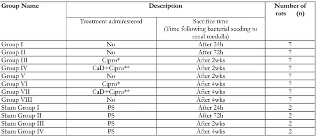
Picture 2. Pyelonephritis morphology in a rat treated with ciprofloxacin and CaD after 4 wks (Hematoxylin and eosin, X40).
Fibrosis scores of groups were illustrated in Table 2. Fibrosis was not observed in Groups I and II and Shams. In rats treated with Cipro+CaD (Group IV),
1657
the fibrosis score was significantly the lowest among the rest of the groups terminated after 2wks (Table 1) (Group III, V; p=0.03 and p=0.04, respectively). However, there was no significant difference between fibrosis scores of Group IV and the groups treated with Cipro and Cipro+CaD and terminated
after 4wks (Groups VI and VII) (p>0.05); but the extent of fibrosis was narrower than the untreated group in which rats were sacrificed after 4wks from E Coli injections (Group VIII) (p=0.03). Fibrosis scores among other groups were not significantly different (p>0.05) (Picture 3).
Table 2. Pyelonephritis and fibrosis scores among groups Group
Median (Upper-lower value) mean±SD* Pyelonephritis score Fibrosis score
Group I 1.0 (1.0-1.0) 1.0 ± 0.0 0.0 (0.0-0.0) 0.0 ± 0.0 Group II 2.0 (1.0-3.0) 2.0 ± 0.8 0.0 (0.0-0.0) 0.0 ± 0.0 Group III 2.0 (1.0-3.0) 2.0 ± 0.6 3.0 (1.0-4.0) 2.9 ± 0.9 Group IV 1.0 (1.0-2.0) 1.3 ± 0.5 2.0 (1.0-2.0) 1.6 ± 0.5 Group V 3.0 (1.0-3.0) 2.4 ± 0.8 3.0 (1.0-4.0) 2.7 ± 1.1 Group VI 2.0 (1.0-2.0) 1.6 ± 0.5 2.0 (1.0-4.0) 2.4 ± 1.0 Group VII 2.0 (1.0-2.0) 1.7 ± 0.5 2.0 (1.0-4.0) 2.7 ± 1.3 Group VIII 2.0 (1.0-2.0) 1.6 ± 0.5 3.0 (2.0-4.0) 3.3 ± 0.8 Sham Group I 0.0 (0.0-0.0) 0.0 ± 0.0 0.0 (0.0-0.0) 0.0 ± 0.0 Sham Group II 0.0 (0.0-0.0) 0.0 ± 0.0 0.0 (0.0-0.0) 0.0 ± 0.0
Sham Group III 0.0 (0.0-0.0) 0.0 ± 0.0 0.0 (0.0-0.0) 0.0 ± 0.0
Sham Group IV 0.0 (0.0-0.0) 0.0 ± 0.0 0.0 (0.0-0.0) 0.0 ± 0.0
* SD: standard derivation.
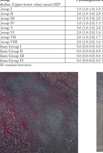
Picture 3: Fibrosis morphology in a rat treated with ciprofloxacin and CaD after 4 wks ( Masson’s trichrom, X40).
TGFb1 scores in terms of groups are shown in Table 3. There was no significant difference between Group I and II (p>0.05). However, the TGFb1 scores of both groups were significantly lower than the groups terminated after 2 and 4wks (Groups III-VIII) (p<0.05). Nevertheless, the extent of TGFb1 staining was not statistically different among the rest of the pyelonephritis groups (Picture 4).
Picture 4: TGFb morphology in a rat treated with ciprofloxacin and CaD after 4 wks (X40).
HGF-beta scores according to groups were given in Table 3. HGF-beta scores of Group I and II were statistically comparable; but both were lower than the figures of other groups (Group III-VIII) (p<0.01). The extent of HGF-beta staining of Group IV; the group of Cipro+CaD treated rats sacrificed after 2 wks was scored the highest of all groups. In comparison with the scores of HGF-beta of Groups I-III and Group IV, the differences were statistically significant (p<0.05); however, compared with the
rest of the groups, the higher values observed in Group IV was not significant (p>0.05) (Picture 5). Similarly, there was not a significant difference among other groups (p>0.05).
Picture 5: HGFb morphology in a rat with no treatment given after 2 wks (X40).
Figures about bFGF scores were represented in Table 3. Scores of Group I and II were lower than the ones of all other groups (p<0.01), however the scores of both groups were not significantly different
(p>0.05). There was not a significant difference among other groups, likewise (p>0.05) (Picture 6). There was no correlation between pyelonephritis, fibrosis, TGFb1, HGF-beta and bFGF scores among all pyelonephritis groups (p>0.05).
Picture 6: bFGF morphology in a sole ciprofloxacin treated rat after 4 wks (X40).
Table 3. Scores of TGFb, HGF-beta and bFGF among Groups Group
Median (Upper-lower value) mean±SD* TGFb HGF-beta bFGF Group I 0.0 (0.0-1.0) 0.4 ± 0.5 0.0 (0.0-1.0) 0.1 ± 0.5 0.0 (0.0-2.0) 0.7 ± 1.0 Group II 0.0 (0.0-1.0) 0.1 ± 0.4 0.0 (0.0-1.0) 0.3 ± 0.5 0.0 (0.0-1.0) 0.1 ± 0.4 Group III 1.0 (1.0-3.0) 1.4 ± 0.8 2.0 (0.0-3.0) 1.6 ± 1.0 2.0 (1.0-2.0) 1.6 ± 0.5 Group IV 2.0 (0.0-3.0) 1.7 ± 1.1 3.0 (2.0-3.0) 2.6 ± 0.5 2.0 (1.0-3.0) 2.0 ± 0.8 Group V 1.0 (1.0-4.0) 1.7 ± 1.1 2.0 (1.0-3.0) 2.1 ± 0.7 2.0 (1.0-3.0) 1.7 ± 0.8 Group VI 1.0 (1.0-3.0) 1.7 ± 1.0 2.0 (1.0-3.0) 2.0 ± 0.6 2.0 (2.0-3.0) 2.3 ± 0.5 Group VII 1.0 (1.0-2.0) 1.1 ± 0.4 2.0 (1.0-3.0) 1.9 ± 0.9 3.0 (1.0-4.0) 2.4 ± 1.1 Group VIII 2.0 (1.0-3.0) 2.1 ± 0.9 2.0 (1.0-3.0) 2.3 ± 0.8 2.0 (1.0-3.0) 1.9 ± 0.7 Sham Group I 0.0 (0.0-0.0) 0.0 ± 0.0 0.5 (0.0-1.0) 0.5 ± 0.7 0.0 (0.0-0.0) 0.0 ± 0.0 Sham Group II 0.5 (0.0-1.0) 0.5 ± 0.7 1.0 (1.0-1.0) 1.0 ± 0.0 0.5 (0.0-1.0) 0.5 ± 0.7 Sham Group III 0.5 (0.0-1.0) 0.5 ± 0.7 0.5 (0.0-1.0) 0.5 ± 0.7 0.5 (0.0-1.0) 0.5 ± 0.7 Sham Group IV 0.0 (0.0-0.0) 0.0 ± 0.0 0.0 (0.0-0.0) 0.0 ± 0.0 0.5 (0.0-1.0) 0.05± 0.7
* SD : standard derivation.
DISCUSSION
Urinary tract infection (UTI) is frequently diagnosed in childhood; unless diagnosed early and managed properly, renal scarring and finally chronic kidney disease (CKD) might be due15. Renal scarring
secondary to UTI has been a leading factor for
paediatric nephrology admissions yet1. E Coli has
been reported to be the major responsible bacteria in UTI16. Hence, an uropathogenic E Coli strain (ATCC
25922) was used in this study12,13.
Acute pyelonephritic changes characterized by interstitial PMNL infiltration, oedema and tubulitis
1659
are the evidences of active infection17. Acute signs of
UTI were distinctly observed in the first 72 h after E Coli injection; becoming more prominent in rat kidneys after 72 h than the first 24 h (Group II vs. Group I), with the aggravation of active infection. However, chronic pyelonephritis findings (mononuclear cell infiltration and tubular atrophy) were determined in kidney specimens of rats, sacrificed after 2-4 wks. in our study. (Groups III-VIII). Rugo et al. revealed the peak time of PMNL infiltration to be on the 3rd day and stated the soaring
mononuclear cell infiltration after 5 days following bacterial seeding in their pyelonephritis model18.
Bıyıklı et al. observed chronic signs of pyelonephritis in rats, sacrificed after 1st wk.; but acute changes in 24
h following intra-renal bacterial injection in their experimental model12. The results of this study were
comparable with the findings of Rugo and Bıyıklı; regarding the fact that chronic signs of pyelonephritis were prominent after 5-7 days after bacterial inoculation12,18.
Studies highlighted the importance of early antibiotic treatment in pyelonephritis; stating that being late to start antibiotic treatment after 72h from bacterial inoculation would not avoid renal scarring19. Renal
scarring has been reported to be associated with the complicated inflammatory process including the release of lysosomal enzymes and free oxygen radicals triggered by infection; rather than bacterial growth itself19-21. This study determined no difference
between the rats having no treatment within 72h of bacterial injection to kidneys and the ones, antibiotic treatment started only after 72 h following bacterial inoculation (Groups II, III, V-VIII); regarding to pyelonephritis scores. In this regard, we claim that starting antibiotics lately; after 72 h following bacterial seeding to kidney could not avoid the progression from acute infection to chronic pyelonephritis and finally to renal fibrosis and scarring. Our findings were compatible with the results of Yagmurlu et al. indicating that lately started antibiotic treatment in UTI was inefficient in preventing hazards of renal scarring19.
Among the rats sacrificed after 2 wks following bacterial inoculation to renal medulla, the lower pyelonephritis score of rats treated with Cipro+CaD (Group IV), compared to the rest of the groups terminated at the second week, constituting of rats with no treatment or treated with an antibiotic solely (Group V and III respectively) might be the result of a sort of synergy between CaD and Cipro. In
addition, a complicated process including capillary obliteration and tissue ischemia due to intravascular granulocyte and platelet aggregation, release of super-oxides and toxic enzymes following reperfusion with bacterial killing causes interstitial tissue devastation and scarring21-23. In this regard; a recent research in
an experimental model of obstructive jaundice revealed that CaD had hepatoprotective effects on liver damage associated with its antioxidant properties24. Hence; other sorts of effects of CaD
which were out of scope of this research; including inhibiting platelet aggregation and thrombus formation, decreasing blood hyper-viscosity, in addition; anti-oxidant characteristics over free oxygen radicals might be associated with the lower pyelonephritis and fibrosis extent detected after 2nd
wks. in Cipro+ CaD administered rats21-23 (Group
IV).
Moreover; there have been reports claiming favourable impacts of CaD over a variety of complex molecular and cellular mechanisms in the pathogenesis of pyelonephritis and subsequent renal interstitial fibrosis - a common pathway to chronic kidney disease. Li et al. reported that CaD, attenuated renal interstitial fibrosis by improving the imbalance between synthesis and degradation of extracellular matrix via discoidin domain receptor 2 activation in a ureteral obstruction model25. Wang et al., recently
published that CaD supressed renal interstitial fibrosis by promoting anti-apoptotic effects through Sirt1/p53 signaling pathway26. CaD was also reported
to attenuate diabetes-induced endothelial dysfunction and chronic inflammation in diabetic nephropathy via reducing inflammatory and endothelial dysfunction markers27. However; none of the
markers mentioned above was investigated in this study.
Despite not statistically significant, the lower pyelonephritis score in Group IV; compared to the scores of groups terminated after 4 wks. (Groups VI-VIII) might be associated with insufficient treatment time with Cipro+CaD. Our findings indicated that use of CaD with antibiotics might have a favourable effect over severity of pyelonephritis regarding to dosage and period of treatment. However optimum treatment dose and period should be determined. Neither pyelonephritis nor fibrosis was diagnosed in Sham groups; indicating the trauma due to needle puncture to inoculate bacteria to kidneys was not conducive to renal fibrosis. In addition; the lower fibrosis score in Cipro+CaD administered rats
(Group IV) among rats sacrificed after 2 wks compared to other groups terminated following 2 wks (Group III and V; p=0.03 and p=0.04 respectively) might be associated with the relative lower pyelonephritis scores in Group IV in proportion to the figures in Group III and Group V (p=0.03 an p=0.01 respectively) (Table 2). Additionally; CaD might have acted likewise by supressing collagen IV retention and “tissue inhibitor of metalloproteinase-1” (TIMP-1) expression; however investigating such an efficacy was out of the scope of this study28. Dong et al. reported therapeutic
effect of CaD related to collagen IV and TIMP-1 in diabetic model following a treatment for 12wks28.
Although favourable effects of CaD associated with TGFb1 and bFGF were reported in a variety of clinical or experimental models; no significant relation between CaD and TGFb1 or bFGF was determined in this study9,10.
It has been well known that TGFb is a major fibrosis-promoting growth factor in renal fibrosis pathogenesis6. Far lower TGFb1 expressions (almost
no expression observed) were seen in the first 72 h, when no fibrosis was seen (Group I and II). Nevertheless, interstitial staining observed after 2nd
and 4th wks. (Groups III-VIII) might confirm the
impact of TGFb1 in renal fibrosis6,29. Considering no
difference between TGFb1 scores was observed among groups with fibrosis (Groups III-VIII) (excluding Groups I and II); no significant efficacy of CaD was detected on tissue TGFb1 staining in this pyelonephritis model. However, CaD might supress renal fibrosis development regarding to dose and treatment duration, or CaD might have had some other sort of impacts on the scarring process as discussed previously24-27.
Likewise, interstitial HGF-beta expression was prominent in rats sacrificed on the 2nd and 4th wks. of
bacterial injection. This might be considered in the complex fibrotic process as a contrary impact to TGFb112. Significant HGF-beta staining was seen in
pyelonephritis rather than in Sham groups; indicating its potential role in post-infectious inflammation. Determining the highest HGF-beta score on the 2nd
wk. in rats treated with Cipro+CaD. (Group IV) might recall a potential favourable effect of CaD over HGF-beta expression. However; although, the differences regarding to HGF-beta scores of Group IV in comparison with the figures of the groups I, II and III were significant; the HGF-beta expression in Group IV was not significantly higher than the
staining scores of the groups terminated after 4 wks. (Groups VI-VIII). Obviously, HGF-beta expression was increased in our pyelonephritis model. Indicating its role and other potential impacts of related cytocines in chronic pyelonephritis and scarring pathogenesis, bFGF interstitial expression was seen in rat kidney specimens in the 2nd and 4th wks.
following bacterial inoculation. Although there are reports stating inhibitory impacts of CaD on bFGF; we could not determine such an impact10.
The major limitation of the study might be the lack of an exactly determined dosage or treatment period for CaD in a pyelonephritis model. However; to our knowledge; the state of this study as being a pioneering research on CaD effectiveness in an experimental pyelonephritis model might be an excuse. Secondly; the therapeutic mechanisms underlying the favourable effect of CaD against pyelonephritis and renal fibrosis after 2 wks and the reasons of discontinuation of this effect could not be elucidated properly.
In conclusion, accelerated expressions of major factors casting in renal fibrosis pathogenesis, examined in this study were observed in collaboration after 72 h following bacterial inoculation to the kidney. This pyelonephritis rat model revealed that delayed antibiotic administration solely, could not ameliorate renal scarring. CaD, when administered with Cipro has alleviated pyelonephritis and fibrosis after 2wks; rather than sole antibiotic treatment. However its activity could not proceed after the drug was discontinued. Anti-oxidative characteristics or other sort of features of CaD not investigated in this model might be associated with its favourable efficacy; rather than its previously reported impacts on TGFb1 and bFGF. We claim that CaD might alleviate pyelonephritis and scarring, depending on dosage and treatment period and further studies are needed to determine optimum treatment dose and duration. The complex nature of the renal fibrosis pathology and the alluring idea of reversing renal scars seem to keep on appealing researchers temptation.
Yazar Katkıları: Çalışma konsepti/Tasarımı: BŞ; Veri toplama: BŞ,
AKB, AN, AA; Veri analizi ve yorumlama: BŞ, AKB, AN, AA; Yazı taslağı: BŞ; İçeriğin eleştirel incelenmesi: BŞ, GG, AKB, AN, AA; Son onay ve sorumluluk: BŞ, GG, AKB, AN, AA; Teknik ve malzeme desteği: BŞ, GG, AKB; Süpervizyon:BŞ, AKB, AN, AA; Fon sağlama (mevcut ise): yok.
Etik Onay: Bu çalışma Çukurova Üniversitesi Tıp Bilimleri Deney
Hayvanları Araştırma Merkezi Adana Çukurova Üniversitesi Hayvan Etik Kurulu tarafından “B.30.2.CKO.0.5L.00.00-128” doğrulama numarası ile onaylanmıştır.
1661
Çıkar Çatışması: Yazarlar çıkar çatışması beyan etmemişlerdir. Finansal Destek: Yazarlar finansal destek beyan etmemişlerdir. Author Contributions: Concept/Design :BŞ; Data acquisition: BŞ, AKB, AN, AA; Data analysis and interpretation: BŞ, AKB, AN, AA; Drafting manuscript: BŞ; Critical revision of manuscript: BŞ, GG, AKB, AN, AA; Final approval and accountability: BŞ, GG, AKB, AN, AA; Technical or material support: BŞ, GG, AKB; Supervision: BŞ, AKB, AN, AA; Securing funding (if available): n/a.
Ethical Approval: This study was approved by the Animal Ethics
Committee of Cukurova University Affliated to Cukurova University Medical Sciences Experimental Animals Research Center Adana with the verification number “B.30.2.CKO.0.5L.00.00-128”.
Peer-review: Externally peer-reviewed.
Conflict of Interest: Authors declared no conflict of interest. Financial Disclosure: Authors declared no financial support
REFERENCES
1. Eddy AA. Molecular basis of renal fibrosis. Pediatr Nephrol. 2000;15:290-301.
2. Eddy AA. Overview of the cellular and molecular basis kidney fibrosis. Kidney Int Suppl (2011). 2014;4:2-8.
3. Genovese F, Manresa AA, Leeming DJ, Karsdal MA, Boor P. The extracellular matrix in the kidney: a source of novel non-invasive biomarkers of kidney fibrosis? Fibrogenesis Tissue Repair. 2014;7:4. 4. Guan X, Nie L, He T, Yang K, Xiao T, Wang S et al.
Klotho supresses renal tubulo-interstitial fibrosis by controlling basic fibroblast growth factor-2 signalling. J Pathol. 2014;234:560–72.
5. Strutz F. The role of FGF-2 in renal fibrosis. Front Biosci. 2009;(Suppl 1):125-31.
6. Youhua Liu. Renal fibrosis: New insights into the pathogenesis and therapeutics. Kidney Int. 2006;69:213–17.
7. Xu J, Yu T-T, Zhang K, Li M, Shi H-J, Meng X-J et al. HGF alleviates renal interstitial fibrosis via inhibiting the TGF-β1/SMAD pathway. Eur Rev Med Pharmacol Sci. 2018;22:7621-7.
8. Zhou Y, Qi C, Li S, Shao X, Mou S, Ni Z. Diabetic nephropathy can be treated with Calcium Dobesilate by alleviating the chronic inflammatory state and improving endothelial cell function. Cell Physiol Biochem. 2018;51:1119-33.
9. Liu X, Liu X. Effect of calcium dobesilate on nephropathy in type 2 diabetic rats. Huazhong Univ Sci Technolog Med Sci. 2005;25:36-8.
10. Cuaves P. Treatment of basal cell carcinoma with dobesilate. J Am Acad Dermatol. 2005;33:526-7. 11. World Medical Association Declaration of Helsinki.
Adopted by the 18th World Medical Assembly, Helsinki 1964 as amended by the 64th World Medical Assembly, Fortaleza, Brazil, October 2013.
12. Bıyıklı NK, Tuğtepe H, Çakalağaoğlu F, İlki A Alpay H. Downregulation of the expression of bone morphogenic protein 7 in experimental pyelonephritis. Pediatr Nephrol. 2005;20:1230-6. 13. Kavukçu S, Soylu A, Türkmen M, Sarıoğlu S,
Büyükgebiz B, Güre A. The role of vitamin A in
preventing renal scarring secondary to pyelonephritis. BJU Int. 1999;83:1055-9.
14. Palomar R, Mayorga M, Ruiz JC, Cuevas J, Rodrigo E, Cotorruelo JG et al. Markers of fibrosis in early biopsies of renal transplants. Transplant Proc. 2005;37:1468-70.
15. Kehr KK, Leichter HE. Urinary tract infection. In Clinical Pediatric Nephrology (Eds Kher KK, Makker SP): 1st Ed., New York: Mc Graw Hill. 1992;277-321.
16. Simsek B. The prevalence of bacterial strains isolated from urine cultures and their antibiotic susceptibility patterns among children with urinary tract infection. 1st Euresion Pediatric Congress and VIIth Kosova
Pediatric School Abstract Book,17-20 October 2019, Kosova, OP-06, p.48.
17. Heptinstall RH (editor). Pyelonephritis: pathological features. In Pathology of The Kidney. 4th ed., Boston:
Little Brown, 1992;1489-561.
18. Rugo HS, O’Hanley P, Bishop AG, Pearce MK, Abrams JS, Howard M et al. Local cytokine productionin a murine model of Escherichia Coli pyelonephritis. J Clin Invest. 1992;89:1032-9. 19. Yağmurlu A, Boleken ME, Ertoy D, Özsan M,
Gökçora IH, Dindar H. Preventive effect of pentoxyfilline on renal scarring in rat model of pyelonephritis. Urology. 2003;61:1037-41.
20. Matsumato T, Mitzunoe Y, Ogata N. Antioxidant effect on renal scarring following infection of mannose-sensitive –pilliated bacteria. Nephron. 1992;60:210-5.
21. Roberts Ja, Kaack MB, Fussell EN. Immunology of pyelonephritis. VII. Effect of allourinol. J Urol. 1986;136:960.
22. Roberts JA, Roth JK, Dominique G. Immunology of pyelonephritis in the primate model. VI. Effect of complement depletion. J Urol. 1983;129:193. 23. Shimamura T. Mechanisms of renal tissue destruction
in an experimental acute pyelonephritis. Exp Mol Pathol. 1981;34:34.
24. Unal Y, Tuncal S, Kosmaz K, Kucuk B, Kımet K, Cavusoglu T. The effect of Calcium Dobesilate on liver damage in experimental obstructive jaundice. J Invest Surg. 2019;32:238-44.
25. Li X, Bu X, Yan F, Wang F, Wei D, Yuan J et al. Deletion of discoidin domain receptor 2 attenuates renal interstitial fibrosis in a murine unilateral ureteral obstruction model. Ren Fail. 2019;41:481-8.
26. Wang Y, Zuo B, Wang N, Li S, Liu C, Sun D. Calcium dobesilate mediates renal interstitial fibrosis and delay renal capillary loss through Sirt1/p53 signaling pathway. Biomed Pharmacother. 2020;132:110798. 27. Zhou Y, Qi C, Shao X,Mou S, Ni Z. Diabetic
nephropathy can be treated with Calcium Dobesilate by alleviating the chronic inflammatory state and improving endothelial cell function. Cell Physiol Biochem. 2018;51:1119-33.
28. Dong J, Liu X, Liu S, Li M, Xu Y, Cui B. Effects of calcium dobesilate on glomerulusTIMP1 and collagen
IV of Diabetic rats. Huazhong Univ Sci Technolog
Med Sci 2005;25:416-26. 29. Bottinger EP, Letterio JJ, Roberts AB. Biology of TGF-beta in knockout and transgenic mouse models. Kidney Int. 1997;51:1355-60.