© 2018 Turkish Journal of Plastic Surgery | Published by Wolters Kluwer - Medknow
40
Abstract
Case Report
I
ntroductIonClear cell acanthoma (CCA) is a rare skin lesion originating from epidermal keratinocytes. While it has also been reported in young patients, it is more commonly seen in middle and older ages regardless of gender.[1-3] CCA can develop as solitary or multiple lesions and most often involves the distal extremities. CCA lesions are periodic acid–Schiff (PAS) positive as a result of intracellular glycogen accumulation. Clinically, it is hard to distinguish whether the lesion is benign or malignant; therefore, the best approach would be a total excision of the lesion.
c
aser
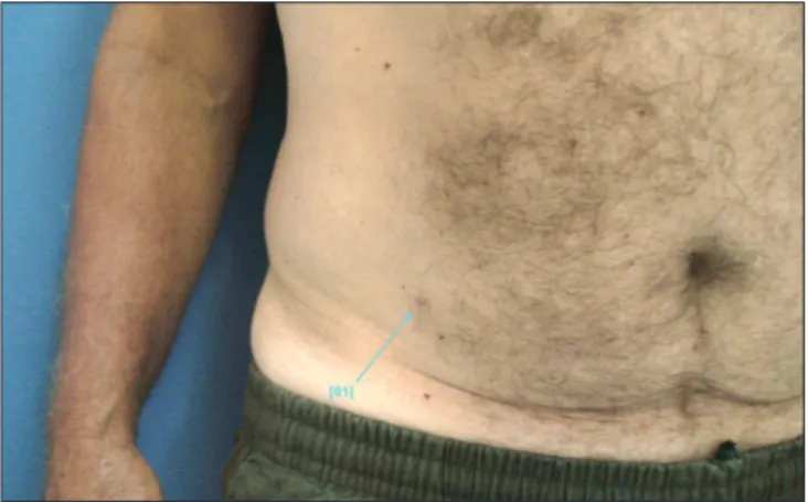
ePortA 62-year-old male presented to our clinic with a wound in the left abdominal region that persisted for 2 years [Figure 1]. Dermatological examination showed a sharply demarcated erythematous nodular lesion measuring 0.6 cm × 0.4 cm [Figure 2]. Excisional biopsy and histopathological examination were planned with a diagnosis of dermatofibroma. The patient was informed about the surgical procedure, and his verbal consent was obtained for publishing his case. The lesion was excised under local anesthesia and with 3-mm negative surgical margin. The skin defect was primarily closed after hemostasis was achieved. No wound-healing problems were observed following the surgery and sutures were removed after 14 days. Histopathological examination reported CCA.
d
IscussIonCCA is a rare skin lesion originating from the epidermal keratinocytes. Although it has been reported also in young patients, it is more commonly seen in middle and older ages regardless of gender.[2,3] Typically, CCAs are brown-to-red colored, domed, solitary, or nodular lesions of 3–20 mm in diameter[2-4] [Figures 1 and 2].
While it generally involves the distal extremities, it can also, although rarely, occur at atypical localizations.[5] While its cause is not exactly known, it is thought to be a benign epidermal tumor; however, there are views that regard the lesion as reactive inflammatory dermatosis.[6-8]
In differential diagnosis, the lesion can be clinically mistaken for dermatofibroma, pyogenic granuloma, irritated seborrhoeic keratosis, keratoacanthoma, actinic keratosis, malignant skin lesions (basal cell carcinoma, squamous cell carcinoma, and malignant melanoma),[2-9] plaque psoriasis, eccrine poroma,[10] viral warts, or metastatic cancers.[11] Ohnishi and Watanabe[8] emphasized its resemblance to psoriasis,[12] lichen planus, and discoid lupus erythematosus.[8]
Clear cell acanthoma (CCA) is an uncommon, benign, and slow progressing lesion originating from epidermal keratinocytes. Lesions are not gender specific and usually diagnosed at 50–60 years of age. It is generally represented in the lower extremities. CCA was first described by Degos et al. as “Degos acanthoma” for a lesion presented in the lower extremity. Clinically, it is hard to distinguish whether the lesion is benign or malignant. Final diagnosis can be made histopathologically. Biopsy material is periodic acid–Schiff positive. While CAA commonly presents itself in the lower extremities, our patient had a CAA in his abdominal region.
Keywords: Abdominal lesion, clear cell acanthoma, Degos acanthoma
Address for correspondence: Dr. Atilla Adnan Eyüboglu,
Department of Plastic, Reconstructive and Aesthetic Surgery, School of Medicine, Baskent University, Ankara, Turkey. E‑mail: [email protected]
Access this article online
Quick Response Code:
Website:
http://www.turkjplastsurg.org
DOI:
10.4103/tjps.tjps_13_18
This is an open access article distributed under the terms of the Creative Commons Attribution-NonCommercial-ShareAlike 3.0 License, which allows others to remix, tweak, and build upon the work non-commercially, as long as the author is credited and the new creations are licensed under the identical terms.
For reprints contact: [email protected]
How to cite this article: Eyüboglu AA, Togral AK, Akçay EY,
Ertas NM. Clear cell acanthoma in an unexpected location. Turk J Plast Surg 2018;26:40-2.
Clear Cell Acanthoma in an Unexpected Location
Atilla Adnan Eyüboglu, Arzu Karatas Togral1, Eda Yilmaz Akçay2, Nilgün Markal Ertas
Departments of Plastic, Reconstructive and Aesthetic Surgery, 1Dermatology and 2Pathology, School of Medicine, Baskent University, Ankara, Turkey [Downloaded free from http://www.turkjplastsurg.org on Monday, May 6, 2019, IP: 10.232.74.26]
Eyüboglu, et al.: Clear cell acanthoma
Turkish Journal of Plastic Surgery ¦ Volume 26 ¦ Issue 1 ¦ January-March 2018 41 CCA can develop as single or multiple lesions and typically are
seen to be 3–20 mm lesions localized to the distal extremities. Diversely, there are also giant cell, polypoid/pedicled, pigmented, eruptive, atypical, or cystic types reported in the literature.[13] Giant CCAs are larger than 40 mm. In the literature, there are also reports of 40–60 mm lesions localized to the feet,[14] hips,[15] or the perineum.[16]
Polypoid CCA was first described by Petzelbauer and Konrad[17] in 1990. Lesions of 4–30 mm in diameter can localize to regions such as the femur, leg, neck, scalp, or nipple. Pigmented CCA was first described by Fanti et al.[18] in 1990. The presence of melanocyte and melanin pigments observed under a light microscope in clear cells had given the lesion its brownish appearance.
There are more than 20 lesions in the literature described for an eruptive-type acanthoma with diameters ranging from 1 to 10 mm and mostly localized to the leg. CCA cases localized to the arm and the torso are also described.[19]
Atypical-type and originally benign-type lesions are thought to be able to undergo malignant changes. Parson and Ratz[20] have described “squamous cell carcinoma in situ arising within CCA.” Typical cystic-type CCA is defined by the presence of hair follicles. There is one case reported by Hamaguchi and Penneys[21] in the suprapubic region. In our case, the lesion was in the abdominal region.
Histopathological findings are keys in diagnosing cases of CCA. The lesion is characterized by clear cytoplasm due to its glycogen content. It stains PAS positive as a result of intracellular glycogen accumulation. The lesion is composed of squamous epithelium which usually present with acanthotic psoriasiform hyperplasia in the epithelium and is sharply demarcated from the lateral border [Figure 3]. Neutrophil leukocytes can be noted in the parakeratotic layer on its surface. Dilated vascular structures and inflammatory cells can be observed in the papillary dermis. The cells contained in the lesion are separated from the adjacent epithelium by sharply demarcated contours. The cells contained in the lesion have transparent cytoplasm and stain pale in H and E [Figure 4]. Although an eruptive-type CCA was reported by García-Gavín et al.[22] to show spontaneous regression in the course of treatment, CCAs have a predilection for growth. Treatment choices depend on the size, localization, and number of lesions, as well as the experience and capability of the surgeon. Surgical excision is the preferred treatment. Mohs surgery, curettage, electrofulguration, cryotherapy, and carbon dioxide laser are the alternative treatment methods. CCA can be cured after the lesion is accepted to be benign and excised along its contours. There are few cases reported for recurrence after excision.[23]
c
onclusIonWhile its exact etiology remains unknown still today, diagnosis of CCA can be challenging, in that it resembles a number of
skin lesions, both benign and malignant. Recent studies suggest that the lesion is caused by inflammation. Definitive diagnosis is based on histopathological findings. Treatment options can vary based on the condition of the lesion and the preferences of the treatment team. Posttreatment follow-up is recommended due to malignancy potential.
Declaration of patient consent
The authors certify that they have obtained all appropriate Figure 1: Clear cell acanthoma in the left abdominal region
Figure 3: The lesion, composed of squamous epithelium presenting
with acanthotic psoriasiform hyperplasia, is sharply demarcated from the lateral border (H and E, ×50)
Figure 2: Nodular lesion of 0.6 cm × 0.4 cm in abdomen
Eyüboglu, et al.: Clear cell acanthoma
Turkish Journal of Plastic Surgery ¦ Volume 26 ¦ Issue 1 ¦ January-March 2018
42
patient consent forms. In the form the patient(s) has/have given his/her/their consent for his/her/their images and other clinical information to be reported in the journal. The patients understand that their names and initials will not be published and due efforts will be made to conceal their identity, but anonymity cannot be guaranteed.
Financial support and sponsorship Nil.
Conflicts of interest
There are no conflicts of interest.
r
eferences1. Degos R, Delort J, Cıvatte J, Poıares Baptısta A. Epidermal tumor with an unusual appearance: Clear cell acanthoma. Ann Dermatol Syphiligr (Paris) 1962;89:361-71.
2. Brownstein MH, Fernando S, Shapiro L. Clear cell acanthoma: Clinicopathologic analysis of 37 new cases. Am J Clin Pathol 1973;59:306-11.
3. Fine RM, Chernosky ME. Clinical recognition of clear-cell acanthoma (Degos’). Arch Dermatol 1969;100:559-63.
4. Burg G, Würsch T, Fäh J, Elsner P. Eruptive hamartomatous clear-cell acanthomas. Dermatology 1994;189:437-9.
5. Williams RE, Lever R, Seywright M. Multiple clear cell acanthomas-treatment by cryotherapy. Clin Exp Dermatol 1989;14:300-1.
6. Park SY, Jung JY, Na JI, Byun HJ, Cho KH. A case of polypoid clear cell acanthoma on the nipple. Ann Dermatol 2010;22:337-40.
7. Yamasaki K, Hatamochi A, Shinkai H, Manabe T. Clear cell acanthoma developing in epidermal nevus. J Dermatol 1997;24:601-5.
8. Ohnishi T, Watanabe S. Immunohistochemical characterization of keratin expression in clear cell acanthoma. Br J Dermatol 1995;133:186-93. 9. Arida M, English JC 3rd, Mully TW. Giant clear-cell acanthoma with
keratoacanthoma-like changes: A case report. Dermatol Online J 2006;12:11.
10. Mckee PH, Calonje E, Granter SR. Tumors of the sweat glands. Pathology of the Skin with Clinical Correlations. 3rd ed. Philadelphia:
Elsevier-Mosby; 2005. p. 1166-7.
11. Inalöz HS, Patel G, Knight AG. Polypoid clear cell acanthoma: Case report. J Eur Acad Dermatol Venereol 2000;14:511-2.
12. Zedek DC, Langel DJ, White WL. Clear-cell acanthoma versus acanthosis: A psoriasiform reaction pattern lacking tricholemmal differentiation. Am J Dermatopathol 2007;29:378-84.
13. Morrison LK, Duffey M, Janik M, Shamma HN. Clear cell acanthoma: A rare clinical diagnosis prior to biopsy. Int J Dermatol 2010;49:1008-11. 14. Roytman M, Frumkin A, Everett MA. Giant clear cell acanthoma. J Am
Acad Dermatol 1987;17:513-4.
15. Langtry JA, Torras H, Palou J, Lecha M, Mascaro JM. Giant clear cell acanthoma in an atypical location. J Am Acad Dermatol 1989;21:313-5. 16. Kim CY, Kim NG, Oh CW. Multiple reddish weeping nodules on the
genital area of a girl. Giant clear cell acanthoma (CCA). Clin Exp Dermatol 2010;35:e67-9.
17. Petzelbauer P, Konrad K. Polypous clear cell acanthoma. Am J Dermatopathol 1990;12:393-5.
18. Fanti PA, Passarini B, Varotti C. Melanocytes in clear cell acanthoma. Am J Dermatopathol 1990;12:373-6.
19. Innocenzi D, Barduagni F, Cerio R, Wolter M. Disseminated eruptive clear cell acanthoma – A case report with review of the literature. Clin Exp Dermatol 1994;19:249-53.
20. Parsons ME, Ratz JL. Squamous cell carcinoma in situ arising within clear cell acanthoma. Dermatol Surg 1997;23:487-8.
21. Hamaguchi T, Penneys N. Cystic clear cell acanthoma. J Cutan Pathol 1995;22:188-90.
22. García-Gavín J, González-Vilas D, Montero I, Rodríguez-Pazos L, Pereiro MM, Toribio J, et al. Disseminated eruptive clear cell acanthoma with spontaneous regression: Further evidence of an inflammatory origin? Am J Dermatopathol 2011;33:599-602.
23. Hashimoto T, Inamoto N, Nakamura K. Two cases of clear cell acanthoma: An immunohistochemical study. J Cutan Pathol 1988;15:27-30. Figure 4: The cells contained in the lesion have transparent adjacent
cytoplasm and stain pale in H and E