A highly ef
ficient and faithful MDS patient-derived
xenotransplantation model for pre-clinical studies
Yuanbin Song
1
, Anthony Rongvaux
2,3
, Ashley Taylor
1
, Tingting Jiang
4
, Toma Tebaldi
1,5
,
Kunthavai Balasubramanian
1
, Arun Bagale
1,6
, Yunus Kasim Terzi
1,7
, Rana Gbyli
1
, Xiaman Wang
1,8
,
Xiaoying Fu
1,9
, Yimeng Gao
1
, Jun Zhao
4
, Nikolai Podoltsev
1
, Mina Xu
4
, Natalia Neparidze
1
, Ellice Wong
10
,
Richard Torres
11
, Emanuela M. Bruscia
12
, Yuval Kluger
4,13,14
, Markus G. Manz
15
,
Richard A. Flavell
2,16
& Stephanie Halene
1
Comprehensive preclinical studies of Myelodysplastic Syndromes (MDS) have been elusive
due to limited ability of MDS stem cells to engraft current immunode
ficient murine hosts.
Here we report a MDS patient-derived xenotransplantation model in cytokine-humanized
immunode
ficient “MISTRG” mice that provides efficient and faithful disease representation
across all MDS subtypes. MISTRG MDS patient-derived xenografts (PDX) reproduce
patients
’ dysplastic morphology with multi-lineage representation, including erythro- and
megakaryopoiesis. MISTRG MDS-PDX replicate the original sample
’s genetic complexity and
can be propagated via serial transplantation. MISTRG MDS-PDX demonstrate the cytotoxic
and differentiation potential of targeted therapeutics providing superior readouts of drug
mechanism of action and therapeutic efficacy. Physiologic humanization of the hematopoietic
stem cell niche proves critical to MDS stem cell propagation and function in vivo. The
MISTRG MDS-PDX model opens novel avenues of research and long-awaited opportunities
in MDS research.
https://doi.org/10.1038/s41467-018-08166-x
OPEN
1Section of Hematology, Department of Internal Medicine and Yale Comprehensive Cancer Center, Yale University School of Medicine, New Haven, CT,
USA.2Department of Immunobiology, Yale University School of Medicine, New Haven, CT, USA.3Fred Hutchinson Cancer Research Center, Program in
Immunology, Clinical Research Division, and Department of Immunology, University of Washington School of Medicine, Seattle, WA, USA.4Department of
Pathology, Yale University School of Medicine, New Haven, CT, USA.5Laboratory of Translational Genomics, Centre for Integrative Biology (CIBIO),
University of Trento, Trento, Italy.6University of New Haven, New Haven, CT, USA.7Department of Medical Genetics, Faculty of Medicine, Baskent
University, Ankara, Turkey.8Department of Hematology, The Second Affiliated Hospital of Xi’an Jiaotong University, Xi’an, People’s Republic of China.
9Department of Laboratory Medicine, Shenzhen Children’s Hospital, Shenzhen, People’s Republic of China.10Section of Hematology/Oncology, VA Medical
Center, West Haven, CT, USA.11Department of Laboratory Medicine, Yale University School of Medicine, New Haven, CT, USA.12Department of Pediatrics,
Yale University School of Medicine, New Haven, CT, USA.13Interdepartmental Program in Computational Biology and Bioinformatics, Yale University, New
Haven, CT, USA.14Program of Applied Mathematics, Yale University, New Haven, CT, USA.15Hematology, University Hospital and University of Zurich,
Zurich, Switzerland.16Howard Hughes Medical Institute, Yale University, New Haven, CT, USA. Correspondence and requests for materials should be
addressed to R.A.F. (email:richard.fl[email protected]) or to S.H. (email:[email protected])
123456789
M
yelodysplastic syndrome (MDS) is a group of
hetero-geneous disorders of the hematopoietic stem cell
characterized by recurrent genetic aberrations in genes
of essential pathways, including transcription factors, epigenetic
regulators, cohesin complex genes, DNA repair genes, and key
factors of the spliceosome (see refs.
1,2and reviewed in ref.
3).
Long-term hematopoietic stem cells (HSCs) cannot be expanded
in culture and only rare MDS cell lines exist
4–6, creating an unmet
need for in vivo models of primary MDS. Xenotransplantation of
primary human MDS stem cells into currently available
immu-nodeficient mice, such as NOD-scid Il2rg
−/−(NSG), has
demon-strated limited success with low efficiency and transient
engraftment, skewing towards the lymphoid lineage, and
engraft-ment mostly restricted to the injected tibial bone when aided by
co-injection of human mesenchymal stem cells (MSCs)
7–10. Human
cytokines provided by constitutive, transgene-driven expression in
the NSG-SGM3 model (overexpressing human stem cell factor
(SCF), granulocyte-monocyte-colony-stimulating factor
(GM-CSF), and interleukin-3 (IL3) from a cytomegalovirus promoter),
improve myeloid differentiation and cellular proliferation, yet stem
cell maintenance is impaired
11–15. This limitation is overcome
transiently by co-injection of autologous human MSCs
16or by
creation of an ossicle from human MSCs that provides an
improved human stem cell environment
17. These latter two
approaches have limited applicability in pre-clinical studies that
require a highly efficient, high-throughput approach.
We here present a novel highly efficient MDS
xeno-transplantation
model,
in
humanized
immunodeficient
“MISTRG” mice, expressing humanized M-CSF, IL3/GM-CSF,
SIRP alpha, and Thrombopoietin in the Rag
−/−, IL2Rγ
−/−genetic background from their endogenous murine loci. MISTRG
mice have previously been shown to be highly permissive for
human hematopoiesis and support robust reconstitution of
human lymphoid and myelo-monocytic cellular systems
18,19. We
demonstrate that primary healthy bone marrow- (BM) and MDS
BM-derived CD34
+cells from lower-risk (International
Prog-nostic Scoring System (IPSS) low- and intermediate 1) and
higher-risk (intermediate 2 and high) MDS, defined by the
number of cytopenias, blast percentage in BM, and cytogenetic
abnormalities, efficiently engraft in MISTRG mice and give rise to
multi-lineage hematopoiesis and specifically to myelo-, erythro-,
and mekagaryopoiesis. We demonstrate that MDS
patient-derived MISTRG xenotransplants (MDS MISTRG PDX)
sup-port the MDS stem cell across all MDS subtypes, replicate the
patients’ MDS immunophenotype and dysplastic features,
faith-fully reproduce the clonal complexity of the disease at time of
diagnosis and along disease progression, and are ideally suited for
the testing of targeted therapeutics. Thus, given the high
multi-lineage engraftment efficiency for normal and MDS HSCs and the
histologic and clonal
fidelity, MISTRG PDX represent a
sig-nificant advancement over currently available
xenotransplanta-tion models and an ideal in vivo pre-clinical model for MDS.
Results
MISTRG engraft healthy adult bone marrow-derived CD34
+HSPCs. Adult CD34
+hematopoietic stem and progenitor cells
(HSPCs) engraft with significantly lower efficiency in
immuno-deficient mice compared to human fetal liver- or cord
blood-derived CD34
+cells
18. However, the majority of myeloid
malignancies and in particular MDS occur in the aging adult with
quantitative and qualitative limitations to the stem cell population
of interest. We transplanted healthy BM-derived CD34
+cells
from adult patients, in whom BM involvement by their
under-lying disease was excluded (see Supplementary Table 1),
intra-hepatically into newborn NSG and MISTRG mice irradiated with
maximum tolerated doses for each strain (Fig.
1a)
18. The
max-imum tolerated radiation in NSG mice is limited due to the
inherent DNA repair defect conferred by the scid mutation
20,21.
Samples were CD34 enriched or CD3 depleted (Supplementary
Figure 1a), and further purged of mature T cells by pre-treatment
with the humanized anti-CD3 antibody OKT3 for prevention of
graft versus host disease
22. Highest available rather than a
fixed
cell number were injected as equal split-donor grafts into NSG
and MISTRG mice to maximize engraftment for each primary
sample.
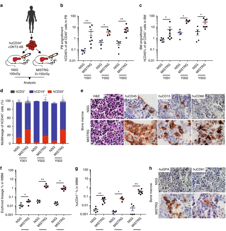
Analysis consisting of complete blood counts and histology
(representative subset), and
flow cytometry of peripheral blood
(PB), BM, and spleen, was performed at least 12 weeks post
transplantation, with >85% survival for both NSG and MISTRG
recipient mice to planned analysis (Supplementary Figure 1b).
Flow cytometric analysis consisted of assessment of overall
human leukocyte engraftment (huCD45) as a function of all
(murine and human) leukocytes as well as assessment for human
erythroid and megakaryocytic engraftment within the murine and
human CD45 negative fraction. Erythroid and megakaryocytic
lineage engraftment based on CD45 negativity and high
transferrin receptor (huCD71)/glycophorin A (huCD235) or
huCD41 expression, respectively, were quantitated as % of all
single live cells in whole BM (Supplementary Figure 1c).
MISTRG mice show significantly higher huCD45
+engraft-ment in PB and BM than NSG mice (Fig.
1b, c) and support
enhanced differentiation towards myelopoiesis (Fig.
1d) over
lymphopoiesis, rectifying a key difference between human and
mouse hematopoiesis. CD3
+T cells are efficiently depleted with
OKT3 treatment of the graft and represent only a minor fraction.
Histologically, myeloid cells express the mature myeloid markers
huCD15 and huCD68. As previously described
18,23, expression of
human GM-CSF and macrophage colony-stimulating factor
(M-CSF) enhance myeloid maturation with differentiation towards
mature granulocytes and macrophages (Fig.
1e and
Supplemen-tary Figure 1d) with repopulation of bone marrow as well as
spleen and non-hematopoietic tissues, such as liver
(Supplemen-tary Figure 1e).
Interestingly, MISTRG bone marrows show significantly higher
numbers of erythroid progenitor cells (CD71
bright, GPA
+) (Fig.
1f,
h) as well as human CD41+ megakaryocytes and platelets
(Fig.
1g, h).
In summary, MISTRG mice support superior healthy adult BM
xenografts with tri-lineage representation.
MISTRG efficiently support all risk MDS PDX with
multi-lineage output. NSG mice have represented a major breakthrough
in xenotransplantation studies due to the lack of mature murine T,
B, and functional natural killer (NK) cells
24and the presence of the
Sirpα gene polymorphism, allowing enhanced binding of the
mSirpα to human CD47
25,26. However, engraftment of MDS
BM-derived CD34
+HSPCs remains a challenge, despite several
altera-tions to NSG mice and the transplantation protocol
7–10,12–14,16. We
engrafted MDS CD34
+(or CD3-depleted) BM cells into NSG and
MISTRG recipients as split-donor grafts, as in Fig.
1a. To avoid a
priori exclusion of lower-risk MDS samples or patient samples with
low cell numbers, CD34
+cell injections for different samples
ranged from 0.5 × 10
5to 1 × 10
6cells per recipient mouse, while
maintaining the same cell number for all recipients within each
experiment (for detailed patient and sample information see
Sup-plementary Table 1).
We engrafted a total of 10 low- and intermediate 1 risk and 8
intermediate 2 and high-risk MDS samples (Fig.
2
and
Supplementary Figure 2). MISTRG consistently resulted in higher
engraftment than NSG for all MDS subtypes in peripheral blood
(top row) and bone marrow (bottom row) (Fig.
2a–c). Only 2 out
of 29 samples (MDS with multi-lineage dysplasia (MLD) Y006,
MDS with excess blasts 2 (EB-2) Y018), injected at <1 × 10
5CD34
+cells/mouse displayed BM engraftment levels <1% in
MISTRG. The engraftment persisted until the time of analysis,
>12 weeks post transplantation, without development of
compromising anemia or thrombocytopenia in recipient mice
(Supplementary Figure 3a-c) or differences in survival between
a
g
h
d
e
f
b
c
Y001 Y002 Y003
0 20 40 60 80 100 Mul ti lineage o f hCD4 5 + cells ( % ) hCD33+ hCD19+ hCD3+
*
*
*
n.s.*
*
**
*
*
NSG MISTRG NSG MISTRG NSG MISTRG NSG MISTRG NSG MISTRG NSG MISTRG NSG MISTRG NSG MISTRG NSG MISTRG NSG MISTRG NSG MISTRG NSG MISTRG NSG MISTRG NSG MISTRG NSG MISTRGY001 Y002 Y003
**
*
**
0.01 0.1 1 10 100 PB engraftment hCD45% o f all CD4 5 + c ells in P B*
*
*
Y001 Y002 Y003
0.01 0.1 1 10 100 BM engraftment hCD45% o f all CD4 5 + c ells in B M
Y001 Y002 Y003
*
**
*
0.001 0.01 0.1 1 10 100 Erythroid lineage % in WBMY001 Y002 Y003
**
*
**
0.001 0.01 0.1 1 10 100 huCD4 1 + % i n W B M huCD15 huCD45 H&E huCD68 NSG MISTRG Bone marrow huCD61 huGPA NSG MISTRG Bone marrow MISTRG 2×150cGy NSG 100cGy huCD34+ +OKT3 AB AnalysisFig. 1 Enhanced engraftment of adult healthy bone marrow (BM)-derived CD34+hematopoietic stem and progenitor cells (HSPCs) in human
cytokine-knockin MISTRG mice.a Universal experimental setup. Human BM-derived CD34+HSPCs were pre-incubated with anti-CD3 antibody (OKT3) and
injected intrahepatically into newborn (D2–3) NSG or MISTRG mice conditioned with the respective maximum tolerated irradiation doses (NSG 100 cGy,
MISTRG 2 × 150 cGy). Mice were analyzed 10–17 (healthy BM), 13–30 (myelodysplastic syndrome (MDS)), and 9−24 (acute myeloid leukemia (AML))
weeks post transplantation.b, c Comparison of overall human CD45+engraftment in peripheral blood (PB) and BM in NSG versus MISTRG mice. Individual
mice are represented by symbols.d Relative distribution of myeloid CD33+(red), B-lymphoid CD19+(blue), and T-lymphoid CD3+(gray) cells as % of
human CD45+cells in NSG vs. MISTRG mice.e BM histology of representative NSG and MISTRG mice from (d). Hematoxylin and eosin (H&E) and
immunohistochemistry (IHC) stains for huCD45, huCD15, huCD68 in NSG (top) and MISTRG BM (bottom row) (scale bars 10µm, original magnification
60×).f, g Comparison of erythroid and megakaryocytic lineage engraftment in BM of NSG and MISTRG mice. h BM histology of representative NSG and
MISTRG mice from (d). H&E and IHC stains for huCD235 and huCD61 as in (e). For detailed sample information see Supplementary Table 1. In (c, d, e, f, g)
MISTRG and NSG mice (Supplementary Fig 1b), interestingly
with similar engraftment in female and male mice of the
respective strains (Supplementary Figure 3d), not seen with
engraftment in adult NSG mice in previous studies
27. As
described previously for normal hematopoiesis, CD34
+cells
from MDS bone marrow give rise to myeloid predominant grafts,
while NSG mice give rise to lymphoid-predominant grafts
(Fig.
2d–f). Expression of human M-CSF, GM-CSF, and IL-3
MDS/MPN; MDS-EB-1 MDS-SLD/MLD/RS/5q- MDS-EB-2 PB engraftment hCD45% of all CD45 + cells P B BM engraftment hCD45% of all CD45 + cells B M Multilineage distribution Multilineage of hCD45 + cells (% )
a
d
e
f
b
c
* ** ** 0.01 0.1 1 10 100 n.s. n.s. n.s. n.s. ** *** *** * * ** NSG MISTRG NSG MISTRG NSG MISTRG NSG MISTRG NSG MISTRG NSG MISTRG NSG MISTRG NSG MISTRG NSG MISTRG 0.01 0.1 1 10 100 * * n.s. 0.01 0.1 1 10 100 0.01 0.1 1 10 100 ** * * * 0.01 0.1 1 10 100g
h
0 20 40 60 50 100 150 200 250 huCD34+ cells (×103) huCD45 + engraftment in BM (%) rp:0.42 P: 1.7e–06 Y= 0.12X–2.97 MISTRG rp: 0.39 P: 1.1e–04 Y= 0.02X–1.06 NSG % of all mice****
NSG MISTRG 0 20 40 60 80 100 % Engraftment > 0.01% <1% % Engraftment 1 – 10% % Engraftment >10% NSG MISTRG NSG MISTRG NSG MISTRG NSG MISTRG NSG MISTRG NSG MISTRG NSG MISTRG NSG MISTRG NSG MISTRG NSG MISTRG NSG MISTRG NSG MISTRG NSG MISTRG NSG MISTRG NSG MISTRG hCD33 + hCD19 + hCD3 + 0 20 40 60 80 100 0 20 40 60 80 100 n.s. n.s. n.s. n.s. n.s. n.s. n.s. n.s. n.s. n.s. n.s. * n.s. n.s. n.s. NSG MISTRG NSG MISTRG NSG MISTRG NSG MISTRG NSG MISTRG NSG MISTRG NSG MISTRG NSG MISTRG NSG MISTRG NSG MISTRG NSG MISTRG NSG MISTRG NSG MISTRG NSG MISTRG NSG MISTRGY004 Y006 Y009 Y011 Y007 Y008 Y010 Y013 Y014 Y015 Y017 Y018 Y022 Y023 Y025
Int-1 Int-1 Int-2 Int-1 Low Low Low n/a Int-1 Int-1 High High Int-2 Int-2 Int-2 RS-SLD RS-MLD RS-T 5q– MLD RS-MLD MDS/MPN MDS-EB-1 MDS-EB-2 *** *** *** **** **** **** **** *** * * NSG MISTRG NSG MISTRG NSG MISTRG 0 20 40 60 80 100 ** *** *** n.s. n.s. n.s. n.s. ** *** * * ** ** ** *** * * ** ** * * * *** *** * * * *** *** 0.01 0.1 1 10 100 NSG MISTRG NSG MISTRG NSG MISTRG
Y004 Y006 Y009 Y011 Y007 Y008 Y010 Y013 Y014 Y015 Y017 Y018 Y022 Y023 Y025
Int-1 Int-1 Int-2 Int-1 Low Low Low n/a Int-1 Int-1 High High Int-2 Int-2 Int-2 RS-SLD RS-MLD RS-T
further enhances maturation of MDS-derived myeloid cells with
differentiation profiles close to the patients’ phenotypes
(repre-sentative example given in Supplementary Figure 2a).
When plotting engraftment in all mice against injected CD34
+cell number, it is evident that a minimum number of 1 × 10
5CD34
+cells/mouse was required for reliable engraftment
(Fig.
2g). Interestingly, increasing cell numbers resulted in
improved engraftment in MISTRG while engraftment in NSG
recipients remained limited. Although all recipients engrafted
above 0.01%, the minimum engraftment threshold set in several
studies, for the purpose of pre-clinical modeling a higher
engraftment threshold may prove advantageous. When
compar-ing all split-donor graphs, engraftment of >1% was achieved in
85% of MISTRG and in 52% of NSG mice. Importantly,
engraftment levels of >10%, more likely to reliably afford
pre-clinical studies, were achieved in 53% of MISTRG but in less than
10% of NSG mice (Fig.
2h).
Importantly, we here show for the
first-time engraftment of
primary adult MDS-derived erythropoiesis and megakaryopoiesis.
Analysis of the CD45
negpopulation (Supplementary Figure 1c)
revealed significant contribution by human erythropoiesis (defined
by huCD71
brightand huCD235 positivity among CD45
negcells) and
megakaryopoiesis (huCD41
+among CD45
negcells) in
immuno-deficient mice, with significantly higher representation in MISTRG
mice for all subtypes of MDS (Fig.
3a, b). CD3 depletion of primary
MDS BM samples, similar to CD34 enrichment, resulted in similar
engraftment levels in PB and BM (Supplementary Figure 2,b, c),
myeloid predominant grafts in MISTRG mice (Supplementary
Figure 2d), and significant erythropoietic and megakaryocytic
development (Supplementary Figure 2e, f).
Importantly, MISTRG mice revealed erythroid differentiation
as evident by progressive acquisition of glycophorin A expression
and downregulation of transferrin receptor expression with
maturation. As an example, a patient’s BM aspirate
(MDS-EB-2, Y025) with significant erythroid hyperplasia (Fig.
3d, top) is
shown. Cytospins of sorted erythroblasts of engrafted primary
MISTRG and to a lesser extent of engrafted NSG revealed
erythroid precursors with signs of dysplasia, such as binuclear
forms (Fig.
3d, bottom). Importantly, BM histology revealed
prominent development of huCD235
+erythroid progenitors in
MISTRG mice (Fig.
3e), confirmed by flow cytometric
determi-nation of huCD71
posand huCD235
pos(gated on CD45
neg,
mTer119
negand huCD45
negcells) erythroid development as
shown in Fig.
3f with limited erythroid development in NSG
mice. This significant support of erythropoiesis in MISTRG mice
is not unique to MDS, but also evident in xenografts from healthy
BM- (Supplementary Figure 4a) and human umbilical cord
blood-derived CD34+ HSCPs (Supplementary Figure 4b–f).
Importantly, erythroid lineage representation is present in
secondary MDS xenograft recipients (Supplementary Figure 4g–
i), suggesting that it is derived from the MDS stem cell.
To assure that xenografts are derived from the malignant MDS
clone we performed mutational analysis by targeted exome
sequencing of patient samples and corresponding murine
cell-depleted patient-derived xenografts. Presence of corresponding
driver mutations at equivalent variant allele frequencies (VAFs)
confirmed engraftment of MDS-derived hematopoiesis
(Supple-mentary Table 2).
In summary, MISTRG mice support superior long-term
engraftment of clonal MDS with representation of mature
myeloid lineages and importantly MDS-derived erythro- and
megakaryopoiesis.
MISTRG replicate MDS heterogeneity and myeloid dysplasia
and clonal evolution. Although several murine models of MDS
have been generated, the
finding of dysplasia is rare and
fre-quently subtle (reviewed in ref.
28). Currently available
xeno-transplantation models have not been shown to replicate
myelodysplasia, the essential diagnostic criterion for MDS, nor to
support development of erythro- and megakaryopoiesis, two of
the three principal cell lines affected in MDS
29,30.
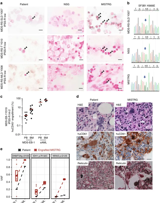
Mutations in the RNA splicing factor SF3B1 are
pathogno-monic for MDS with ring sideroblasts (RS). To date, no model
exists that allows studying the development of RS. Sf3b1 mutant
mice do not develop RS
31and despite successful protocols for
erythroid differentiation in vitro, development of RS has not been
described.
We engrafted three low-risk MDS samples with RS with single
and multi lineage dysplasia(RS-SLD, RS-MLD) and
with
thrombocytosis (RS-T)) to evaluate dysplasia in human MDS
xenografts. All three patient samples successfully engrafted in
MISTRG mice (Fig.
2a) with development of erythropoiesis
(Fig.
3a). Iron stain of patient bone marrow and MISTRG
xenografts, but not of NSG xenografts, revealed RS (Fig.
4a).
Sanger sequencing of the patient’s BM DNA as well as DNA from
engrafted NSG and MISTRG recipient mice confirmed presence
of the SF3B1 K666E mutation and engraftment of the mutant
MDS clone.
To determine whether MISTRG MDS xenografts could
replicate megakaryocytic dysplasia, we engrafted a sample with
marked MK dysplasia. MISTRG mice efficiently engrafted with
sample Y019 (MDS-EB-1 with normal karyotype, Fig.
4c, left)
displayed numerous dysplastic megakaryocytes and reticulin
fibrosis, faithfully replicating the patient’s MDS dysplastic
features (Fig.
4d). MISTRG mice engrafted with the same
patient’s secondary acute myeloid leukemia (sAML) sample
obtained at the time of disease progression (Fig.
4c; Y028, sAML,
NK) did not show these features. Targeted exome sequencing of
the MDS xenografts confirmed derivation from the patient’s
DNMT3a-mutant MDS clone (Fig.
4e). Interestingly, an isocitrate
dehydrogenase 1 (IDH1) mutation was identified in several of the
MISTRG mice (VAF 18–32%) engrafted with the patient’s initial
Fig. 2 Enhanced engraftment of lower- and higher-risk myelodysplastic syndrome (MDS) in MISTRG mice. a–c Analysis of huCD45 engraftment was
performed as detailed in Fig.1a at >12 weeks post transplantation.a Analysis of MDS-5q-, -SLD-, -MLD-, and -MLD-RS-engrafted NSG and MISTRG mice.
b Analysis of MDS/MPN and MDS-EB-1-engrafted NSG and MISTRG mice. c Analysis of MDS-EB-2-engrafted NSG and MISTRG mice. MISTRG afford
significantly higher engraftment than NSG in lower- and higher-grade MDS. d–f Relative distribution of myeloid CD33+(red), B-lymphoid CD19+(blue),
and T-lymphoid CD3+(gray) cells as % of human CD45+cells in NSG vs. MISTRG mice. Stacked bar graphs represent means ± S.E.M. Mann–Whitney
test; n.s. not significant, *p < 0.05, **p < 0.01, ***p < 0.001, ****p < 0.0001. g Split-donor huCD45+BM engraftment in NSG (black) versus MISTRG (red)
mice plotted against CD34+cell number injected/mouse. Individual mice are represented by symbols. Linear regression, Pearson's correlations and
p values of % engraftment to CD34+cell number in NSG (r = 0.39, p < 0.0001) vs. MISTRG (r = 0.42, p < 0.0001) are displayed. h Percentage of
transplanted mice with huCD45+bonemarrow (BM) engraftment levels >0.01% < 1%, 1–10%, and >10% for split-donor grafts in NSG (59/111, 44/111, and
8/111, respectively) and MISTRG (20/154, 51/154, and 83/154, respectively) mice (Fisher’s exact test, ****p < 0.0001 for NSG vs. MISTRG). For detailed
patient sample information see Supplementary Table 1. SLD single lineage dysplasia; MLD multi lineage dysplasia; RS Ringsideroblasts; MPN myeloproliferative neoplasm
MDS diagnosis sample (MDS-EB-1, Fig.
4e). This IDH1 mutation
was not reported in the patient at the time of MDS diagnosis, but
present at the time of disease progression to sAML (sAML, VAF
24%, Fig.
4e). Re-sequencing detected the IDH1 R132C mutation
in the MDS diagnosis sample at a VAF of ~1% (Fig.
4e, middle
panel, Supplementary Tables 1 and 2). Interestingly, in the sAML
engrafted MISTRG mice, a new NRAS G12S mutation defined the
dominant clone, again detectable in the patient’s sAML at a VAF
0.001 0.01 0.1 1 10 100 ** ** *** 0.001 0.01 0.1 1 10 100 0.001 0.01 0.1 1 10 100 n.d. *** *** **** * NSG MISTRG NSG MISTRG NSG MISTRG NSG MISTRG NSG MISTRG NSG MISTRG NSG MISTRG NSG MISTRG NSG MISTRG NSG MISTRG NSG MISTRG NSG MISTRG NSG MISTRG NSG MISTRG NSG MISTRG NSG MISTRG NSG MISTRG NSG MISTRG NSG MISTRG NSG MISTRG NSG MISTRG NSG MISTRG NSG MISTRG NSG MISTRG NSG MISTRG NSG MISTRG NSG MISTRG NSG MISTRG NSG MISTRG NSG MISTRGY004 Y006 Y009 Y0 11 Y007 Y008 Y010 Y013 Y014 Y015 Y017 Y018 Y022 Y023 Y025
Low Int-1 Int-2 Int-1 Low Low Low n/a Int-1 n/a High High Int-2 Int-2 Int-2
RS-SLD RS-MLD RS-T 5q– MLD RS-MLD MDS/MPN MDS-EB-1 MDS-EB-2 n.d. *** *** **** **** 0.001 0.01 0.1 1 10 ** ** *** 0.001 0.01 0.1 1 10 n.s. ** ** ** * ** *** 0.001 0.01 0.1 1 10 n.s. ** ** * *** ** * MDS/MPN; MDS-EB-1 MDS-SLD/MLD/RS/5q– MDS-EB-2 huCD71 APC huCD235a PE 0 103 104 105 0 103 104 105 0 103 104 105 0 103 104 105 0 103 104 105 0 103 104 105 CD71hi CD235neg CD71hi CD235pos CD71dim/neg CD235pos huCD235a PE huCD71 APC NSG MISTRG BM aspirate NSG Patient Cytospin MISTRG huCD45 H&E H&E NSG MISTRG
NSG MISTRG NSG MISTRG NSG MISTRG
** ** * 0.001 0.01 0.1 1 10 100
d
e
a
b
c
f
ErythroidhuCD71 and huCD235 % in WBM
Megakaryocytic huCD41 + % in WBM hCD45 – mCD45 – mTer119 – 4.04 1.88 93.9 0.18 9.60 1.56 76.2 12.7 hCD45 – mCD45- mTer119 –
% lineage of human cells
huCD71hi huCD235a– huCD71hi huCD235a+ huCD71dim/– huCD235a+ huCD45 huCD235 huCD61 huCD61 huCD235
Fig. 3 Erythroid and megakaryocytic lineage representation in myelodysplastic syndrome (MDS) MISTRG xenografts. a Analysis of human erythroid
lineage output in NSG versus MISTRG mice engrafted with lower- and higher-risk MDS (as in Fig.2) via determination of CD71+/−huCD235−/+
expression in hu/muCD45−muTer119−bone marrow (BM).b Analysis of human megakaryocytic lineage output (MK and platelets) in NSG versus
MISTRG mice (engrafted as in Fig.2) via determination of huCD41+in hu/muCD45−BM.c NSG and MISTRG xenografted with MDS-EB-2 (Y025) BM
with inverted myeloid/erythroid ratio.d Patient BM aspirate (top) and sorted human erythroblasts from engrafted NSG and MISTRG BM (bottom) (for
overall engraftment see Fig.2c, Y025).e Representative BM histology from representative NSG and MISTRG recipients engrafted >1% stained with
hematoxylin and eosin (H&E), huCD45, huCD235, and huCD61.f Representativefluorescence-activated cell sorting (FACS) plots of erythroid lineage
differentiation based on huCD71 and huCD235 expression in huCD45−muCD45−mTer119−cells (huCD71hihuCD235a−(pro-erythroblasts (EB)),
MISTRG NSG Patient T Q MDS-RS-SLD Y007 NSG MISTRG
a
b
SF3B1 K666E MDS-RS-T Y010 IPSS-R low MDS-RS-MLD Y008 IPSS-R low MDS-RS-SLD Y007 IPSS-R lowc
PB BM PB BM MDS-EB-1 sAML 0.01 0.1 1 10 100 Patient MISTRG MK MK MK MK MK MK huCD61 H&E Reticulin huCD61 H&E Reticulind
e
Patient Engrafted MISTRGDNMT3Ap.I310S IDH1 p.R132C NRAS p.G12S
0.0 0.2 0.4 0.6 0.8 1.0 VAF G I K/E I V T G I K/E I V Q T G I K/E I V Q
MDS-EB-1Y019 IPSS-R Int-2
huCD45 + engraftment (%) MDS-EB-1 sAML MDS-EB-1 sAML MDS-EB-1 sAML
Fig. 4 MISTRG replicate myelodysplasia and clonal evolution upon disease progression. a NSG and MISTRG were engrafted with low and int-1 riskSf3B1
mutant myelodysplastic syndrome (MDS) with ring sideroblasts (see Figs.2,3) and patient and NSG and MISTRG xenografts were stained with Prussian
blue iron stain (scale bars 10µm, original magnification 60×). b SF3B1 mutation was verified in the patient’s and representative NSG and MISTRG
xenografts by Sanger sequencing.c–e MISTRG engrafted with consecutive MDS-EB-1 and secondary acute myeloid leukemia (sAML) samples from the
same patient (Y019 and Y028, respectively).c Overall (huCD45+) engraftment in peripheral blood (PB) and bone marrow (BM). Individual mice are
represented by symbols, with means ± S.E.M.d Histology from MDS-EB-1 (Y019) diagnostic BM and representative engrafted MISTRG BM. Hematoxylin
and eosin (H&E) and huCD61 stains reveal human megakaryocytic dysplasia and reticulin stain reveals bone marrowfibrosis (high-power magnification
scale bars 20µm). e Targeted exome sequencing results from MISTRG xenografted with same patient’s primary MDS-EB-1 diagnosis samples and sAML at
the time of disease progression. For each mutation, variant allele frequencies (VAFs) are shown for the patient (black) and representative MISTRG (red) mice with engraftment levels >1%. Mean VAF values between MDS-EB-1 and sAML are connected by lines
<5% in addition to the dominant DNMT3a and IDH1 mutations.
RAS mutations have been described as a potential mechanism of
resistance to mutant IDH inhibitor treatment
32and identification
of these mutant clones in a pre-clinical MDS PDX may thus guide
the use of pre-emptive combination regimens.
In addition to erythroid and megakaryocytic dysplasia, we
noted a functional difference of myeloid cells in normal BM,
MDS, and AML engrafted MISTRG mice (Supplementary
Figure 4j). While healthy BM xenografts engraft BM, liver, and
spleen and give rise to resident tissue macrophages in all three
tissues (Supplementary Figure 5a), in age-matched
patient-derived MDS xenografts these populations are mostly absent
from the spleen and the liver, consistent with a functional defect
of the myeloid lineages in MDS (Supplementary Figure 5b). This
is in stark contrast to myeloid leukemia, where immature blasts
infiltrate spleen and liver (Supplementary Figure 5c).
In summary, we here present the
first MDS PDX model that
replicates myelodysplasia and that affords the study of MDS
erythropoietic and megakaryopoietic defects. In addition, we
show that MISTRG MDS PDXs may predict clonal evolution
upon disease progression.
The MISTRG humanized niche allows propagation of MDS
HSCs via serial transplantation. HSCs are critically dependent
on the stem cell niche. MDS HSCs are dysfunctional and their
in vitro and in vivo propagation has been elusive to date. We
hypothesized that cytokine humanization of the HSC niche would
afford homing and engraftment of primary patient-derived MDS
long-term HSCs in MISTRG mice capable of serial repopulation.
Human thrombopoietin is essential for stem cell function
33,34.
IL3, GM-CSF, or M-CSF are not directly implicated in stem cell
maintenance, but via their role in myeloid cell development, such
as BM macrophages, they may indirectly supply additional niche
signals
35,36. We assessed human versus murine cytokine
expres-sion in MDS (Supplementary Figure 6a) and murine MISTRG
and NSG BM-derived mesenchymal stromal cell (MSC) cultures
(Supplementary Figure 6b). Human and MISTRG MSCs but not
NSG MSCs express human THPO, GM-CSF, and M-CSF instead
of their murine counterparts (Supplementary Figure 6c) at
phy-siologic levels similar to human MSCs (Supplementary
Fig-ure 6d). IL3, as expected, is not expressed in MSCs
37.
We next determined whether MISTRG mice engraft human
HSC via phenotypic
38,39and functional assays. CD34
+cells
localize along the trabecular bone in MISTRG bone marrow
(Fig.
5a and Supplementary Figure 6e). In addition to the overall
increased engraftment (Fig.
2
and Supplementary Figure 2),
MISTRG support phenotypic MDS HSCs as evident by
flow
cytometric analysis (Fig.
5b, c). The clonality of these MDS grafts
was verified by targeted exome sequencing in representative mice
(Supplementary Figure 6f, g and Supplementary Table 2).
Phenotypic identification of HSCs is insufficient to prove stem
cell engraftment. Long-term engraftment (≥16 weeks) and
functional assessment in the form of secondary engraftment are
critical. Previous studies have shown successful secondary
transplantation of AML
40,41and more recently of chronic and
juvenile myelomonocytic leukemia
in NSG and NSG-SGM3
mice
42but no study has shown successful serial transplantation of
MDS.
We therefore tested secondary transplantation of a higher and
lower-risk MDS samples according to our standard protocol
(Fig.
5d). Primary NSG and MISTRG recipient mice were
maintained for
≥16 weeks. At the time of analysis, BM was
enriched for human cells via bead depletion of murine CD45
+and Ter119
+cells (Supplementary Figure 6h, i) and transplanted
intrahepatically into equal numbers of irradiated newborn mice
of the respective strains. Secondary recipient mice were analyzed
≥12 weeks (unless otherwise noted) post 2° transplantation. For
all samples tested, primary MISTRG recipient mice showed
significantly higher overall engraftment levels (Figs.
2,
5e, i and
Supplementary Figure 7a). These superior engraftment outcomes
are also reflected in the significantly higher phenotypic stem cell
frequency in 1° and 2° MISTRG compared to 1° and 2° NSG
grafts (Fig.
5f and Supplementary Figure 7c, d, g) accompanied by
myeloid predominant multi-lineage output (Fig.
5g, j and
Supplementary Figure 7b, h, i) as well as erythro- and
mega-karyopoiesis (Supplementary Figure 4g–i). While engraftment
levels were significantly lower for NSG mice, MDS clonality of
primary and secondary grafts in MISTRG and NSG recipients
was confirmed by targeted exome sequencing or cytogenetic
analysis as indicated (Fig.
5h and Supplementary Figure 7e, j, k).
The similar VAFs between MISTRG and NSG mice suggest that B
cells, which predominate in NSG mice in MDS xenografts, are
derived from the MDS clone.
Overall, these data show that MISTRG not only provide
superior engraftment in primary recipients but also in serial
transplantation, with propagation of clonal MDS stem cells that
give rise to tri-lineage hematopoiesis with myeloid predominance
and representation of hallmarks of dysplasia. This may at last
fill
the unmet need for MDS PDXs for the study of MDS disease
mechanism and the development and testing of novel therapies.
MISTRG MDS PDXs are ideally suited for pre-clinical
mod-eling of targeted therapeutics. Targeted therapeutics provide
novel opportunities for the treatment of MDS, but to date have
failed to cure the disease. Recently, inhibitors of mutant IDH1/2
have entered clinical trials, and early data suggest that they result
in blast differentiation and hematopoietic remissions, but fail to
abrogate the mutant clone in the majority of patients
32,43,44.
While transgenic murine models can provide proof of principle
data, patient-derived xenografts are critical to evaluate efficacy
against complex clonal hematopoietic malignancies such as MDS
and are likely to hasten development of valuable combination
therapies.
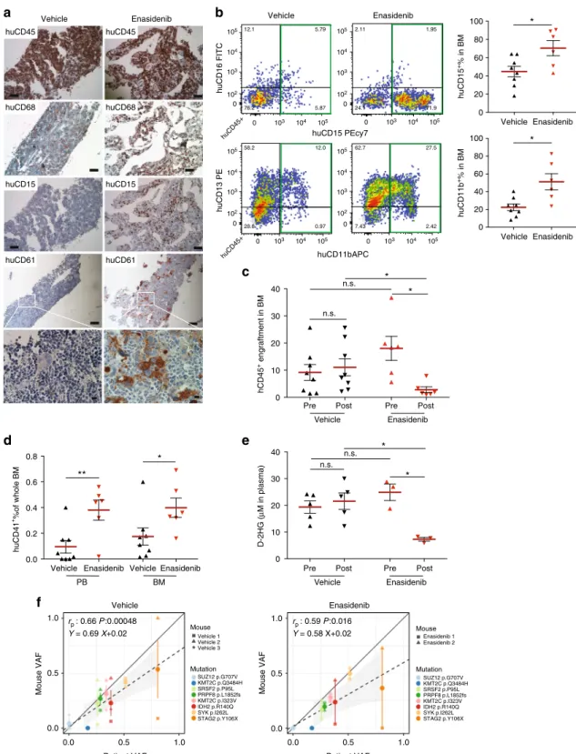
We transplanted MISTRG mice with IDH2
R140Q-mutant
MDS-EB-2 CD34
+cells (Y021, Supplementary Table 1) and
treated engrafted mice with either vehicle or enasidenib via oral
gavage for 30 days. Mice were assigned to enasidenib or vehicle
16 weeks post transplantation based on equal engraftment levels
as determined by BM aspiration (pre). Activity of enasidenib was
verified in vitro via measurement of 2-hydroxy-glutarate (2-HG)
levels in IDH2-wild-type (WT) and -mutant (MUT) expressing
human erythroid leukemia (HEL) cell lines (Supplementary
Figure 8a) and primary AML (Supplementary Figure 8b, Y029,
Y031). Enasidenib efficiently suppressed 2-HG production and
inhibited proliferation of IDH2
R140Q- and IDH2
R172K-mutant
but not IDH2-wild-type AML cell lines and IDH2
R140Q-mutant
primary AML compared to vehicle and WT AML
(Supplemen-tary Figure 8c). Enasidenib treatment resulted in differentiation of
IDH2 mutant myeloid blasts (Supplementary Figure 8d).
Treatment with enasidenib, but not vehicle, resulted in myeloid
differentiation in the IDH2
R140QMDS-EB-2-engrafted MISTRG
mice (Fig.
6a, hCD68 and hCD15 and Fig.
6b). Overall
engraftment levels were significantly reduced in
enasidenib-treated animals when compared to pre-treatment and
vehicle-treated mice (Fig.
6c). Of note, enasidenib-treated mice also
exhibited increased numbers of CD41
+platelets in PB and
clustering megakaryocytes in BM but no difference in the
erythroid lineage compared to vehicle-treated mice. (Fig.
6a
(huCD61), Fig.
6d and Supplementary Figure 8e, f, g). Plasma
2-HG levels in vivo, elevated pre-treatment and in vehicle-treated
MISTRG mice, were significantly suppressed after administration
of enasidenib (Fig.
6e). Variant allele frequencies of mutations
identified in the patient were represented in all MISTRG mice
and not significantly altered by enasidenib treatment (Fig.
6f,
Supplementary Table 2).
MISTRG PDX represent the
first MDS pre-clinical model that
allows to test not only for cytotoxic but also for differentiating
effects of targeted therapeutics and capturing multi-faceted
responses relevant to clinical success.
Patient NSG MISTRG
HSC HSC HSC
MDS-EB-2 Y023 IPSS-R Int-2
huCD90 APC huCD90 APC
NSG MISTRG 0.0 0.5 1.0 1.5 2.0 * NSG MISTRG
MDS-EB-1 Y014 IPSS-R Int-1
mCD45– muTer119– Primary engraftment Secondary engraftment BM MNC NSG 1×100cGy NSG 1×100cGy MISTRG 2×150cGy MISTRG 2×150cGy BM NSG MISTRG NSG MISTRG PB BM NSG to NSG MISTRG to MISTRG NSG to NSG MISTRG to MISTRG PB * * **** **** 1 0.01 0.1 10 100 NSG MISTRG NSG to NSG NSG to NSG MISTRG to MISTRG MISTRG to MISTRG 0.0 0.1 0.2 0.3 0.4 0.6 0.8 * * NSG MISTRG 0 20 40 60 80 100 hCD33+ **** **** ** ** hCD19+ hCD3+ 0.0 0.5 1.0 0.0 0.5 1.0 Mouse VAF Mouse M1 M2 M3 M4 N1 N2 Mutation EGFR p.Q820E KMT2C p.D341V NOTCH2NL p.F203L SRSF2 p.P95R SMC3 splicing TET1 p.L166F ASXL2 p.C1225S BRCA2 p.I1364L SMO p.G388R SYK p.I262L Y = 0.64X+0.16 rp : 0.71 P : 2.7e−07 0.0 0.5 1.0 0.0 0.5 1.0 Mouse VAF Mouse M4.1 M4.2 N2.1 N2.2 Mutation EGFR p.Q820E KMT2C p.D341V NOTCH2NL p.F203L SRSF2 p.P95R SMC3 splicing TET1 p.L166F ASXL2 p.C1225S BRCA2 p.I1364L SMO p.G388R SYK p.I262L NSG MISTRG 0 20 40 60 80 100 hCD33+ huCD19+ huCD3+ 0.1 1 10 100 NSG MISTRG *** *** *** *** *** **** n.s.
huCD45RA PE cy7 huCD45RA PE cy7
Gated on huCD45+ Lin– huCD34+ huCD38–
huCD45RA PE cy7 MDS/MPN Y013 huCD45 + engraftment (%) % HSC of huCD45 + cells in BM Multilineage of hCD45 + cells (%) 1° recipients (22 wks) 2° recipients (15 wks)
1° recipients 2° recipients 1° recipients 2° recipients MDS patient huCD34+ +OKT3 Ab huCD45 huCD34 huCD34 huCD45 % HSC of hCD45 + cells
Multilineage distribution of hCD45+ cells (%)
huCD45+ engraftment in BM (%) 2° recipients (6 wks) 2° recipients (6 wks) 1° recipients (21 wks) 1° recipients (21 wks) MISTRG to MISTRG MISTRG to MISTRG NSG to NSG NSG to NSG MDS-RS-SLDY007 IPSS-R low n.s. n.s. Secondary Primary Patient VAF Patient VAF Y = 0.61X+0.17 rp : 0.64 P : 2.9e−08
a
d
b
c
e
f
g
h
i
j
0 0 103 103 102 104 104 105 0 103 104 105 0 103 104 105 105 huCD90 APC 0 103 104 105 0 103 102 104 10520.7 1.97 50.2 27.1 0 7.62 10.5 81.9 4.10 2.95 9.84 83.1Discussion
MDS is a disease of the hematopoietic stem cell and studies of
MDS have been hampered by the inability to expand HSCs in
general and MDS stem cells in particular. There is an unmet need
for an in vivo pre-clinical model to accelerate development of
novel treatments for a disease where allogeneic stem cell
trans-plantation currently represents the only cure. Mouse models only
partially recapitulate the genetic and epigenetic complexity of
patients’ MDS. Prior xenotransplantation studies have allowed
identification of the MDS HSC
38,39, but have been hampered by
preferential engraftment of the remnant normal hematopoiesis
10,
transient engraftment
9, and low efficiency with low engraftment
levels of only a subset of samples
38,39,45. Cytokine humanization
via transgenic expression in the NSG-SGM3 mice, while
advan-tageous in AML
12and other myeloid malignancies
42, impairs
stem cell function
11,14and provides limited advantages over NSG
mice for MDS engraftment
15,45. Co-injection of human MSC may
provide transient support to MDS HSC
16,45and generation of a
human niche via growth of human MSC-derived ossicles may
afford improved engraftment of HSCs
17,46and difficult-to-engraft
leukemias
46, but applicability in pre-clinical models at a large
scale is likely limited due to technical complexity.
MISTRG mice were engineered to express key
non-crossreactive human cytokines from the endogenous murine
loci in place of their murine counterparts, thereby providing
temporally and spatially physiologic expression of human
cyto-kines. In addition, lack of murine cytokines reasonably provides
additional benefit to human hematopoiesis by rendering the stem
and progenitor niches in the BM less hospitable to murine HSPC.
This is likely to be particularly critical to adult HSCs that have
markedly lower proliferative and self-renewal capacity than their
fetal liver and umbilical cord blood counterparts (reviewed in
ref.
47). In addition, MDS stem cells frequently fail to give rise to
colony-forming units in vitro, a manifestation of their defective
proliferative and differentiation capacity. Research material from
human BMs is limited and worse in aging marrows that are
characterized by progressively lower cellularity.
Here, we present for the
first time a highly efficient and versatile
xenotransplantation model for MDS. We show that MISTRG mice
can be engrafted with as few as ~1.5 × 10
5MDS BM-derived
HSPCs. Higher engraftment levels clearly improve MISTRG utility
as a model. Over 80% of MISTRG mice engraft when a threshold
of 1% BM huCD45
+cells is set. More remarkably, over 50% of
MISTRG mice, compared to fewer than 10% of NSG mice, engraft
above a threshold of 10% BM huCD45
+cells. In addition,
MISTRG mice persistently show improved myeloid representation
and differentiation, as evident by both
flow cytometry and
histo-logic evaluation.
Study of adult erythropoiesis and megakaryopoiesis in in vivo
models has been elusive to date. MDS is characterized by
cyto-penias in the peripheral blood, left-shifted myeloid maturation,
and erythroid and megakaryocytic dysplasia
3. Very little is known
about the causes of the phenotypic heterogeneity in MDS and
genotype–phenotype studies would greatly advance our
mechanistic understanding of this complex entity. To date,
immunodeficient mouse models have supported erythro- and
megakaryopoiesis solely from fetal liver- and cord blood-derived
HSPCs
48,49, further enhanced by mutation of the murine ckit
receptor conferring impaired function to murine stem cells
50–52and likely murine erythropoietic progenitors
53. None of these
models have supported erythro- and megakaryopoiesis from
adult HSPCs. We propose that while cytokine humanization
directly enhances overall engraftment and myeloid maturation,
lack of the corresponding murine cytokines impairs murine
hematopoiesis, thereby synergistically promoting human HSPC
competitiveness in the mouse niche. As a result, we here show for
the
first-time development of both erythropoiesis and
mega-karyopoiesis from healthy adult and MDS BM in a murine host.
This may be further aided by introduction of the murine c-kit
mutation
into
MISTRG
51,54.
Intriguingly,
MDS-engrafted
MISTRG mice replicate the patient’s erythroid and
mega-karyocytic dysplasia, with development of ring sideroblasts and
dysplastic megakaryocytes with reticulin
fibrosis, making
MISTRG MDS PDXs uniquely suited to assess MDS-associated
abnormalities in all three myeloid lineages.
We have previously reported that MISTRG life span is limited
in fetal liver engraftment due to destruction of murine RBC and
platelets by human macrophages
18. Interestingly, MDS-engrafted
MISTRG lack significant development of cytopenias and their life
span is similar to that of engrafted NSG mice. One possible
explanation is the lower engraftment compared to fetal liver
HSPC, yet mice engrafted with normal adult CD34
+cells with
similar engraftment levels to MDS-engrafted mice show evidence
of hemophagocytosis by human macrophages
23. In contrast to
normal CD34
+engrafted MISTRG, MDS PDX lack
human-derived tissue macrophages in spleen and liver, confirming a
functional defect of MDS-derived mature myeloid cells that likely
also lack in vivo hemophagocytic activity. Interestingly, MDS
Fig. 5 MISTRG support phenotypic and functional clonal myelodysplastic syndrome (MDS) stem cells with long-term multi-lineage engraftment potentialin serial transplantation.a Representative immunohistochemistry (IHC) for huCD45 and huCD34 distribution in NSG (ofn = 5) and MISTRG (of n = 12)
bone marrow (BM) engrafted with MDS-EB-1 (Y014; scale bars for low-powerfield: 100 µm, original magnification 10×; high-power field: 10 µm,
original magnification 60×). b, c MISTRG engraft phenotypic MDS stem cells. b Representative fluorescence-activated cell sorting (FACS) plots and
c quantification of hematopoietic stem cell (HSC) representation (lin−CD34+CD38−CD45RA−CD90+of huCD45+) of corresponding patient (high-risk
MDS-EB2, Y023), and NSG and MISTRG xenografts.d–j MISTRG engraft functional MDS stem cells. d Secondary xenotransplantation experimental setup.
e–h Primary and secondary transplantation of MPN/MDS sample with 3% blasts (Y013) comparing e overall huCD45+engraftment in peripheral blood
(PB) and BM,f phenotypic HSC % in BM, and g relative distribution of myeloid CD33+(red), B-lymphoid CD19+(blue), and T-lymphoid CD3+(gray) cells
as % of human CD45+cells in NSG vs. MISTRG mice. In scatter plots individual mice are represented by symbols with means ± S.E.M.; symbols for
corresponding 1° and 2° recipient mice are color coded; statistics represent Mann–Whitney test; n.s. not significant, *p < 0.05, **p < 0.01, ***p < 0.001,
****p < 0.0001. Stacked bar graphs represent means ± S.E.M. Mann–Whitney test; n.s. not significant, *p < 0.05, **p < 0.01, ***p < 0.001, ****p < 0.0001.
h Clonality was determined in representative primary and secondary MISTRG recipients with engraftment levels >1% via targeted exome sequencing.
Variant allele frequencies (VAFs) in primary and secondary recipients were plotted against the corresponding patient’s. Individual mice are represented
by symbol shape and mutations are color coded. Linear regression, Pearson's correlations, andp values between patient and xenograft VAF are displayed.
i, j Primary and secondary transplantation of a low-risk MDS-RS-SLD sample (Y007) comparing i overall engraftment in PB and BM and j multi-lineage representation in BM of primary and secondary NSG and MISTRG recipients. Individual mice are represented by symbols with means ± S.E.M.; symbols for
corresponding 1° and 2° recipient mice are color coded; statistics represent Mann–Whitney test; n.s. not significant, *p < 0.05, **p < 0.01, ***p < 0.001,
c
f
d
e
a
Vehicle Enasidenibb
Vehicle Enasidenib* Enasidenib Vehicle Enasidenib Vehicle 0 20 40 60 80 100 * 0 20 40 60 80 100 0.0 0.5 1.0 0.0 0.5 1.0 Mouse VAF Mouse Vehicle 1 Vehicle 2 Vehicle 3 Mutation SUZ12 p.G707V KMT2C p.Q3484H SRSF2 p.P95L PRPF8 p.L1852fs KMT2C p.I323V IDH2 p.R140Q SYK p.I262L STAG2 p.Y106X Vehicle huCD16 FITC huCD15 PEcy7 huCD11bAPC Y = 0.58 X+0.02 rp : 0.59 P :0.016 0.0 0.5 1.0 0.0 0.5 1.0 Mouse VAF Mouse Enasidenib 1 Enasidenib 2 Mutation SUZ12 p.G707V KMT2C p.Q3484H SRSF2 p.P95L PRPF8 p.L1852fs KMT2C p.I323V IDH2 p.R140Q SYK p.I262L STAG2 p.Y106X Enasidenib huCD45 huCD68 huCD61 huCD61 huCD15 huCD68 huCD45 huCD13 PE n.s. n.s. * * 0 10 20 30 40 Enasidenib PB Enasidenib Vehicle Vehicle BM * ** Enasidenib Vehicle
Pre Post Pre Post
Enasidenib Vehicle
Pre Post Pre Post
n.s. n.s. * * 0.0 0.2 0.4 0.6 0.8 0 10 20 30 40 huCD15 +% in BM huCD11b +% in BM huCD45+ huCD45+ 0 102 103 104 105 2.11 1.95 24.1 71.9 12.1 5.79 76.2 5.87 62.7 27.5 7.43 2.42 58.2 12.0 28.8 0.97 0 102 103 104 105 0 0 102 103 103 104 105 0 103 104 105 0 103 104 105 0 103 104 105 104 105 0 102 103 104 105 huCD15 hCD45 + engraftment in BM D-2HG ( µ M in plasma) huCD41 +%of whole BM
Patient VAF Patient VAF
Y = 0.69 X+0.02 rp : 0.66 P :0.00048
Fig. 6 MISTRG replicate granulocytic and megakaryocytic differentiation in response to inhibition of mutant isocitrate dehydrogenase 2 (IDH2) in vivo. a In
vivo treatment of mutant IDH2 R140Q in MDS-EB-2 (Y021)-engrafted MISTRG mice with the IDH2MUTinhibitor enasidenib. Representative histologic
images of vehicle-treated (n = 8, left) and enasidenib-treated (n = 6, right) mice engrafted with MDS-EB-2 (Y021). Immunohistochemistry (IHC) stains for
huCD45, huCD68, huCD15, and huCD61 (scale bars 100µm, original magnification 10×; high-power field 10 µm, original magnification 60×). b
Representativefluorescence-activated cell sorting (FACS) plots showing myeloid maturation in response to enasidenib and quantitation of huCD15+and
huCD11b+expression in vehicle- versus enasidenib-treated MISTRG mice.c Comparison of human engraftment in bone marrow (BM) from vehicle-treated
(n = 8) and enasidenib-treated (n = 6) MISTRG mice. d Quantitation of huCD41+expression in peripheral blood (PB) and BM from vehicle-treated (n = 8)
and enasidenib-treated (n = 6) MISTRG mice. e Quantitation of D-2-HG in plasma of pre- and post-administration of vehicle or enasidenib. Individual mice
are represented by symbols with mean ± S.E.M.; statistics represent Mann–Whitney test; n.s. not significant, *p < 0.05, **p < 0.01 for aggregate NSG vs.
MISTRG.f Representation of variant allele frequencies (VAFs) of driver mutations in vehicle-treated (left) or enasidenib-treated (right) MISTRG (y-axis)
plotted against the patient’s VAFs (x-axis). Individual mice represented by symbol shape, mutations color coded. Linear regressors, Pearson's correlations,
blasts, unlike in AML, do not infiltrate non-hematopoietic tissues,
functionally distinguishing MDS also from AML in MISTRG
PDX.
Cytokine humanization does not alter the lack of mature
human red blood cells (RBCs) and the low human platelet
per-centage in the peripheral blood as also shown previously
18,23.
Administration of human erythropoietin has shown no benefit in
this regard
54as the defect lies in RBC and platelet destruction by
the murine innate immune system. Thus, modulation of the
murine innate immune system will be necessary to promote
mature human cell persistence in peripheral blood, transiently
achieved
by
administration
of
liposome-encapsulated
clodronate
49,55.
MDS is a clonal hematopoietic stem cell disorder and reliable
engraftment of the malignant HSC is essential for high-quality
pre-clinical studies of disease biology and response to
ther-apeutics. While phenotypic evidence of HSC can suggest their
presence, functional assays are critical. Serial transplantation
represents the gold-standard functional hematopoietic stem cell
assay. We here show successful serial transplantation of MDS into
MISTRG secondary recipients with faithful representation of the
clonal composition and lineage representation of the parental
patients’ BMs in primary and secondary recipients. Importantly,
MISTRG mice allow the expansion of xenografts from one
pri-mary into several secondary recipients, essential for pre-clinical
modeling and therapeutic testing.
We interrogated the utility of the MISTRG MDS PDX model in
the testing of targeted therapeutics, specifically inhibition of
mutant IDH2. Early clinical studies have shown that enasidenib,
an oral inhibitor of mutant IDH2, results in differentiation of
mutant myeloblasts without abrogation of the mutant clone in the
majority of patients. We here show for the
first time, in an in vivo
MDS PDX model, differentiation towards dysplastic
mega-karyocytes and myeloid maturation with preservation of the
clonal composition of the graft. The MISTRG MDS PDX model is
ideally suited for the systematic study of targeted therapeutics
alone and in combination with other agents. Concurrent targeted
exome sequencing may allow predicting ideal combination
regi-mens for individual patients.
In summary, we here present a highly efficient, faithful MDS
PDX model, ideally suited for the study of MDS biology, the
development of novel treatment approaches, and adaptation of
patient-specific regimens in the era of precision medicine.
Methods
Human progenitor cell isolation. Peripheral blood, BM, and umbilical cord blood
were obtained with donor’s written consent. All human studies were approved by
the Yale University Human Investigation Committee and by the West Haven Veterans Affairs Human Investigation Committee.
Human BM, cord blood, and peripheral blood samples wereficolled (GE
Healthcare, Munich, Germany) and mononuclear cells cryopreserved within 24 h after collection in fetal bovine serum/10% dimethyl sulfoxide. Samples were CD34 enriched with the CD34-Microbead-Kit or T cell depleted via negative selection with the CD3-Microbead-Kit (Miltenyi-Biotech, Bergisch-Gladbach, Germany). CD34-enriched or CD3-depleted HSPCs were incubated with a murine anti-human CD3 antibody (clone Okt3, BioXCell, NH, USA) at 5 µg/100 µl for 10 min at room temperature prior to injection into mice.
Generation and analysis of MISTRG PDXs. All animal experiments were approved by the Institutional Animal Care and Use Committee of Yale University.
Mouse breeding and xenografting: MISh/hTRG mice with homozygous knockin
replacement of the endogenous mouse Csf1, Il3, Csf2, Tpo, and Sirpa with their human counterparts were bred to MITRG mice to generate human cytokine
homozygous and hSIRPA heterozygous mice18,19. MISTRG mice have been
deposited at Jackson laboratory. Mice will be available via MTA and requests should be sent to [email protected]. NSG mice were obtained from Jackson
laboratory. MISh/mTRG (labeled MISTRG throughout the study) and NSG mice
were maintained on continuous treatment with enrofloxacin in the drinking water
(0.27 mg/mL, Baytril, Bayer Healthcare). Newborn MISTRG or NSG mice (1 to 3 days of age) were sublethally irradiated (X-ray irradiation with X-RAD 320
irradiator; MISTRG 2 × 150 cGy 4 h apart, NSG 1 × 100 cGy). Equal numbers of split-donor MDS BM CD34-selected or CD3-depleted (as indicated) were injected intrahepatically in a volume of 20 µL into with a 22-gauge Hamilton needle (Hamilton, Reno, NV). Mice were analyzed at least 12 weeks post transplantation and only sooner if moribund. For secondary transplantation, human cells were isolated from primary recipient BMs and depleted of murine cells via negative
selection of murine CD45+and Ter119+cells by magnetic labeling with
biotin-anti-muCD45 (clone 30-F11, Biolegend, San Diego, CA) and muTer119 (clone TER-119, Biolegend) and BD IMag Streptavidin Particles (BD Biosciences, San Jose, CA).
Flow cytometric analysis. Engraftment of human CD45+cells and their stem cell,
progenitor, and mature myeloid, lymphoid, and erythroid or megakaryocytic
subsets were determined byflow cytometry using antibody panels detailed in
Supplementary Table 3. In brief, cells were isolated from engrafted mice, blocked with human/murine Fc block, and stained with indicated combinations of anti-bodies. Data were acquired with FACSDiva on a LSR Fortessa (BD Biosciences) equipped with 5 lasers and analyzed with FlowJo V10 software.
Histologic analysis. Tissues werefixed in Bouin’s Fixative solution (RICCA
Che-mical Company, TX, USA) and embedded in paraffin. Femurs were decalcified with Formic Acid Bone Decalcifier (Decal Chemical, NY, USA). Paraffin blocks
were sectioned at 4μm and stained with hematoxylin and eosin (H&E) or
antigen-specific antibodies routinely used in the Yale Clinical Pathology and Yale Pathology Tissue Services (Supplementary Table 4). Images were acquired using Nikon Eclipse 80i microscope. Bone marrow aspirate smears were stained with Prussian Blue Iron stain per standard protocol.
All animal experimentations were performed in compliance with Yale Institutional Animal Care and Use Committee protocols.
Targeted exome capture and sequencing and analysis. DNA was digested using the QIAamp DNeasy blood and tissue DNA extraction kits (Qiagen), according to the manufacturer’s recommendations. Purity and concentration of the extracted DNA was measured using NanoDrop 1000 spectrophotometer (Thermo Scientific) and Quant-iT PicoGreen dsDNA Assay Kit (Life Technologies, Carlsbad, CA) for all samples.
A library of coding exons and intron–exon boundaries of 142 genes (see Supplemental Table 6) known to carry mutations in myeloid malignancies and cancers was prepared using the HaloPlex target enrichment kits and HaloPlex HS Target Enrichment System (Agilent Technologies, Santa Clara, CA) according to the manufacturer’s instructions. In brief, approximately 200 ng of DNA was fragmented using restriction enzymes proprietary to the kit. For mixed human/ mouse samples isolated from MISTRG mice total DNA (human/mouse) input was calculated with the endpoint of 200 ng human DNA input based on engraftment
percentage of huCD45+muCD45−cells determined byflow cytometry. Probes
with sequence indexes were hybridized to the targeted DNA fragments. Each probe is designed to hybridize to both ends of a targeted DNA restriction fragment resulting in their circularization. The biotinylated probe-DNA fragment hybrids were retrieved with magnetic streptavidin beads. Small fragments of <150 bp and unligated probes were removed from the mix by AMPure purification (Agencourt Bioscience, Beverly, MA). Circular molecules were ligated and enriched DNA
fragments were amplified with universal primers. Quality of the libraries was
verified using the Tape Station 4200 (Agilent) and input DNA estimated using a library quantification kit (Kapa Biosystems, Wilmington, MA, USA). For samples Y013, Y014, Y016, Y019, Y021, Y022, Y028, Y029, and their engrafted NSG and MISTRG mice, a second-generation enrichment kit was used with Agilent’s improved high-sensitivity technology with addition of molecular barcodes to each probe. Sequencing was performed on Illumina HiSeq 2000 using 74 base pairs paired-end reads, HiSeq 4000 using 100 base pairs paired-end reads, or MiSeq
using 250 base pairs paired-end reads. Reads werefiltered by Illumina CASAVA
1.8.2 software, and trimmed at the 3’ end using FASTX v0.0.13. To remove potential mouse contamination, each read pair was aligned to a concatenated genome of human (GRCh37) and mouse (mm10) reference genome by Burrows-Wheeler Aligner v0.7.5a. Only read pairs that were specifically aligned to human reference genome were extracted for the downstream analysis. Local realignment was performed around putative and known insertion/deletion (INDEL) sites using RealignerTargetCreator (Genome Analysis Toolkit: GATK v3.1.1) and applied base quality recalibration using GATK. MuTect v.1.1.4 and Strelka v.1.0.14 were applied to call somatic single-nucleotide variants and indels, respectively. Whole-exome sequencing data from 10 external normal blood samples were pooled to serve as reference normal cohort for somatic variant calling by MuTect and Strelka. In each
sample, low confidence somatic calls were removed by applying the following
filters: (i) variants with total coverage <50, (ii) with a ratio of mutant allele frequency (MAF) in tumor versus normal <5, and (iii) variant base quality <20. Variants that were considered likely to be germline because they were listed in any of the following datasets, dbSNP, ESP6500, 1000Genome, or Exac01, or had MAF <0.02 in the tumor samples were excluded from further analysis. Recurrent
(N > 5 cases) annotated variants in COSMIC v64 and Clinvar (http://www.ncbi.
nlm.nih.gov/clinvar/) were white-listed. At last, only non-synonymous variants were kept. To extract the allele frequency of the variants, all non-synonymous