FUNCTIONAL CHARACTERIZATION OF LOC115098: A
NOVEL GENE CONSERVED IN EUKARYOTIC GENOMES
A THESIS SUBMITTED TO
THE DEPARTMENT OF MOLECULAR BIOLOGY AND GENETICS AND THE INSTITUTE OF ENGINEERING AND SCIENCE OF
BILKENT UNIVERSITY
IN PARTIAL FULFILLMENT OF THE REQUIREMENTS FOR THE DEGREE OF MASTER OF SCIENCE
BY
ESRA KARAKÖSE JANUARY 2007
ii
I certify that I have read this thesis and that in my opinion it is fully adequate, in scope and in quality, as a thesis for the degree of Master of Science.
Assist Prof. Ayşe Elif Erson
I certify that I have read this thesis and that in my opinion it is fully adequate, in scope and in quality, as a thesis for the degree of Master of Science.
Assist Prof. Kamil Can Akçalı
I certify that I have read this thesis and that in my opinion it is fully adequate, in scope and in quality, as a thesis for the degree of Master of Science.
Assist Prof. Uygar Halis Tazebay
Approved for the Institute of Engineering and Science
Director of Institute of Engineering and Science Prof. Mehmet Baray
iii
ABSTRACT
FUNCTIONAL CHARACTERIZATION OF LOC115098: A NOVEL GENE CONSERVED IN EUKARYOTIC GENOMES
Esra Karaköse
M.Sc. in Molecular Biology and Genetics Supervisor: Assist Prof. Uygar H. Tazebay
January 2007, 76 Pages
Loc115098 is a newly identified gene in our laboratory. The gene was firstly discovered in an unrelated study in which the cis-acting regulatory elements of a neighboring gene NIS (Sodium-Iodide Symporter) were investigated. It is composed of 4 exons. According to the computational predictions, it has rather interesting features such as having a composition of mostly charged amino acids, carrying a nuclear localization signal and being well conserved throughout the eukaryotic kingdom. Our aim in this study was to understand the function of this new gene. Firstly we confirmed that it is expressed in several different tissues, later we conducted a subcellular localization study whose results led us to hypothesize that the function of loc115098 could be related to cell cycle regulation. According to our hypothesis we decided to utilize a lower eukaryote carrying a human homologue of loc115098 which we selected to be Aspergillus nidulans. It was a very useful model organism for our study not only because it carries a very similar homologue of loc115098 but also it is an easily handled organism which is widely used for genetic studies. Firstly we compared the expression profile of loc115098 in several cell cycle mutants of A. nidulans as well as a wild type strain, the results indicated that the expression of loc115098 decreases in the restrictive temperature of cell cycle mutant strains. We also conducted subcellular localization studies in A. nidulans and the results indicated that it was mostly cytoplasmic and sometimes perinuclear. Our other aim was to utilize A. nidulans as a model for our knock-out experiments. We designed a knock-out system by the help of a sophisticated method called Double-Joint PCR. Our first attempts supported that the knock-out of loc115098 was likely to cause lethality in the organism and therefore we improved our system to conditionally knock out the gene. Our preliminary results indicated that the knock out of loc115098 causes cell death and we suspect that it could be due to a damage in one of the crucial metabolic events for cell viability such as mitosis. Our attempts to functionally characterize the function of loc115098 are currently going on.
iv
ÖZET
ÖKARYOTİK CANLILARDA KORUNMUŞ OLAN LOC115098 GENİNİN İŞLEVSEL TANIMLANDIRILMASI
Esra Karaköse
Moleküler Biyoloji ve Genetik Yüksek Lisans Derecesi Tez Yöneticisi: Yard. Doç. Dr. Uygar H. Tazebay
Ocak 2007, 76 Sayfa
Loc115098 ilk kez tarafımızdan bulunmuş bir gendir. İlk defa bu genin komşusu olan NIS (Sodyum-Iyot Simporter) geninin anlatımından sorumlu cis kontrol bölgelerinin aydınlatılması amacıyla yapılan bir çalışma sırasında bulunmuştur. Bu gen 4 eksondan meydana gelmektedir. Bioınformatik analizlerden faydalanarak yaptığımız araştırmalar bu genin oldukça ilginç özelliklere sahip olduğunu gösterdi, örneğin proteinin yapısı genel olarak pozitif yüklü amino asitlerden oluşuyor, çekirdek sinyali taşıyor ve ökaryotik organizmalarda oldukça korunmuş bir şekilde bulunuyor. Bizim bu çalışmadaki amacımız bu genin işlevini anlamaktı. Bu nedenle öncelikle farklı dokularda bu genin ifadesi olduğunu teyid ettik. Sonra bu proteinin hücre içersindeki konumunu anlamak için çalışmalar yaptık. Sonuçlar, bu proteinin hücre döngüsü kontrolüyle ilgili bir işleve sahip olabileceği hipotezini kurmamızı sağladı. Bunun üzerine çalışmalarımızı sürdürmek amacıyla loc115098 geninin homoloğunu taşıyan daha düşük bir ökaryot olan Aspergillus nidulans’ı seçtik. Bu organizma bizim çalışmalarımız açısından oldukça kullanışlıydı çünkü hem loc115098 geninin çok benzer bir homoloğunu taşıyordu hem de bir model organizma olarak genetik araştırmalarda sıklıkla kullanılıyordu. Öncelikle loc115098 geninin anlatımını çeşitli hücre döngüsü mutasyonlarına sahip olan A. nidulans ırklarında ve doğal (wild type) ırkta karşılaştırmalı olarak inceledik. Bu çalışmanın sonucu bize mutant olan ırkların yaşamalarına müsaade etmeyen sıcaklık derecelerinde loc115098 ifadesinin azaldığını gösterdi. Aynı zamanda bu organizma ile de LOC115098 proteinin hücre içindeki konumlanışını araştırdık ve sonuç bize bu proteinin çoğunlukla sitoplazmada bazen de çekirdeğin etrafını saracak şekilde bulunduğunu gösterdi. Ayrıca Double-Joint PCR adı verilen oldukça sofistike bir metoddan faydalanarak knock-out çalışmalar yaptık ve ilk elde ettiğimiz sonuçlar bize bu genin, hücrenin yaşamsal olaylarından sorumlu bir işleve sahip olabileceğini gösterdi çünkü bu genin knock-out edildiği durumlarda canlının yaşadığı örneklere hiç rastlamadık. Loc115098 geninin işlevini aydınlatmaya dair çalışmalarımız halen devam ediyor.
v
All truths are easy to understand once they are discovered;
the point is to discover them.
vi
ACKNOWLEDGEMENTS
Fistly, I would like to thank my supervisor Dr Uygar Tazebay for his continious help, support and friendship during my days in Ankara and in Athens.
I would also like to express my gratitude to my second supervisor Dr Georges Diallinas who was not only a great supervisor but also a nice friend whenever necessary.
I would like to thank the members of Molecular Biology and Genetics Department at Bilkent University for the nice working environment especially to Dr Hani Alotaibi for his valuable technical guidance.
I would like to thank Dr Diallinas’ group in the University of Athens for the lovely working atmosphere in the lab and their immense technical support. Furthermore I would like to thank Dr Ioannis Serafimidis for both his contributions to the project and his company.
I would also like to express my deep gratitude to my family for their existence and moral support during my entire life.
Lastly I would like to express that I am very grateful to all my friends who never left me alone during this long journey and always stood by me.
This project was supported by a 8-month scholarship for research from the Greek Ministery of Education.
vii
TABLE OF CONTENTS
ABSTRACT ...III ÖZET ... IV DEDICATION PAGE ...V ACKNOWLEDGEMENTS ... VI TABLE OF CONTENTS... VII LIST OF TABLES... XI LIST OF FIGURES ... XII ABBREVIATIONS……….XIV1. INTRODUCTION ...1
1.1. General knowledge about Hypothetical Proteins...1
1.2. How to Study the Hypothetical Proteins ...2
1.2.1. The bioinformatics tools ...2
1.2.2. Biochemical and genomic approaches to study hypothetical proteins...3
1.3 General Knowledge about ‘Loc115098’ ...4
1.3.1. Identification of loc115098 as an expressed gene...4
1.4. Model Organisms to Study Gene Function ...4
1.4.1. General Knowledge about Model Organisms...5
1.4.2. Features and Characteristics of Aspergillus nidulans...6
1.4.2.1. Life Cycle of Aspergillus nidulans...8
1.4.2.2. Cell cycle Mutant Strains of Aspergillus nidulans ...9
1.4.3. Why we selected Aspergillus nidulans...12
viii
2. MATERIALS AND METHODS ...14
2.1. Bioinformatics ...14
2.1.1. Data bases ...14
2.1.2. DNA analysis ...14
2.2. Plasmids and Cloning...15
2.2.1. Site directed mutagenesis...18
2.3. Primers ...19
2.4. Cell Culture...20
2.4.1. Cell lines ...20
2.4.2. Growth Media ...20
2.4.3. Transfection ...20
2.4.4. cDNA synthesis and RT-PCR...20
2.5. Aspergillus nidulans handling ...21
2.5.1. Strains ...21 2.5.2. Growth Media ...21 2.5.2.1. Complete Medium (CM)...21 2.5.2.2. Minimal Medium (MM)...21 2.5.2.3. Fructose Medium (FM)...21 2.5.2.4. Sucrose Medium (SM)...21 2.5.3. Growth tests ...22 2.5.4. Transformation...23 2.5.5. DNA Extraction...24 2.5.6. RNA Extraction...24 2.5.7. Southern Blotting ...25 2.5.8. Northern Blotting ...26 2.6. Immunofluorescence (IP) ...27 2.7. Microscopy ...28 3. RESULTS...29
ix
3.1.1. The identification of loc115098 ...29
3.1.2. Features...30
3.1.3. Post Translational Modifications ...30
3.1.4. Biological Significance of Loc115098 ...31
3.2. Analysis of Loc115098 Expression in Different Tissues...31
3.2.1. Breast cell lines ...32
3.2.2. HCC cell lines ...32
3.2.3. Human brain tissues and Mouse tissues ...33
3.2.4. Zebrafish tissues...34
3.3. Subcellular Localization of Loc115098 in MCF-7 Cell Line ...34
3.3.1. Construction of pFLAGLOC and pLOCFLAG plasmids ...34
3.3.2. Ectopically expressed loc115098 is likely to involve in mitosis ...35
3.3.3. Localization studies with anti-Loc115098...36
3.4. Aspergillus nidulans Expression Data ...37
3.4.1. Human and Aspergillus protein alignment confirms the conservation...37
3.4.2. Different strains of Aspergillus nidulans indicate different expression patterns ...38
3.4.2.1. The growth difference between the wild type and cell cycle mutant strains...38
3.4.2.2. The Northern Blot Assays confirm the different expression pattern at different temperatures...39
3.5. Subcellular Localization of AN0879.3 in Aspergillus nidulans mycelia...41
3.5.1. The subcellular localization of AN0879.3 in several strains of A. nidulans ...41
3.6. Overexpression Studies in Aspergillus nidulans ...42
3.6.1. The outcomes of overexpression of AN0879.3 ...43
3.7. Knock-out Studies of AN0879.3 ...45
3.7.1. Strategy for the knock-out construct by Double-Joint PCR ...45
x
4. DISCUSSION...51
4.1. The subcellular localization studies ...52
4.2. The overexpression study ...55
4.3. The knock-out studies ...55
4.4. Future perspectives ...56
xi
LIST OF TABLES
2.1. The restriction enzymes utilized in this study………..16
2.2. The oligonucleotides utilized in this study………..19
2.3. The PCR polymerase enzymes utilized in this study………...21
xii
LIST OF FIGURES
1.1. The SEM pictures of Aspergillus nidulans………7
1.2. Different cultures of A. nidulans grown on petri dishes………7
1.3. Microscopic appearance of A. nidulans……….7
1.4. Life cycle of A. nidulans………9
1.5. The cell cycle mutants of A. nidulans………..10
1.6. NimA5 and BimE7 mitotic mutants of A. nidulans……….11
2.1. Vector maps of the plasmids used in localization studies………17
2.2. Vector map of pGEM–T Easy plasmid………....18
3.1. Vista plot of conservation levels of Loc115098 in mouse and rat………...29
3.2. The expression levels of Loc115098 in mammary gland cell lines……….32
3.3. The expression levels of Loc11098 in hepatic cell lines……….32
3.4. The expression levels of Loc115098 in human brain tissues and rat tissues…...33
3.5. The expression levels of Loc115098 in zebrafish tissues………34
3.6. The cloning strategy of pLOCFLAG represented by gel pictures……...………35
3.7. Subcellular localization of N and C termini FLAG epitope tagged Loc115098 in MCF7 cell lines ………..36
3.8. Subcellular localization of Loc115098 detected by anti-locus115098 antibody………...37
3.9. The alignment of human Loc115098 and its A. nidulans homologue AN0879.3………38
3.10. The growth tests of all A. nidulans strains we utilized for this study…………39 3.11. The expression pattern of Loc115098 in wild type and cell cycle mutants of A.
xiii
nidulans………...40
3.12. The subcellular localization of C-terminus GFP tagged AN0879.3…………..42 3.13. The cloning strategy of pALCLOC represented by gel pictures ………..43 3.14. The results of overexpression studies indicated by Southern blots and growth tests……….44 3.15. Schematic explanation of Double-Joint PCR (DJ-PCR)………...46 3.16. The results of DJ-PCR experiments………...47 3.17. The preliminary results obtained from conditional knock-out studies………..50 4.1. Schematic explanation of mitosis in higher and lower eukaryotes………..54 4.2. In-situ hybridization for Loc115098 in Zebrafish embryos……….54
xiv
ABBREVIATIONS
3D: 3 dimensions
AlcA: alcohol dehydrogenase A
A. nidulans: Aspergillus nidulans
BLAST: Basic Local Alignment Search Tool Bim: blocked in mitosis
BSA: Bovine serum albumin Bp: base pair
cAMP: cyclic adenosine monophosphate cDNA: complementary DNA
cGMP: cyclic guanosine monophophate CM: complete medium
C-terminus: carboxy-terminus
DAPI: 4,6-diamidino-2-phenylindole, dihydrochloride DEPC: diethylpyrocarbonate
dCTP: deoxycytidine triphosphate dH2O: distilled H2O
dNTP: deoxynucleotide triphosphate DNA: deoxyribonucleic acid
EDTA: ethylenediaminetetraacetic acid EtBr: ethidium bromide
xv FBS: Fetal Bovine Serum
G1: Gap 1 G2: Gap 2
GAPDH: Glyceraldehyde 3-phosphate dehydrogenase GFP: Green Fluorescent Protein
HCl: hydrochloric acid Loc: locus Kb: kilobase µg: microgram µl: microliter Mb: Megabase ml: milliliter MM: minimal medium mRNA: messenger RNA N-terminus: amino-terminus NaCl: sodium chloride NaOH: sodium hydroxide
NCBI: National Center for Biotechnological Information Nim: Never in mitosis
NIS: Sodium Iodide Symporter Nud: nuclear distribution OD: optic density
ORF: open reading frame pBKS: pBlueScript vector PBS: phosphate buffer saline
xvi PCR: Polymerase Chain Reaction
PEG: polyethylene glycol pGEM-T: pGEM-Teasy vector RNA: ribonucleic acid
rRNA: ribosomal RNA rpm: revolutions per minute
RT-PCR: Revers transcriptase-PCR S: Synthesis
SDS: sodium dodecyl sulfate
SEM: Scanning Electron Microscope SM: sucrose medium
Sud: suppressor of bim TAE: tris-acetate-EDTA
TRIS: 2-amino-2-hydroxymethyl-1,3-propanediol Wt: wild type
1
1. INTRODUCTION
1.1. General knowledge about Hypothetical Proteins
The completion of Human Genome Project (Human Genome Sequencing Consortium, 2004), revealed that the expected number of genes in the human genome is neither around 100000 as it was expected in some text books nor around 30000 (Lander et al., 2001) as expected in others. More recent studies have indicated that this number varies between 20000 and 25000. Even though we are able to perform such estimations we are still far from understanding the human genome in its all aspects. Indeed it is well-known that coding sequences, as well as many non-coding regulatory regions tend to keep their sequences conserved during evolution. Although there are certain efforts to understand the function of non-coding regulatory conserved elements at the level of entire genome, the identification, functional annotation and systemic classification of the coding sequences remain incomplete.
Until today the DNA sequences of more than 100 species have been completed and one of the most surprising outcomes of genome research is that roughly 20-40% of genes in newly sequenced genomes do not have statistically significant matches to functionally annotated sequences and they are annotated as ‘hypothetical proteins’ (Eisenstein et al., 2000).
We believe that one of the most important questions of the today’s genome research is to assign annotations to the genes whose biological function is yet to be understood. The most common approaches and several methods will be mentioned in the following sections.
2 1.2. How to Study the Hypothetical Proteins
Since the annotation of hypothetical proteins encoding genes is crucial for the future of genomic studies, the tools which are generated for this scope is a hot topic in the field, too. There are indeed several different approaches to study the hypothetical proteins and it is possible to classify them as ‘the bioinformatics tools’ and ‘the biochemical and genomic tools’.
1.2.1. The bioinformatics tools
There are several different approaches to functionally identify new genes. However the most accurate and effective annotations which have been performed up to date are based on sequence homology. Among them BLAST and PSI-BLAST are the most widely used tools (Altschul et al., 1997). However more advanced tools which have been used by several groups exist and the number of these tools keep growing everyday. Some of these methods will be named here. One of the most reliable tools is ‘The Rosetta Stone Technique’ which is based on protein-domain fusion events (Marcotte et al., 1999) (Enright and Spratt, 1999). There are others which are based on chromosomal proximity information. The rational behind this technique is that when two genes appear as a neighboring gene pair in the genomes of several distantly related organisms, meaning that they form a conserved gene cluster. This suggests the possibility that these genes might be functionally related (Overbeek et al., 1999). Another technique is based on the phylogenetic profile method. Basically this method indicates the presence or the absence of the gene of interest in each organism by an entry of 1 or 0. Thus the functional linkage is established when the two genes have similar phylogenetic profiles, they show a correlated pattern of inheritance across the genomes examined (Marcotte, 2000). Some other methods also combine the ones mentioned above (Zheng et al., 2002) in which they explain the genomic functional annotation using co-evolution profiles of gene clusters. Finally, by using specific amino acid frequency and the periodicity at the proteome level, functionally unknown proteins can be characterized (Fujishima et al., 2003).
3
bioinformatics analyses; BLAST. Moreover, we also focused on the biochemical and genomic analyses of our gene of interest.
1.2.2. Biochemical and genomic approaches to study hypothetical proteins
Although there is no certain method to follow in order to conduct biochemical and genomic studies of the hypothetical proteins, the general forward and reverse genetics methods are applicable. Firstly the confirmation of the expression of these genes is essential. After this confirmation, further analyses can be performed depending on the future scope of the study. The overexpression and disruption of the gene of interest are two main approaches in order to observe how crucial the protein is for the organism. As long as there is no domain similarity with previously annotated proteins, any method aiming to identify the protein-protein interaction such as immunoprecipitation or yeast two hybrid methods would be useful to find the targets of the hypothetical proteins. Moreover some localization studies by using immunofluorescence can be conducted to observe the subcellular localization of the hypothetical proteins. All these experiments can also be performed in time or growth dependent manners meaning that different developmental stages of a given organism could be investigated in terms of gene expression or the localization studies can be performed in a time dependent manner and the difference can be observed. Moreover the interaction of the protein of interest with other proteins may also vary according to the developmental or metabolic activities.
On the other hand the disruption of the gene of interest could also be a prominent approach. In case of any phenotypic changes or lethality occurs this would bring new insights to the study. Moreover, according to the bioinformatics analyses, some mutations can be inserted to check the possible outcomes of the mutation of those sequences.
Other more sophisticated approaches such as crystallizing the protein and exploring the 3D structure are possible. However these methods are generally considered to be more complicated and require higher technology and longer periods of commitment.
4 1.3 General Knowledge about ‘Loc115098’
1.3.1. Identification of loc115098 as an expressed gene
Loc115098 was firstly identified in an unrelated study where cis-acting transcriptional elements of the NIS (Sodium-Iodide Symporter) gene were being analyzed. The focus of that study was to analyze a putative transcriptional control element localized in the 3’ downstream of NIS gene by using a tool called VISTA (Negre et al., 2005). In this study 10 conserved putative regions were identified and we firstly discovered the 5’ regulatory region of Loc115098 as one of these putative regions and then the Loc115098 itself was discovered by our group (Hani Alotaibi et
al, unpublished data). The gene could be found in University of California Santa
Cruz database (Kent et al., 2002) with the accession number of LOC115098.
1.4. Model Organisms to Study Gene Function
Model systems have always been useful in basic research. They have contributed to the creation and development of basic biological knowledge for many years. Especially in the last decades due to the rapidly expanding genetic research area revealing the genomes of different species, model organisms have gained even more importance. The common evolutionary heritage makes it possible to use genetically tractable organisms to model important aspects of human medical disorders such as cancer, birth defects, neurological dysfunctions, reproductive failure, malnutrition and aging in systems amenable to rapid and powerful experimentation (Spradling et al., 2006).
Utilizing model organisms will help us identify common genes, proteins and biological processes. This will not only be useful for unrevealing the complications of human genetic disorders but also establish a better understanding of the roles of the common mechanisms which take part in all fundamental life processes, including development, physiology, growth, behavior and aging. Moreover the studies with model organisms provide information which can not practically be obtained in any other way. Mostly due to practical difficulties to utilize and manipulate higher
5
organisms and sometimes due to the ethical issues, the idea of using a model system has always been very useful.
1.4.1. General Knowledge about Model Organisms
The number of model organisms which are used for genetic, biochemical and molecular studies are growing rapidly. The most commonly used organisms include rat (Rattus norvegicus), mouse (Mus musculus), zebrafish (Danio rerio), the fruit fly (Drosophila melanogaster), the budding yeast Saccharomyces cerevisiae, the amphibian Xenopus tropicalis, the nematode Caenorhabditis elegans, cellular slime mold Dictyostelium discoideum and finally the ascomycete Aspergillus nidulans. They are all useful to study different functions, for instance the C. elegans is mostly utilized for neurobiological and developmental studies whereas the S. cerevisiae is a good model to study the control of cell division. According to the aim of the study and the laboratory facilities, the most easily handled and the most appropriate organism can be selected from a big variety.
Taking fungi into consideration, it is obvious that they have a lot of features which make them useful as experimental genetic organism. Many of the fungi that are commonly used for genetic studies have most or all of the following features:
• A haploid phase for mutagenesis and screening.
• A dikaryon or heterokaryon phase for complementation and epistasis analysis. • The ability to grow on defined media for selection and screening of nutritional and drug-resistance markers.
• The ability to grow at wide temperature ranges which facilitates the isolation of conditional lethal mutants.
• Heterothallic strains for out-crossing.
• Homothallic derivatives to allow haploid screens for mutants that are defective in fundamentally diploid processes (for example, meiosis).
• Small genomes (12–50 Mb) to facilitate mutant recovery and gene cloning. • The ability to carry out tetrad analysis, in which all four meiotic products are held together in an ascus. (Casselton and Zolan, 2002)
6
1.4.2. Features and Characteristics of Aspergillus nidulans
Aspergillus nidulans is an ascomycetes belonging to the subphylum of Euascomycetes (Ascomycota). However the filamentous ascomycota is called pyrenomycetes. The classification can be made as following according to Micheli ex Link, 1809: Kingdom: Fungi Phylum: Ascomycota Order: Eurotiales Family: Trichocomaceae Genus: Aspergillus Species: nidulans
The yeasts which are unicellular ascomycotan fungi are considered to be the best organisms for the study of basic eukaryotic genetics (Hedges, 2002). The multicellular (filamentous) ascomycotan fungi have apparently larger genomes but they are as useful and practical to study basic eukaryotic genetics.
Aspergillus nidulans first appeared in the literature as an alternative model to
Neurospora crassa. It was firstly introduced by Guido Pontecorvo in the 1950s
(PONTECORVO et al., 1953). Like all other ascomycetes, A. nidulans also has a sac formation which is called ascus. The nuclear fusion and meiosis take place in the ascus. This eventually leads to the formation of sexual spores so called ascospores. The ascospores are formed within the ascus by an enveloping membrane system, which packages each nucleus with its adjacent cytoplasm and provides the site for ascospore wall formation. These membranes are derived from the ascus plasma membrane. This larger structure bursts and releases the thick-walled haploid ascospores which are resistant to adverse environments, but when they come across to the right conditions they start germinating and form the new haploid fungus. The hyphea is one filament of A. nidulans carrying several nuclei.
7
Figure1.1. The pictures of A. nidulans which were taken by SEM (Scanning Electron microscope). Both pictures show how A. nidulans look like in its natural environment.
Figure1.2. Different cultures of Aspergillus nidulans. Both pictures show how A. nidulans grows under laboratory conditions. It might have different color mutants as it is indicated in one of the pictures
Figure1.3. In the first picture the conidial heads which are short and columnar are shown. Conidiophores are usually short, brownish and smooth-walled. Conidia are globose and rough-walled. In the second picture the cleistothecium of A.
8
1.4.2.1. Life Cycle of Aspergillus nidulans
The life cycle of the fungal mycelium of A. nidulans consists of sexual and asexual cycles. A. nidulans is a web of branched filaments (hyphae) of connected compartments or cells, each containing several nuclei. This mycelium, or homokaryon, which develops from a single haploid spore, differentiates into many identical asexual spores known as conidia or conidiospores (the asexual cycle). A.
nidulans is homothallic, which means that it is self fertile, but crosses can be initiated
by hyphal fusions between homokaryons with genetically different nuclei. The resulting heterokaryons are not stable, but can be forced to maintain a balanced ratio of the component nuclei by including complementing auxotrophic mutations in the parental nuclei and forcing growth without the corresponding supplements. A.
nidulans can also reproduce sexually. In the fruiting body, which produces the sexual
spores, a pair of nuclei that is destined for meiosis divides in synchrony to form a mass of cells known as the ascogenous hypha. These hyphae are highly branched and each tip cell becomes an ascus (a specialized cell) in which the two haploid nuclei fuse. The diploid nucleus undergoes meiosis followed by a post-meiotic mitosis, which results in the formation of eight haploid ascospores. The fruiting body, called the cleistothecium, can hold tens of thousands of ascospores, which are released into the environment when the cleistothecium bursts open. In addition to an asexual cycle and sexual cycle, a parasexual cycle offers the genetic benefits of meiosis achieved through a mitotic route (PONTECORVO and KAFER, 1958). The parasexual cycle is initiated when haploid nuclei fuse in the vegetative cells of a heterokaryon and continue to divide mitotically. Crossing over might occur between homologues and random chromosome loss restores the haploid chromosome number, which is eight in
A. nidulans. These events can be used to map gene orders and assign new genes to
9
Figure1.4. The life cycle of A. nidulans shown schematically. The sexual cycle is shown in purple, the asexual cycle in orange and the parasexual cycle in green. (Casselton and Zolan, 2002)
1.4.2.2. Cell cycle Mutant Strains of Aspergillus nidulans
The A. nidulans cell cycle mutants have long been a good model to study gene function. The first mitotic mutants were screened by Ron Morris in 1975. He carried out the classical microscopic screen in which the morphology of the germinating conidiospore was used as the identifying phenotype. As the conidiospore germinates, the germ tube elongates to generate the first hyphal compartment, into which the conidiospore’s nucleus moves and divides. The mitosis mutants were those in which the nuclear division (but not cell elongation) was affected at the restrictive temperature. In this screen the idea was to isolate the mutants as temperature-sensitive conditional-lethals because otherwise the cells which cannot go through
10
mitosis would immediately die. The permissive temperature was 32ºC and the restrictive temperature was 42ºC. Basically 3 classes of mutants were isolated in this study: nim (never in mitosis), bim (blocked in mitosis), nud (nuclear distribution). (See Figure 1.5)
Figure1.5. The schematic representation of cell cycle mutants and how they were obtaine. The permissive temperature is 32ºC and the restrictive temperature is 42ºC. (Casselton and Zolan, 2002)
The mutant strains we use in this study are nim, bim and sud mutants. The NimA protein is a kinase that controls the entry into mitosis (Osmani et al., 1987). Thus the
nimA mutants are arrested in late in G2 with duplicated spindle pole bodies, the fungal equivalent of the centrosome (Oakley et al., 1983). Further genetic analyses
11
showed that the interphase arrest of nimA5 mutants was bypassed by bimE7 mutation, with double mutants entering an aberrant mitotic state characterized by abnormalities of the nuclear envelope, which remains intact during mitosis in fungi, and the failure to form bipolar spindle (Osmani et al., 1991). In other words the
bimE7 mutation causes nuclei to accumulate and persist in a pre-anaphase mitotic state with tightly condensed chromatin and a normal-appearing spindle apparatus at restrictive temperature(James et al., 1995). (See Figure 1.6)
Figure1.6. The nimA5 and the bimE7 are shown in the picture. In the nimA5 mutant the cell cycle arrest occurs at late G2 whereas in the bimE7 mutant mid-mitotic arrest occurs. (O'Connell et al., 2003)
The sud mutant has also the background of bimD6 mutation. The protein which is encoded by bimD shares sequence motifs with known DNA binding transcription factors (May et al., 1992a). The bimD6 mutant germlings at restrictive temperature block in anaphase and eventually undergo nuclear fragmentation. In other words the
bimD6 mutation confers a heat-sensitive mitotic defect characterized by a failure of chromosomes to correctly attach to the spindle microtubules, resulting in an increase in chromosome loss. The extragenic suppressors of bimD6 (sud) were isolated and characterized before (Holt and May, 1996b). Moreover it was discovered that in addition to their ability to suppress bimD6, sud genes are themselves required for successful nuclear division in A.nidulans. The germlings harboring sud mutations with the bimD6 mutation undergo normal rounds of interphase and mitosis at 37ºC. After a shift to 25ºC for 8 hours, the approximate length of one cell cycle, nuclear morphology of all sud strains strikingly alters. The nuclei are not seen as delimited organelles but stretch about the hyphal interior as disorganized masses of chromatin. (Holt and May, 1996a).
12 1.4.3. Why we selected Aspergillus nidulans
There were several reasons which led us to choose the Aspergillus nidulans as our experimental model. First of all, the homologues counterpart of loc115098 in the Aspergillus genome had 34.4% identity with the human homologue. (From now on the Aspergillus homologue will be called AN0879.3.) This high conservation rate would make our results more reliable. Furthermore Aspergillus nidulans is a non pathogenic and easily handled model organism for genetic, molecular and biochemical studies. It grows on very cheap and simple growth media.
Saccharomyces cerevisiae could have been another alternative for this study since this organism is equally easy and cheap to handle. Interestingly this organism does not have a homologue of the protein of interest, therefore the idea was dismissed. In addition to these advantages of A. nidulans mentioned above, it was also important that all the methods such as genetic transformation and targeted gene replacement were described in the 80’s and 90’s which obviously made all manipulations easier because the establishment of such studies and protocols is also quite time consuming and hard to deal with. Moreover the A. nidulans has always played an important role in the genetic dissection of the cell cycle. And last, but not the least, the experience and expertise of Dr. Diallina’s laboratory was an extra stimulus for us to conduct our experiments on A. nidulans.
1.4.5. Aim
Our aim in this study was firstly to characterize the loc115098 gene. For this purpose we designed experimental approaches for both the human cell lines and the filamentous fungus A. nidulans. Our initial observations which will be explained in the ‘Results’ pointed that Loc115098 could be involved in mitosis and therefore we concentrated on the cell cycle regulation and the possible participation of Loc115098 into this process. Since loc115098 is a newly identified gene, its features, function and any related data were still elusive. Our aims in this study were focusing on the identification of such data by utilizing as many tools and approaches as possible. Therefore we concentrated on a) localization, b) over expression, c) knock-out studies as well as the bioinformatics analyses of this new gene. The idea
13
behind all these approaches was firstly to understand the characteristics of Loc115098 and to check its expression profile and correlate it with the data coming from the bioinformatics analyses. In addition to this, we sought to identify the subcellular localization of this protein and to observe any phenotypical changes caused by the overexpression and/or the knock-out of the gene.
The subcellular localization studies were mainly focusing on the determination of the localization of Loc115098 protein in the cell. It might be crucial to understand where this protein localizes in the cell because it might give us a clue about the possible Loc115098 interacting proteins. In the overexpression studies; our aim was to observe the possible differences in the phenotype of the cells which were caused by the increase in loc115098 or AN0879.3 expression. The results coming from this experiment would lead us to conduct some microscopic or biochemical analysis to observe and understand these changes in a better way. Lastly, our knock-out strategy was set in order to be able to observe the possible outcomes of the deletion of AN0879.3.
Certainly, all the results obtained from our already designed approaches would bring new aspects for the current project. This study was a part of our long term attempt to functionally characterize loc115098.
14
2. MATERIALS AND METHODS
2.1. Bioinformatics 2.1.1. Data bases
The accession number of human loc115098 in the NCBI database (http://www.ncbi.nlm.nih.gov) is NM138442. The same sequence can also be obtained from Genome Browser database at the University of California Santa Cruz (Kent et al., 2002) with the accession number of LOC115098. The Zebrafish
loc115098 can also be found in NCBI with the accession number of BC066532. The
A.nidulans copy of loc115098 which we call AN0879.3 can be found in the
A.nidulans database prepared by Broad Institute:
(http://www.broad.mit.edu/annotation/genome/aspergillus_nidulans/Home.html). The accession number for Loc115098 protein is AN0879.3; the mRNA data can also be reached and the exons and the introns of the gene can optionally be viewed. In order to find the homologues of loc115098 in different organisms generally the NCBI Blast (Altschul et al., 1990; Altschul et al., 1997) search was utilized.
2.1.2. DNA analysis
Once the sequences of different organisms were obtained, they were not only used for further studies such as amplification and cloning but also for performing some bioinformatical sequence comparisons. For these comparisons mainly the ClustalW (Higgins, 1994) (http://www.ebi.ac.uk/clustalw/#) was used.
When loc115098 was firstly identified the annotation for the human sequence was obtained from the Genome Vista database (Couronne et al., 2003), and the sequences were aligned with the mVista too (Frazer et al., 2004) (Hani Alotaibi unpublished).
15 2.2. Plasmids and Cloning
The plasmids which were used for localization studies in human cell line MCF-7 were p3XFLAG-CMV-10 (Sigma) and p3XFLAG-CMV-14 (Sigma). The plasmids which were used for A. nidulans studies were pBKS (Stratagene) and pGEM-T (Promega). All plasmids had ampicillin resistance.
The standard protocol for cloning was to double digest both the plasmids and the inserts which have already been amplified with the appropriate enzyme and reaction conditions. This was followed by a ligation step and later the ligated plasmid was transformed into E.coli DH5α strain. After that the plasmids were rescued from the bacteria. Then several further digestions and PCR analyses were carried out in order to obtain the plasmid carrying the gene of interest.
16
Table2.1. The restriction enzymes which were used for the cloning and plasmid confirmation purposes.
RESTRICTION ENZYMES
Name Temperature Buffers
BamHI 37ºC 10mM Tris-HCl (pH 8.0 at 37°C), 5mM MgCl2, 100mM KCl, 0.02% Triton X-100, 1mM 2-mercapthoethanol, 0.1mg/ml BSA BglI SalI 37ºC 50mM Tris-HCl (pH 7.5 at 37°C), 10mM MgCl2, 100mM NaCl, 0.1mg/ml BSA EcoRI 37ºC 50mM Tris-HCl (pH 7.5 at 37°C), 10mM MgCl2, 100mM NaCl, 0.02% Triton X-100, 0.1mg/ml BSA HindIII 37ºC 10mM Tris-HCl (pH 8.5 at 37°C), 10mM MgCl2, 100mM KCl, 0.1mg/ml BSA KpnI 37ºC 10mM Tris-HCl (pH 7.5 at 37°C), 10mM MgCl2, 0.02% Triton X-100, 0.1mg/ml BSA NcoI 37ºC DpnI 37ºC SmaI 30ºC XbaI 37ºC 33mM Tris-acetate (pH 7.9 at 37°C), 10mM magnesium acetate, 66mM potassium acetate, 0.1mg/ml BSA
The p3XFLAG-CMV-10 was basically used for N-terminus tagging of Loc115098. It was firstly double digested by XbaI and HindIII enzymes. The Loc115098 was amplified by the primers called HALNG-F1 and HALNG-R1 and these two fragments were ligated in appropriate conditions and the transformation was carried out. The p3XFLAG-CMV-14 was digested by BglI and HindIII enzymes and the
17
PCR product of Loc115098 was obtained by using the primers called HALNG-F1 and LOC-HN-Ctrev. Later the same ligation and transformation protocol was carried out. Several enzyme digestions were done to confirm the correct plasmid.
Figure2.1. The vector maps of plasmids which were used to construct the N and C-termini FLAG epitope tagged Loc115098. The p3XFLAG-CMV-14 was double digested with BglI and HindIII and Loc115098 was inserted between these 2 sites with the same linkers. The p3XFLAG-CMV-10 was double digested with XbaI and
HindIII and Loc115098 was cloned in between these two sites with the same linkers. The pALC plasmid was kindly provided by Dr V. Sophianopoulou’s group. The plasmid was already carrying the alcA promoter cloned in the polyT site of the plasmid. Thus we inserted the AN0879.3 carrying NcoI linker on both sides into
NcoI digested pALC. After the ligation and E. coli transformation steps the orientation of AN0879.3 in the plasmid was further confirmed by PCR. Later the
argB selection gene was cloned into the same plasmid with EcoRI sites. After all the
cloning steps this plasmid was named pALCLOC. This plasmid was transformed into each strain by the method which is explained in detail in the methods section. Following the transformation the transformants were grown on CM in Petri dishes and they were checked whether or not they were carrying our gene of interest together with alcA promoter in the correct orientation by PCR. The correct
18
orientation meant that the AN0879.3 was intact with the promoter, confirming that it is under the control of this promoter. Later the copy number of these transformants was detected by Southern Blot analyses and the ones carrying multi copy were further studied.
Figure2.2. The vector map of pGEM-T Easy plasmid which was used for overexpression studies.
2.2.1. Site directed mutagenesis
After cloning A. nidulans homologue of loc115098 into the pBSK, the stop codon of AN0879.3 was destroyed by site directed mutagenesis by using the primers called ‘McaP no-stop NcoI FOR’ and ‘McaP no-stop NcoI REV’. For this reaction Phusion High Fidelity DNA polymerase (Finnzymes) was used according to its manual. After the PCR amplification this product was digested by DpnI enzyme for 1,5 hours. After that it was transformed into E.coli DH5α.
19 2.3. Primers
Table2.2. The primers which have been used concerning the current study.
Name Sequence 5' →3' Expected Size
Locus Expression Primers
EKYG F2 GAGGAGGAGGACTCCAAGCTC EKYG R2 CTGGGCTTCCTCAAAGGC F1-ZFYGxp ACCAAAGGCTTTTGGAGG R1-ZFYGxp GTTGGCTTCTTCAAACGC GAPDH-F GGCTGAGAACGGGAAGCTTGTCAT GAPDH-R CAGCCTTCTCCATGGTGGTGAAGA
Locus Cloning Primers
HALNG-F1 GAATTCAAGCTTATGCCCAAGAAGTTCCAG HALNG-R1 GGATCCTCTAGAGCTCACTTGGGGGCATTG HALNG-F1 GAATTCAAGCTTATGCCCAAGAAGTTCCAG LOC-HN-Ctrev GATATCAGATCTGCCTTGGGGGCATTGAAGGGC McaP Forward NcoI CGCCATGGGCGGCAAAAAGGGC
McaP Reverse NcoI CGCCATGGTGCTGAGCGCGAGCTATG 5’ FOR KpnI GCGGTACC-TATCGAATGGCCAATTCC 5’ REV EcoRI GCGAATTC-AGGTGAATGAGGCTTGTC 3’ FOR EcoRI CGGAATTC-TGAGGATTGGAAGGACTG 3’ REV XbaI CGTCTAGA-GTGGATGATAGTGCACGA Pyr4 FOR EcoRI CGGAATTC-AAGCTTAGACTTCAACAA Pyr4 REV EcoRI CGGAATTC-GGATCTTCATCATTCGTC 5 ∆McaP Fo GATTCGCACAGACAGCCATAG 5 ∆McaP Re GTCTTCACCTCGTAGGGGATG 3 ∆McaP Fo CATCCCCTACGAGGTCAAGAC 3 ∆McaP Re CGCTGCATCATAGCTGAGAC CPFP4Fo CACAAGAAACGGACAAGAAGGTCTGAAAAGCTTAGACTTCAACAACCCCACCATC CPFP4Re TAAAACACAATCATTATGTAAACAAATCATGGATCTTCATCATTCGTCGCTTTCGGG CPF5Fo GCAAGTCTTAGCGAGTG CPF5Re GATGGTGGGGTTGTTGAAGTCTAAGCTTTTTCAGACCTTCTTGTCCGTTTCTTGTG CPF3Fo CCCGAAAGCGACGAATGATGAAGATCCATGATTTGTTTACATAATGATTGTGTTTTA CPF3Re GCCAGTCTGTGTGAAGT PNPCRFo CGTGGAACACCTTACTC PNPCRRe GAAACTCTCGCTGGACA
McaP upstream BamHI GCGGATCCTGATCGGGGAGGGTAGCA McaP downstream EcoRI CCGGAATTCTGCTGAGCGCGAGCTATG
McaP no-stop NcoI FOR GCGAGACTCACGAGGTCCATGGTTTGTTTACATAATGATTG McaP no-stop NcoI REV CAATCATTATGTAAACAAACCATGGACCTCGTGAGTCTCGC GFP NcoI linker FOR GGCCATGGTAATGGTGAGCAAGGGCGA
GFP NcoI linker REV CGCCATGGTTACTTGTACAGCTCGTCC
Conditional Knock-out Primers
ALCMcaP For+ GACAAGCCTCATTCACCTGAATTCGCGAGCTACCATCCAATAACCCCCAGCTG
ALCMcaP Rev+ GTTGAAGTCTAAGCTTGAATTCCGGGATGAACCGACATACAGAATAAGTGAC
site directed mutagenesis 745bp 1371bp 3699bp 1985bp 3160bp 6200bp
pLOCGFP Construction Primers Double-Joint PCR Primers Knock-out Cloning Primers pALCLOC Conctruction Primers
1859bp 3013bp 1337bp 2034bp 2149bp 1859bp 4087bp 667bp 318bp 314bp 143bp 672bp
20 2.4. Cell Culture
2.4.1. Cell lines
In order to check the expression profile of Loc115098 several hepatic and mammary gland cell lines were used. The hepatic cell lines were HepG2, Hep40, SNU182, SNU387, SNU398, SNU449, Huh7, and Focus. The mammary gland cell lines are which were used were BT474, BT20, T47D, MCF7, 453, 468, HME, HCC, 231. The cDNAs of these cell lines were all kindly provided by Prof Mehmet Ozturk’s group.
2.4.2. Growth Media
MCF-7 cell line were maintained in high glucose Dulbecco’s modified Eagle’s medium (Gibco) [supplemented with 10% fetal bovine serum (FBS), 1% penicillin/streptomycin (P/S) and 1% L-glutamine (Biochrom)], abbreviated as DMEM, at 37°C in a 5% CO2 incubator.
2.4.3. Transfection
The transfection of MCF-7 cells was carried out with plasmids by using FuGENE reagent (Roche). Cells which were 90%-100% confluence in 100 mm dishes were harvested by trypsinization and washed once with DMEM (not containing the FBS, penicillin/streptomycin and L-glutamine) then resuspended in 10 ml DMEM, and diluted 1:7 in culture medium (3 ml cells in 20 ml medium), diluted cells were then seeded in 24-well plates for transfection; 400 µl cells per well.
2.4.4. cDNA synthesis and RT-PCR
The cDNAs of the cell lines were kindly provided by Dr Mehmet Ozturk’s group whereas the cDNAs of Danio rerio (Zebrafish) were kindly provided by Dr Ozlen Konu’s group and the cDNAs from human brain tissues were again kindly provided by Dr Cengiz Yakicier’s group. In order to perform cDNA synthesis 2µg of RNA was used and the reaction was done according to the Revert-Aid First Strand cDNA Synthesis Kit (Fermentas) manual. The rat tissue RNAs were isolated by Dr Hani Alotaibi and the cDNA synthesis was performed by using 2µg of these RNAs. After
21
the PCR amplifications the PCR products were resolved on 2% agarose gels stained with ethidium bromide and visualized using the Gel Doc-2000 supported with the Multi-Analyst Ver.1.1 image analysis software (Bio-Rad).
Table2.3. The PCR Polymerase enzymes which have been used for this study.
Company Name Reagents
Stratagene Platinum Pfx DNA Polymerase 1xbuffer,0.3mM dNTPs each,1mM MgSO4,0,3µM each primer,1Uenzyme
Finnzymes Phusion 1xbuffer,200µM dNTPs each,0,3µM each primer,1Uenzyme FinnzymesDyNAzyme EXT DNA Polymerase 1xbuffer,360µM dNTPs each,0.3µM each primer,3Uenzyme HyTest HyTest Taq Polymerase 1xbuffer,0.3mM dNTPs each,1mM MgSO4,0,3µM each primer,1Uenzyme
Roche Taq DNA Polymerase 1xbuffer,0.3mM dNTPs each,1mM MgCl2,0,3µM each primer,1Uenzyme PCR ENZYMES
2.5. Aspergillus nidulans handling 2.5.1. Strains
Several different strains were used in this study. There were some wild-type strains (pyrG89 paba panto, pyrG89 argB2 paba panto) as well as some cell cycle mutant strains SO6 (nimA5, wA2, pyrG89, cnxE16, choA1, yA2, chaA1, sC12), DBE4 (bimE7, riboA2, pyrG89) which were kindly provided by Dr. S. Osmani and 8-16(1-3) (riboA1, wA3, bimD6, pyroA4, sudD7), 8-16(3-6) (pyrG89, [bimD6?], pyroA4,
sudD7) which were kindly provided by Dr G May and Dr S Osmani.
2.5.2. Growth Media
The complete medium (CM) and minimal medium (MM) for A. nidulans have been described previously by Tilburn et al., 1983; Cove, 1966.
2.5.2.1. Complete Medium (CM) Vitamin solution 10 ml Salt solution 20 ml Casamino acids 10 ml D-glucose 10 gr Bacto-peptone 2 gr Yeast Extract 4 gr Agar 30gr (3%)
22 Vitamin Solution
20 mM p-amino benzoic acid, 1mM Biotin, 0,1M Nicotinic acid, 50 mM Calcium-D-pantothenate, 0,1M Riboflavin, 50mM Pyridoxine, 50mM Thiamine filled with dH2O to 1 liter
Salt Solution
26g KCl, 26g MgSO47.dH2O, 76g KH2PO4, 50ml trace elements, 2ml chloroform filled with dH2O to 1 liter
2.5.2.2. Minimal Medium (MM)
20ml salt solution, 10g D-glucose, pH adjusted to 6.8 and filled with dH2O to 1 liter
2.5.2.3. Fructose Medium (FM)
0,1g fructose, 2ml salt solution, 2g agar, pH adjusted to 6.8 and filled with dH2O to 1 liter
2.5.2.4. Sucrose Medium (SM)
20ml salt solution, 342,3g sucrose, 10g glucose, 2% agar pH adjusted to 6.8 and filled with dH2O to 1 liter
2.5.3. Growth tests
After each transformation and when the phenotypic observations were needed to be made, we performed the growth tests. Basically different strains or different transformants were inoculated on either CM or MM which contained the appropriate oxotrophies and usually grown at different temperatures in parallel to observe the changes.
23 2.5.4. Transformation
Transformations were made with protoplasts of mycelia from the appropriate strains. The transformation was essentially as described by Tilburn et al., 1983 (Tilburn et al., 1983). The routine protocol was adapted from M.Koukaki et al., 2003 (Koukaki et al., 2003). Aspergillus strains were grown on CM for 3-4 days at various temperatures depending on the strain. From these Petri dishes the conidiospores were taken by carefully scraping the dish into 5 ml dH2O containing falcon and they were separated from mycelia by filtration through sterile blutex. Afterwards, 1010-11 conidiospores were inoculated in MM, supplemented with 5mM urea as a nitrogen source, necessary vitamins and either 5mM arginine (argB2 strains) or 10mM uracil and uridine (pyrG89) or both (argB2 and pyrG89) for 3,5-4 (7 hours for nimA5) hours at the appropriate temperature at 130rpm. Upon the initiation of the germination, the conidiospores were collected with centrifugation at 4000rpm for 10 minutes and resuspended in 10 ml of the standard isosmotic buffer (Tilburn et al., 1983). In order to start the protoplastation, Glucanex (Novozymes) was added in the concentration of %90 (m/v) and it was incubated at 30ºC for 1,5 hour to let it act. After the completion of protoplastation conidiospores protoplasts were washed twice in 1M Sorbitol, 10mM Tris-HCl, and 10mM CaCl2 pH 7.4, resuspended in the same buffer at a concentration 108 conidiospores/200µl and distributed into sterile eppendorfs. Each eppendorf was used for a single transformation experiment with 0.5µg of plasmids. After the mixing the appropriate plasmid with the conidia protoplasts, a drop of PEG was added on top and it was chilled on ice for 15 minutes before adding the 500µl PEG. They were incubated at room temperature for 15 minutes and then centrifuged for 10 minutes at 6000rpm. After the centrifugation they were washed with the above mentioned solution, they were again centrifuged at 5000rpm for 4 minutes and later they were plated on SM and allowed to grow for 3-4 days at various temperatures depending on the strain.
24 2.5.5. DNA Extraction
The DNA isolation protocol was previously described by Lockington et al., 1985 (Lockington et al., 1985). The strains were inoculated and grown overnight in 50ml MM containing the appropriate vitamins and urea as a nitrogen source at distinct temperatures depending on the strain at 130rpm. The next day mycelia were collected by filtering through blutex and they were firstly kept at -80ºC for 10 minutes. Later they were homogenized by mortar and pestle in liquid nitrogen. An aliquot of 100-200mg was kept in an eppendorf and 800µl breaking solution (Tris-HCl pH8 0,2M; SDS 1%; EDTA pH8, 1mM) was added to further break the cells. Then it was vortexed vigorously and kept on ice for 20 minutes. Later phenol chloroform extraction was carried out.
2.5.6. RNA Extraction
The strains were inoculated and grown overnight in MM containing the appropriate vitamins and urea as a nitrogen source at distinct temperatures depending on the strain at 130rpm. All the materials that were used for RNA extraction were RNase free. The next day mycelia were collected by filtering through blutex and they were firstly kept at -80ºC for 10 minutes. Later they were homogenized by mortar and pestle in liquid nitrogen. An aliquot of 100mg was kept in an eppendorf and 1ml of trizole was added together with 10mg of glass beads. It was vortexed vigorously and kept at room temperature for 10 minutes. Then it was centrifuged at 4ºC for 15 minutes at 12000rpm. The supernatant was taken into a clean eppendorf and 200µl of chloroform was added, it was shaken by hand and incubated at room temperature for 2 minutes. The centrifugation was repeated with the same conditions. After the centrifugation the upper phase was transformed into a new clean eppendorf. 500µl isopropanol was added on top of it and it was chilled on ice for 10 minutes after shaking the eppendorf well. After the centrifugation at 4ºC for 10 minutes at 12000rpm the supernatant was discarded and the pellet was washed by 1 ml of 75% Ethanol. After the washing step the pellet was dried and resuspended in DEPC treated dH2O.
25 2.5.7. Southern Blotting
The nucleic acid manipulations were done according to the previously prepared protocols by Sambrook et al. After the DNA extraction step, the concentration was measured by Optic Density (OD) measurements. 2,5-3µg of DNA was digested by EcoRI for 2 hours at 37ºC and then more enzyme was added and the digestion was carried out overnight. The next day 1% agarose gel was prepared to run the digested fragments in 1xTAE (242g Tris base, 57.1ml Glacial Acetic Acid, 18.6 g EDTA in 1 lt dH2O). The electrophoresis was done at 58V for 4 hours. After the completion of the electrophoresis the gel picture was taken. The gel was baked at 100% Ultraviolet light (254nm) for 5 minutes and the irrelevant parts of the gel was cut and discarded. Then the baked gel was incubated in denaturing buffer (0,5M NaOH and 1,5M NaCl) for half an hour. Then the gel was incubated in the neutralizing solution (1M Tris-HCl pH 8 and 1,5M NaCl) for half an hour. After these steps the overnight capillary transfer was carried out in alkaline conditions using the sandwich method. It is called sandwich because the gel and the membrane are kept together between two layers of Whatman paper. It is necessary for the capillary movement. 20XSSC (0,3 M Na3C6H5O7.2H2O and 3M NaCl pH 7) was used for the transfer. In the sandwich method, firstly the long Whatman No:2 papers were placed in such a manner that both ends of the paper could be deep in the transfer buffer. The gel was placed on top of the Whatman papers. The edges of the gel were wrapped by plastic wrap so as to let the capillary movement continue through the gel and the membrane. On top of the gel the nylon membrane was placed and it was made sure that there were no air bubbles left in between the gel and the membrane. 2 pieces of Whatman paper were cut in the same size as the membrane and wetted in 2X SSC then they were placed on top of the membrane. The air bubbles were smoothened out by a glass rod. A stack of paper towels (5-8 cm) were placed on the Whatman papers and lastly a 500-g weight was placed on top of all. The tissue papers were changed as they became wet. The transfer was carried out for at least 20 hours. Following the transfer the membrane was cross linked by UV light. Then it was ready for hybridization.
26
EDTA; %20 SDS and dH2O) was used. After placing the membrane in a cylinder with 100ml of Church buffer the pre-hybridization step took place where the membrane was incubated at 65ºC for 2 hours. Meanwhile the [32P]dCTP labeled probe was prepared using a random hexanucleotide-primer kit following the supplier’s instructions (Promega). After the prehybridization, the probe was added in the cylinder and the hybridization was initiated by leaving the membrane for incubation at 65ºC overnight. The next day probe containing church buffer was removed and the washing steps were carried out. The washings were as following: 30 minutes at 65ºC in 2xSSC, 0.1% SDS twice and in the cylinder. After that 30 minutes at 65ºC in 1xSSC, 0.1% SDS in a plastic container. Once the washing was completed the membrane was placed in a lightproof cassette with a film on and it was kept at -80ºC for a period ranging from 3hours to 7 days (depending on how strong the probe is). The development was made manually by using developer and fixer solutions.
2.5.8. Northern Blotting
The nucleic acid manipulations were done according to the previously described protocols by Sambrook et al. Once the RNA extraction was completed the OD measurement was carried out. All the materials were either RNase free or made RNase free manually by adding DEPC treated water or NaOH washings when necessary. Firstly the running buffer (10mM orthophosphate) was prepared which was used both for electrophoresis and the preparation of the agarose gel. 10µg RNA was mixed with the following mixture; for x volume of RNA, 3x volume of 11,5µl DMSO; 2,25µl (0,1M pH7) orthophosphate; 3,3µl glyoxal. This mixture was incubated at 50ºC for one hour and then on ice 5 minutes. After the 1% agarose gel was ready, and the incubation on ice was over, the samples were ready to be loaded after adding the dye (10mM orthophosphate, 2%Bromophenol blue, 50% glycerol) to a final ratio of 1/8 of the total volume. After the loading of the samples, the electrophoresis was carried out. The voltage was adjusted according to the distance between the two electrodes; it was carefully calculated to 3V per centimeter. After letting the samples run for 5 minutes, the peristaltic pump was started to keep the currency stable (maximum 60mA). The samples were allowed to run for 5 hours and
27
once the electrophoresis was over the transfer was ready to be started. The sandwich model was prepared as described in the ‘Southern Blotting’ section. The transfer was carried out in 20X SSC overnight. Next day the membrane was baked under UV for 5 minutes and then it was stained with methylene blue dye (0,5M NaOAc pH: 5.2 with 0,03% methylene blue) for 2 minutes and a picture was taken. After that the membrane was soaked into the destaining buffer (1% SDS; 0,2X SSC) until there was no stain left on the membrane. After the destaining, the membrane was ready to be hybridized. The hybridization was the same as it was described in ‘Southern Blotting’ section. The washings were as following: 10 minutes at 65ºC in 100 ml of 2xSSC, 0.1% SDS twice and in the cylinder. After that, 20 minutes at 65ºC in 200 ml of 1xSSC, 0.1% SDS in a plastic container. Once the washing was completed the membrane was placed in a light-proof cassette with a film on and it was kept at -80ºC for a period ranging from 3hours to 7 days (depending on how strong the probe is). The development was made manually by using developer and fixer solutions.
2.6. Immunofluorescence (IP)
The localization studies which were performed with MCF-7 cell line were carried out as following: Firstly the cells were grown on cover slips in 8-well plates and when they became confluent the cover slips were removed from the cell culture dish and they were treated with 4% paraformaldehyde at room temperature for 15 minutes. After that the cells were treated with 1:1000 TritonX-100 in PBS for 6 minutes at room temperature. The blocking was performed by blocking solution (5% BSA in PBS) for 1 hour at room temperature. Later ANTI-FLAG M2 monoclonal antibody (Sigma) was applied to these cover slips. They were kept in this antibody diluted in blocking solution (20µg/ml) for one hour in a dark place. Afterwards they were washed 3 times in 1xPBS. Then they were kept in anti-Texas Red antibody which was diluted in blocking solution (1:200) for one hour at room temperature in a dark place. The washing step was repeated and then the counter stain HOESCHT (1:1000) was applied for 5 minutes. Following the washing step the cover slips were mounted by glycerol and they were ready for visualization.
28 2.7. Microscopy
In the subcellular localization studies which were performed by using A.nidulans the samples were prepared as following. Initially the cover slips were washed in nitric acid, water and aceton and later poly-L-lysine hydrobromide (Sigma) was applied on each cover slip. Later two of these clean and poly-L-lysine hydrobromide treated cover slips were placed in sterile Petri dishes. Following the addition of the medium into the dish, the appropriate strains were inoculated and they were allowed to grow at either their permissive or restrictive temperature. When the strains were grown according to their growth rate, the cover slips were removed from the petri dish and the fixation and staining step took place according toHarris et al., 1994 (Harris et al., 1994) and May et al., 1992 (May et al., 1992b). After removing the cover slips from the petri dish firstly the fixation buffer (3.7% formaldehyde, 50mM phosphate buffer pH 7, 0.2% TritonX-100) was applied to the cover slips. They were incubated at room temperature in a dark for 20 minutes and then they were then washed in dH2O, later the stains were applied. For the staining of the nucleus DAPI (4,6-diamidino-2-phenylindole, dihydrochloride) and for the staining of the cell wall calcofluor were used. They were incubated in these dyes for 10 minutes and then rinsed in dH2O. The cover slips were then mounted on slides which had 20µl mounting buffer (10% phosphate buffer, 50%glycerol and dH2O) and they were ready for microscopic analyses. The photographs were taken by Axionplan Zeiss phase-contrast fluorescent microscope and appropriate filters.
The photographs of MCF-7 cells were taken by AxioCamMRm Zeiss fluorescent microscope.
29
3. RESULTS
3.1. Computational Data about Loc115098 3.1.1. The identification of loc115098
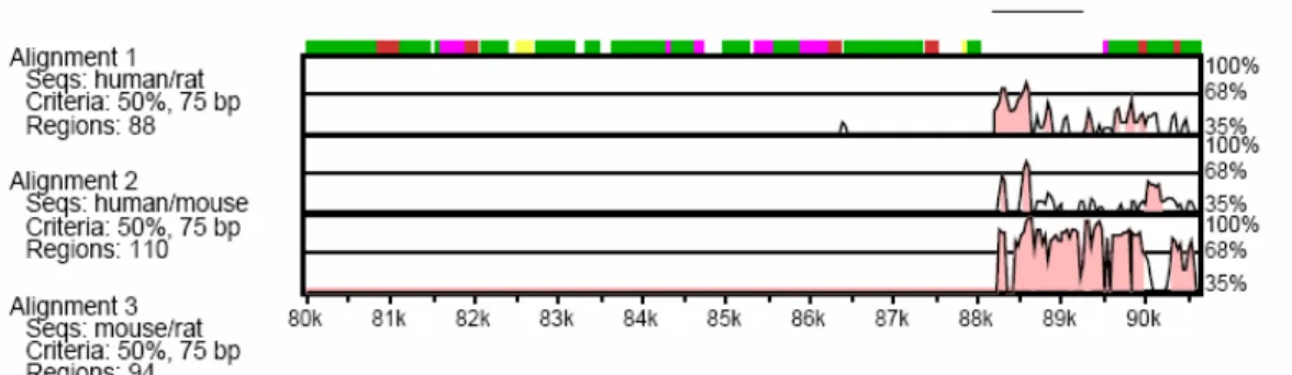
As it was explained in the introduction section, Loc115098 was firstly identified in an unrelated study where cis-acting transcriptional elements of the NIS (Sodium-Iodide Symporter) gene were being analyzed. The focus of that study was to analyze a putative transcriptional control element localized in the 3’ downstream of NIS gene by using a tool called VISTA (Negre et al., 2005). Loc115098 was one of the 10 conserved putative regions identified. (Alotaibi et al, unpublished data). (See Figure 3.1)
Figure 3.1. VISTA plot of conservation levels in a 90 kbp genomic DNA in human, mouse and rat. Only the 10th region is shown here. Percent nucleotide identities between human, mouse and rat DNA sequences are plotted as a function of position along the human sequence. Peaks of evolutionary conservation in overlapping exonic sequences are shaded blue. Aligned regions of more than 50% identity over 75 bases are shaded pink. (Alotaibi et al., unpublished)
30 3.1.2. Features
According to our computational analyses, the human Loc115098 is located in chromosome 19p22. The gene consists of 4 exons and the predicted protein is 223 amino acid-long. It has rather an unusual amino acid composition and it is very hydrophilic. 42% of the protein consists of charged amino acids such as Arginine, Lysine, Aspartate and Glutamate. Therefore it is a very basic protein (pI=10). It bears no domain similarities with any previously known proteins. The conservation of Loc115098 is remarkably high. Our in silico analysis through the databases has proved that it is conserved in the eukaryotic kingdom with the exception of the budding yeast Saccharomyces cerevisiae. Although the similarity ratios differ, there is more than 75% identity in mammals, 50% identity with insects and nematodes and 30-40% identity with plants and ascomycetes.
In the simplest possible model organism A.nidulans, the gene is located on the 8th chromosome, Contig 14: 46513-47339 +. It has the ID number of AN0879.3 and can
be reached at the Broad Institute web page with the same accession number. (http://www.broad.mit.edu/annotation/genome/aspergillus_nidulans/Home.html). The Aspergillus and human homologue of this protein had a 34.4% identity. However the Aspergillus homologue was 222-amino acid long but the composition of the amino acids showed similar features. (See Figure 3.9)
3.1.3. Post Translational Modifications
Aiming to better understand this unusual protein carrying no domain similarities with other proteins, we also searched for the post translational modifications through Expasy/Prosite webpage (http://www.expasy.org/prosite/). The results of this study indicated that both the human and the Aspergillus homologues had several predicted post translational modifications. Both of them had predicted Tyrosine kinase, protein kinase C, casein kinase II, cAMP and cGMP dependent protein kinase phosphorilation sites, N-myristoylation sites. In addition, the Aspergillus copy has an amidation site and an alanine rich region in the C-terminus and the human copy has
31
an extra tyrosine sulfation site. Moreover, they both carry bipartite nuclear targeting sequence in the C-terminus of the protein. All of these post translational modification predictions might be very important and we intend to further investigate these modifications in near future.
3.1.4. Biological Significance of Loc115098
Our bioinformatics analyses have indicated that loc115098 is an evolutionarily conserved gene suggesting that it exists in all eukaryotic kingdom. We believe that, this high percentage of conservation might be pointing at a biological significance. All genes which are very well conserved during evolution have important functions in crucial biological events. Among the most conserved genes, the histones are best known examples. 16S RNA which is a subunit of ribosomal RNA is another example of the conserved genes which also has an essential role for the organism. Along with this proof we hypothesize that loc115098 could be playing an important role in one of the well conserved biological mechanisms such as mitosis.
3.2. Analysis of Loc115098 Expression in Different Tissues
After the identification of loc115098, the aim of the study was initially to confirm that loc115098 is expressed in different tissues of different organisms. For this purpose several tissues were investigated whether or not they expressed the
loc115098 gene.