Value of cardiopulmonary exercise testing in
the diagnosis of coronary artery disease
doi • 10.5578/tt.68200 Tuberk Toraks 2019;67(2):102-107
Geliş Tarihi/Received: 10.01.2019 • Kabul Ediliş Tarihi/Accepted: 14.06.2019
KLİNİK Ç
ALIŞMA
RESEARCH
AR
TICLE
Berna AKINCI ÖZYÜREK1 Şerife SAVAŞ BOZBAŞ2 Alp AYdINALp3 Hüseyin BOZBAŞ4 Gaye ULUBAY2
1 Clinic of Chest Diseases, Ankara Ataturk Chest Diseases and Thoracic Surgery Education and Research Hospital, Ankara, Turkey
1Ankara Atatürk Göğüs Hastalıkları ve Göğüs Cerrahisi Eğitim ve Araştırma
Hastanesi, Göğüs Hastalıkları Kliniği, Ankara, Türkiye
2 Department of Chest Diseases, Ankara Hospital, Baskent Hospital, Ankara, Turkey
2Başkent Üniversitesi Ankara Hastanesi, Göğüs Hastalıkları Anabilim Dalı,
Ankara, Türkiye
3 Department of Cardiology, Ankara Hospital, Baskent University, Ankara, Turkey
3Başkent Üniversitesi Ankara Hastanesi, Kardiyoloji Anabilim Dalı,
Ankara, Türkiye
4 Clinic of Cardiology, Ankara Guven Hospital, Ankara, Turkey 4Ankara Güven Hastanesi, Kardiyoloji Kliniği, Ankara, Türkiye
SUMMARY
Value of cardiopulmonary exercise testing in the diagnosis of coronary artery disease
Introduction: Respiratory and cardiac functions in association with skeletal
and neurophysiologic systems can be evaluated with cardiopulmonary exercise testing (CPET). Compared to treadmill exercise test, CPET provides more comprehensive data about the hemodynamic response to exercise.
Materials and Methods: We aimed to evaluate the relationship with CPET
findings and coronary lesions identified on angiography in patients with angina pectoris who underwent teradmill exercise, CPET and coronary angiography (CAG). By this way we sought to examine the CPET parameters that might be predictive for coronary artery disease (CAD) before diagnostic exercise test results and ischemia symptoms develop. Thirty patients in whom CAG was planned because of symptoms and exercise test results were enrolled in the study. Oxygen consumption (VO2), carbondioxide production (VCO2), minute ventilation (VE), maximum work rate (WR), DVO2/DWR and O2 pulse (VO2/HR) values were calculated. Significant CAD was defined as ≥ 50% narrowing in at least one of the coronary arteries.
Results: The mean age was 60.4 ± 8.9 years ve 21 (65.6%) of subjects were
male. On CAG, CAD was detected in 19 (59.4%) patients. Maximum heart rate, heart rate reserve (HRR), VE/VCO2 measured at anaerobic threshold
Dr. Berna AKINCI ÖZYÜREK
Ankara Atatürk Göğüs Hastalıkları ve Göğüs Cerrahisi Eğitim ve Araştırma Hastanesi, Göğüs Hastalıkları Kliniği, ANKARA - TÜRKİYE e-mail: [email protected]
Yazışma Adresi (Address for Correspondence)
Cite this arcticle as: Savaş Bozbaş Ş, Akıncı Özyürek B,
Aydınalp A, Bozbaş H, Ulubay G. Value of cardiopulmo-nary exercise testing in the diagnosis of corocardiopulmo-nary artery disease. Tuberk Toraks 2019;67(2):102-7.
©Copyright 2019 by Tuberculosis and Thorax. Available on-line at www.tuberktoraks.org.com
INTROdUCTION
Cardiopulmonary exercise testing (CPET) is a useful and detailed test in the evaluation of respiratory, cardiovascular, skeletal and neurophysiologic systems. CPET is especially used to assess cardiac functions and prognosis of heart failure and determine the indication of cardiac transplantation in cardiology (1).
CPET for coronary artery disease (CAD) assessment is an area of growing clinical interest in which different parameters provide both diagnostic and prognostic insight for evaluation and management (2). However ECG stress test (classical effort test) is preferred in clinical practice because need for calibration before each test, requirement of experienced and educated personnel and the increased cost of equipment put CPET on the back burner (3).
Myocardial ischemia with increased workload in the
pressure. However, these findings may not be found in all cases. CPET allows us to evaluate the hemodynamic response to the exercise in more detailed manner compared to the classic effort test. Oxygen uptake (VO2), carbon dioxide production (VCO2), anaerobic threshold (AT), O2 pulse, VO2/WR and respiratory equivalent during anaerobic threshold (VE/VCO2) can be abnormal in cardiovascular diseases (3). Gas analysis can detect myocardial ischemia during exercise with reduced pulse volume and cardiac out-put before the development of ST segment changes or chest pain (3).
In the present study CPET was performed to subjects with chest pain in addition to effort test, and coronary angiography (CAG), and we investigated the relationship between CPET findings and CAG findings detected on coronary angiography. Moreover we aimed to determine the diagnostic parameters for coronary artery disease with CPET in subjects before (AT) and VO2(mL/kg/min) were significantly differed in patients with CAD than those without (p= 0.031; p= 0.041; p= 0.028; p= 0.03 respectively). Peak VO2, VO2/WR and O2 pulse values were higher in patients with normal angiographic results than those with CAD but the difference did not reach to statistical significance.
Conclusion: The findings of our study indicate that among CPET parameters AT VE/VCO2, ATVO2 (mL/kg/dk) and HRR can have predictive value in the diagnosis of CAD. We think that these parameters might be used in the evaluation of patients with angina and dyspnea suspected of CAD. In conclusion parameters obtained during the test that are not influenced by patient’s effort might increase the value of CPET in the diagnosis CAD.
Key words: Coronary artery disease; cardiopulmonary exercise testing; heart rate ÖZET
Kardiyopulmoner egzersiz testlerinin koroner arter hastalığındaki tanısal değeri
Giriş: Kardiyopulmoner egzersiz testi (KPET) solunumsal ve kardiyak işlevlerle birlikte kas-iskelet sistemi ve nörofizyolojik sistemlerin
değerlendirilmesini sağlayan bir yöntemdir. KPET egzersize hemodinamik yanıtı klasik efor testine kıyasla detaylı olarak değerlendir-memizi sağlar.
Materyal ve Metod: Çalışmamızda göğüs ağrısı tanımlayan, efor testi yapılan ve koroner anjiyografi (KAG) planlanan hastalara KPET
yapılarak, KPET bulguları ile KAG’da saptanan lezyon yaygınlığı arasındaki ilişkiyi araştırdık. Böylece efor testinde tanısal EKG bulgu-ları ve iskemiyle ilişkili semptomlar gelişmesini beklemeden, KPET ile koroner arter hastalığı (KAH) için tanısal olan parametreleri belirlemeyi amaçladık. Çalışmamıza semptomları ve efor testi sonuçları ile KAG planlanan 30 hasta dahil edildi. Oksijen kullanımı (VO2), karbondioksit üretimi (VCO2), dakika ventilasyonu (VE), maksimum ulaşılan iş yükü (WR), DVO2/DWR ve O2 pulse (VO2/HR) değerleri hesaplandı. Koroner arterlerden herhangi birinde ≥ %50 darlığın saptanması anlamlı koroner arter hastalığı kabul edildi.
Bulgular: Hastaların ortalama yaşı 60.4 ± 8.9 yıl ve 21 (%65.6)’i erkekti. Koroner anjiyografi ile 19 (%59.4) hastada koroner
arter-lerde stenoz saptandı. KAH saptanan grupta maksimum kalp hızı, kalp hızı rezervi (HRR), anaeorobik eşik (AT)'te ölçülen VE/VCO2 ve VO2 (mL/kg/dk) KAH saptanmayan gruba göre anlamlı farklıydı (p< 0.05). KAH saptanmayan hastalarda VO2 pik, VO2/WR ve O2 pulse değerleri KAH saptanan gruba göre yüksek saptandı, ancak istatistiksel olarak anlamlı bulunmadı (p> 0.05).
Sonuç: Çalışmamız sonucunda AT, VE/VCO2, ATVO2 (mL/kg/dk), maksimum HR ve HRR’nin KAH tanısını destekleyen önemli para-metreler olduğu saptandı. Bu nedenle dispne ve göğüs ağrısı etiyolojisine yönelik değerlendirilen hastalarda bu parapara-metrelerin KAH için tanısal olabileceği düşüncesindeyiz. Sonuç olarak hasta eforundan etkilenmeyen ve tetkik sırasında görüntülenebilen parametre-ler KPET’in KAH tanısında kullanımını artıracaktır.
MATERIALS and METHOdS
This study (project numbered KA04/198) was conducted with the approval of the ethics committee of Başkent University Medical Faculty Research Council.
A written informed consent form was obtained from all subjects before participating in the study.
Patients with chest pain, ST segment changes during the effort test, and scheduled for coronary angiography were included. Thirty patients who underwent CPET before coronary angiography were evaluated. Patients with acute coronary syndrome, newly diagnosed myocardial infarction (< 2 months), congestive heart failure, severe valvular disease, diabetes mellitus requiring insulin use, and orthopedic problems that would limit exercise were excluded from the study. Cardiopulmonary Exercise Testing
The symptom-limited cardiopulmonary exercise test (Ergo-metrics 900, SensorMedics™, Bilthoven, The Netherlands) was performed using the bicycle ergometer. All patients were instructed not to eat at least two hours before the test, and not to drink tea, coffee, cola drinks during that time.
Cardiopulmonary exercise tests were conducted at the same time of day after lunch. Calibration with two standard gas mixtures (26% oxygen + balanced N2, 4% carbon dioxide + 16% oxygen + balanced N2) before each testing was performed.
VO2, VCO2 and minute ventilation (VE), were evaluated by using gas analyzers and the face mask (Rudolph Face Mask for Exercise Testing™; Hans Rudolph Inc., Kansas City, MO).
All patients performed a 3-minute baseline period and a 3-minute warm-up period (warm-up, 0W, 60 rpm) followed by an incremental exercise test with an increased workload of 15 watt/min (4). During CPET, patients were monitored with 12-channel ECG, heart rate, oxygen saturation using a pulse oximeter (via a finger probe- NONIN 8600 pulse oximeter™, Plymouth, Minnesota USA). Blood pressure was measured and recorded manually every two minutes.
The collected data was automatically recorded (Desktop Diagnostics/CPX™; Medical Graphics Corporation, St. Paul, Minn.). The maximum workload
was considered the highest workload that patients could tolerate for at least 20-30 seconds. Anaerobic threshold was determined by V-slope method (5). The peak VO2 was obtained by calculating the average of the values obtained in the last 30 seconds of the incremental exercise test.
VO2/WR and O2 pulse (VO2/HR) values were calculated at maximum exercise.
Effort Test
The effort test was performed by using the Bruce protocol. The target heart rate was calculated as 220-age (years). 85% of this value was recorded as a submaximal target. Typical chest pain during the test, 1 mm or more subduction after 80 ms of J junction in two consecutive ST segments, 10 mmHg or more decrease in systolic blood pressure, and ST segment elevation were evaluated as positive effort test criteria. During and after the test, symptoms, heart rate and blood pressure were recorded, and continuous electrocardiographic monitoring was performed during the test and during the recovery period.
Coronary Angiography
Coronary angiography was performed through the right femoral artery after appropriate preliminary preparation and premedication. With the Judkins technique, selective coronary angiographic images were obtained in right and left oblique positions. Angiograms were evaluated by two experienced cardiologists. Each segment was numerically evaluated according to the percentage reduction in lumen diameter based on the nearest proximal intact coronary artery segment. Significant coronary artery disease was accepted in the presence of a ≥ 50% stenosis in any of the coronary arteries.
Statistical Analysis
Statistical analysis was performed using the Windows SPSS version 10.0 (Statistical Package for the Social Sciences, version 10.0, SPSS Inc, Chicago, IL, USA). The data were expressed as mean ± standard deviation or percentage. Chi-square test or t-test were used in the comparision of the variables between groups. The relationship between variables was evaluated by Spearman rank correlation or Pearson correlation test. P value < 0.05 was considered as significant.
RESULTS
Thirty patients with chest pain and positive effort test (ST segment change or chest pain during the test) who were planned for coronary angiography were included to sthe study. The mean age of the patients was 60.4 ± 8.9 years and 21 (65.6%) were male. The demographics, laboratory results and pulmonary function test parameters of the patients are depicted in Table 1. Complaints of patients were listed in Table 2. Twenty (62.5%) patients reached the maximum heart rate and 22 (73%) patients had ST segment depression during effort test During or after the test 18 (56.3%) patients reported symptoms that could indi-cate coronary artery disease. Results of coronary angiography performed after impaired effort test were evaluted and 14 (46%) patients had stenosis in
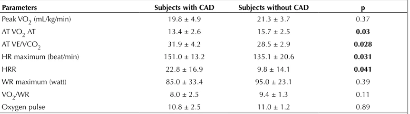
coro-nary arteries. Based on the results of effort test and symptoms CPET was performed before CAG. No com-plications were seen during and after CPET. The CPET parameters of the patients are summarized in Table 3. Results of coronary angiography performed after CPET were evaluated and 19 (59.4%) patients had stenosis in coronary arteries. Grade of coronary artery stenosis is demonstrated in Table 4. CPET parameters of patients with and without CAD were compared. Significant differences were found between patients with and without CAD in terms of maximum heart rate, heart rate reserve (HRR), VE/VCO2 and VO2 (mL/kg/min) (Table 5). Peak VO2 value, VO2/WR and O2 pulse were found to be higher in patients without CAD than patients with CAD, but not at the level of statistical significance (Table 5).
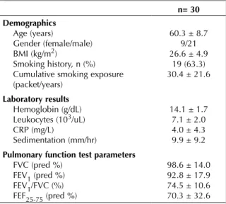
Table 1. Demographic, laboratory and pulmonary function test parameters of study population
n= 30 demographics Age (years) Gender (female/male) BMI (kg/m2) Smoking history, n (%) Cumulative smoking exposure (packet/years) 60.3 ± 8.7 9/21 26.6 ± 4.9 19 (63.3) 30.4 ± 21.6 Laboratory results Hemoglobin (g/dL) Leukocytes (103/uL) CRP (mg/L) Sedimentation (mm/hr) 14.1 ± 1.7 7.1 ± 2.0 4.0 ± 4.3 9.9 ± 9.2 pulmonary function test parameters
FVC (pred %) FEV1 (pred %) FEV1/FVC (%) FEF25-75 (pred %) 98.6 ± 14.0 92.8 ± 17.9 74.5 ± 10.6 70.3 ± 32.6 Data are expressed as the mean ± standard deviation, the median (interquartile range), or frequency counts (percentages), as appropriate.
BMI: Body mass index, CRP: C-reactive protein; FEF25-75: Forced
expiratory flow at 25-75%; FEV1: Forced expiratory volume at first
second; FVC: Forced vital capacity.
Table 2. Symptoms of study population
Symptoms n= 30
Chest pain, n (%) 15 (50.0)
Chest pain and exertional dyspnea n (%) 12 (40.0) Chest pain and palpitation n (%) 2 (6.7)
Table 3. Cardiopulmonary exercise testing (CPET) parameters of study population
CpET parameters n= 30 Resting VO2 (mL/kg/min) 4.4 ± 0.9 Warmup VO2 (mL/kg/min) 11.1 ± 1.9 Peak VO2 (mL/kg/min) 20.3 ± 4.5 Mean VO2 (%) 82.8 ± 18.1 AT VO2 (mL/kg/min) 14.5 ± 3.9 AT VE/VCO2 30.7 ± 5.1 HR maximum (beats/min) 140.9 ± 19.6 WR maximum (watt) 88.6 ± 30.0 VE (BTPS) 59.3 ± 16.8
Exercise duration (min) 6.6 ± 2.6
VO2/WR 8.5 ± 2.2
Oxygen pulse 10.9 ± 2.1
AT: Anaerobic threshold, HR: Heart rate, HRR: Heart rate reserve,
VCO2: CO2 production, VE: Minute ventilation, VO2: Oxygen
uptake, WR: Work rate.
Table 4. Grade of coronary artery stenoses detected in coronary angiography
Coronary artery stenosis n= 19
Minimal CAD, n (%) 6 (31.6)
Single vessel disease, n (%) 5 (26.3) Two-vessel disease n (%) 4 (21.1) Three-vessel disease, n (%) 3 (15.8) Four-vessel disease, n (%) 1 (5.3)
Moreover AT VE/VCO2 and peak VO2 values were negatively correlated (r= -0.49 p= 0.005).
dISCUSSION
CPET is a noninvasive, objective assessment of the body’s response to exercise and can evaluate additional components of the cardiovascular and pulmonary systems. This testing provides useful information on the functional status of patients with heart failure, chronic pulmonary disease and patients with exercise intolerance (6).
In the present study, CPET was performed before angiography and AT VE/ VCO2, AT VO2 (mL/kg/min), maximum HR and HRR were found to be important parameters supporting diagnosis of CAD. Therefore these CPET parameters should be considered as essential parameters for the diagnosis of CAD in clinical use. Furthermore these parameters can be monitored during CPET and do not require additional calculations unlike oxygen pulse or VO2/WR. Peak VO2 is used for diagnosis and prognosis in heart failure and CAD (7). However peak VO2 is affected by patient motivation and threshold values of peak VO2 are not clear (8). In addition, subjects with cardiovascular disease do not achieve the maximal targeted values due to current illnesses. For these reasons, other parameters of CPET independent from patient effort are investigated. Corra et al. evaluated 403 patients with moderately limited functional capacity, and found that VE/VCO2 slope was a strong independent predictor of major cardiac events. In addition, they found that mortality rates of patients with VE/VCO2 slope of ≥ 35 were similar to the mortality rates of patients with low VO2 levels (9).
Corra and co-workers suggested that the prognostic pre-eminence of the VE/VCO2 slope may be linked to an underlying abnormal response to exercise and sympathetic overactivity and neurohormonal imbalance (9).
Van de Veire et al. reported that NT-pro BNP levels were an important independent predictor of VE/ VCO2 in patients with CAD (8). The present study also suggests that AT VE/VCO2 may be used in the diagnosis of CAD as this parameter can be evaluated in subjects those can not reach the maximum exercise level and significant correlation is found between AT VE/VCO2 and CAD. Similarly, low levels of AT VO2 are also important predictors of CAD. Therefore, high AT VE/VCO2 and low AT VO2 values are important parameters in the diagnosis of CAD in patients who can not reach maximum heart rate or have typical ECG findings.
Measurement of cardiac out-put is the best index for the assessment of cardiac functions during exercise. During exercise, cardiac out-put is estimated from HR. The HR gives information about the achievement of the predicted values with age, the patient’s maximal effort and the peak VO2. The predicted maximal HR can be obtained from the formula of 210- (age x 0.65). The maximum HR should exceed 90% of the predicted value. The VO2/WR relationship reflects the metabolic need of the exercise or the oxygen requirement of the work. The normal value is 10 mL/kg/watt. VO2/WR decreases in heart diseases and peripheral artery diseases. Coronary artery disease is associated with an early increase in HR, a decrease in oxygen delivery, and no increase in oxygen consumption (VO2/WR) in parallel with
Table 5. Comparision of CPET parameters between subjects with and without CAD.
parameters Subjects with CAd Subjects without CAd p
Peak VO2 (mL/kg/min) 19.8 ± 4.9 21.3 ± 3.7 0.37 AT VO2 AT 13.4 ± 2.6 15.7 ± 2.5 0.03 AT VE/VCO2 31.9 ± 4.2 28.5 ± 2.9 0.028 HR maximum (beat/min) 151.0 ± 13.2 135.1 ± 20.6 0.031 HRR 22.8 ± 16.9 9.8 ± 14.1 0.041 WR maximum (watt) 85.0 ± 33.4 95.0 ± 23.1 0.39 VO2/WR 8.0 ± 2.5 9.4 ± 1.3 0.11 Oxygen pulse 10.8 ± 2.5 11.0 ± 1.2 0.89
CPET: Cardiopulmonary exercise testing, CAD: Coronary artery disease, AT: Anaerobic threshold, HR: Heart rate, HRR: Heart rate reserve,
increased workload as a result of unmet myocardial oxygen requirement during exercise (10). In the present study, HR was found to be significantly higher in patients with CAD than in patients without CAD, while the VO2/WR value was found to be low. However differences in VO2/WR did not reach statistical significance level. This might be due to the small number of patients.
HRR is interpreted as the difference between the predicted maximum heart rate according to age and the maximum heart rate that the patient achieved during the test. In healthy subjects, the difference is less than 15 beats/min. Heart rate reserve can be increased or decreased or normal in cardiac diseases. HRR is an independent risk factor for mortality in CAD. HRR is usually increased in subjects with respiratory diseases. In our study, the HRR assessed during CPET was higher in patients with CAD than patients without CAD.
CONCLUSION
AT VE/VCO2, AT VO2 (mL/kg/min), maximum HR and HRR independently from subjects efforts are important parameters in the assessment of CAD. Therefore clinical use of CPET in the diagnosis of CAD will increase in near future.
CONFLICT of INTEREST
All authors have no conflict of interests. AUTHORSHIp CONTRIBUTIONS Concept/Design: All of authors. Analysis/Interpretation: All of authors. Data Acquisition: All of authors. Writting: BAO, ŞSB, HB
Critical Revision: BAO Final Approval: BAO, ŞSB
RE FE REN CES
1. Wasserman K, Hansen JE, Sue DY, Whipp BJ, Casaburi R. Principles of exercise testing and interpretation. 3rd ed Vol. 10. Baltimore: Lippincott Williams&Wilkins, 1999. 2. Chaudhry S, Arena R, Bhatt DL, Verma S, Kumar N. A
practical clinical approach to utilize cardiopulmonary exercise testing in the evaluation and management of coronary artery disease: a primer for cardiologists. Curr Opin Cardiol 2018;33:168-77.
3. Belardinellia R, Lacalapricea F, Carleb F, Minnuccib A, Ciancia G, Pernaa GP, et al. Exercise-induced myocardial ischaemia detected by cardiopulmonary exercise testing. European Heart Journal 2003;24:1304-13.
4. Johnson BD, Badr MS, Dempsey JA. Impact of the aging system on the response to exercise. Clin Chest Med 1994;15:229-46.
5. Beaver WL, Wasserman K, Whipp BJ. A new method for detecting anaerobic threshold by gas exchange. J Appl Physiol 1986;60:2020-7.
6. Holmes A, Phillips LM. Cardiopulmonary exercise testing and SPECT myocardial perfusion imaging: Pre-test probability is the key. J Nucl Cardiol 2019;26:107-8. 7. Mancini DM, Eisen H, Kussmaul W, Mull R, Edmunds LH
Jr, Wilson JR. Value of peak exercise oxygen consumption for optimal timing of cardiac transplantation in ambulatory patients with heart failure. Circulation 1991;83:778-86. 8. Van de Veire NR, Van Laethem C, Philippé J, De Winter O,
De Backer G, Vanderheyden M. De Sutter JVE/VCO2 slope and oxygen uptake efficiency slope in patients with coronary artery disease and intermediate peakVO2. Eur J Cardiovasc Prev Rehabil 2006;13:916-23.
9. Corrà U, Mezzani A, Bosimini E, Scapellato F, Imparato A, Giannuzzi P. Ventilatory response to exercise improves risk stratification in chronic heart failure patients with intermediate functional capacity. Am Heart J 2002;143:418-26.
10. Ulubay G. Kardiyopulmoner egzersiz testlerinin klinik kullanımı. Türkiye Klinikleri Arch Lung 2012;13(Suppl).