PAPER
Cite this:Green Chem., 2013, 15, 2566
Received 13th May 2013, Accepted 9th July 2013 DOI: 10.1039/c3gc40885j www.rsc.org/greenchem
E
fficient ammonium removal from aquatic
environments by
Acinetobacter calcoaceticus STB1
immobilized on an electrospun cellulose acetate
nano
fibrous web
Omer Faruk Sarioglu,
aOncay Yasa,
aAsli Celebioglu,
aTamer Uyar*
aand
Turgay Tekinay*
b,cA novel biocomposite material was developed by immobilizing an ammonia-oxidizing bacterial strain, Acinetobacter calcoaceticus STB1, on an electrospun porous cellulose acetate (CA) nanofibrous web. Ammonium removal characteristics of the STB1 immobilized CA nanofibrous web were determined at varying initial ammonium concentrations, and removal rates of 100%, 98.5% and 72% were observed within 48 h for 50 mg L−1, 100 mg L−1and 200 mg L−1samples, respectively. Most of the ammonia is inferred to be converted into nitrogen or is accumulated as bacterial biomass, as only trace amounts of ammonium were converted into nitrite or nitrate. Reusability test results indicate that, at an initial ammonium concentration of 100 mg L−1, bacteria-immobilized CA nanofibrous webs can be reused for at least 5 cycles. SEM images of the STB1/CA nanofibrous web after five cycles of reuse and rigorous washing demonstrate that bacterial biofilms strongly adhere to nanofiber surfaces.
1.
Introduction
Ammonium (NH4+), nitrite (NO2−) and nitrate (NO3−)
consti-tute the most common nitrogenous compounds found natu-rally in aquatic ecosystems, and are formed by atmospheric decomposition, degradation of organic matter, and N2fixation
by certain microorganisms.1 However, human activities have altered the nitrogen content of aquatic environments over the last two centuries, and the accumulated nitrogenous wastes now have a significant effect on the ecosystem.1 Among the
nitrogenous pollutants released to aquatic environments, ammonia is one of the most toxic ones and exists in water as either non-dissociated ammonia (NH3) or the ammonium ion
(NH4+).2,3Ammonia concentrations between 2 and 10 mg L−1
are lethal for many aquatic organisms, and concentrations greater than 1.5 mg L−1are considered unacceptable in drink-ing water by the U.S. Environmental Protection Agency (US EPA).3The main metabolic by-products of ammonia are NO2−
and NO3−both of which are also toxic for aquatic life. The US
EPA regulations dictate that the concentrations of nitrite and nitrate in drinking water should not exceed 1 mg L−1 and 10 mg L−1, respectively, and the sum of nitrite and nitrate con-centrations should be lower than 10 mg L−1.4Therefore, reme-diation of high ammonium and nitrogen concentrations in aquatic systems is necessary for maintaining the quality of water for human or agricultural use. Conventional wastewater treatment systems include biological applications with both autotrophic nitrifiers and heterotrophic denitrifiers under dynamic aerobic and anaerobic conditions.5However, hetero-trophic ammonium removal by a single nitrifier/denitrifier strain has many advantages, such as the simultaneous proces-sing of nitrification and denitrification reactions under equi-valent conditions, and novel bacterial species have been isolated from different aquatic environments for that purpose.5–7
Electrospinning is an emerging nanofiber/nanoweb pro-duction technique and has attracted much attention over the past decade due to its simplicity, versatility and cost-effectiveness.8–10 Electrospun nanofibers and their
nano-fibrous webs display a variety of unique properties, such as large surface-to-volume ratios and highly porous structures, that allow their use as effective matrices in membranes and filters for environmental applications.8–13For instance, the use of immobilized microorganisms on electrospun nanofibrous polymeric mats was recently shown to increase the rate of aUNAM-Institute of Materials Science & Nanotechnology, Bilkent University, Ankara,
06800, Turkey. E-mail: [email protected]; Fax: +90 (312) 266 4365; Tel: +90 (312) 290 3571
bGazi University Life Sciences Research Center, Ankara 06800, Turkey. E-mail: [email protected]; Fax: +90 312 484 62 71; Tel: +90 312 484 62 70 cGazi University, Polatlı Science and Literature Faculty, Ankara, 06900, Turkey
Published on 09 July 2013. Downloaded by Bilkent University on 16/05/2014 07:38:17.
View Article Online
nitrate removal.14 In that study, microalgal cells were e ffec-tively immobilized on electrospun chitosan nanofiber mats in order to generate a hybrid system for nitrate removal.14Nitrate removal by nanofiber-immobilized microorganisms has several advantages over the use of free cells in suspension, including lower space and growth medium requirements, ease of handling, and potential reusability of the same matrix over several treatment cycles.14 Furthermore, immobilization of bacterial cells on polymeric network systems makes them more resistant to harsh environmental conditions, such as metal toxicity or extremes of salinity, temperature and pH.15 Covalent coupling, cross-linking, physical entrapment and the natural process of bacterial adhesion can be used for the immobilization of microorganisms to nanofiber networks.16 Natural adhesion is the most advantageous among these methods, as it enables the formation of biofilms following surface attachment and results in maximal cell viability and biochemical activity.16
We have previously shown that Acinetobacter calcoaceticus STB1 could remove high concentrations of ammonium under heterotrophic conditions.6This strain can successfully remove high concentrations of ammonium in several days, and is non-toxic for aquatic life; hence it can be effectively used for the remediation of aquatic environments.
In the current study, A. calcoaceticus STB1 cells were immobilized on an electrospun cellulose acetate (CA) nano-fibrous web in order to achieve enhanced ammonium removal in aquatic environments. CA was chosen as the fibrous matrix, as it is the most commonly used regenerative cellulose, and is biodegradable and biocompatible, which renders it advan-tageous for biological applications.17 Electrospun CA nano-fibers have been used for filtration, drug delivery, enzyme immobilization, and artificial tissue matrix formation,18–21 and successful growth of fibroblasts and Schwann cells on CA nanowebs has been reported in the literature.22,23 Moreover, CA nanofibers can be converted into more functional and applicable forms by the incorporation of other polymers into the nanofiber mesh or by the chemical conversion of cellulose into a variety of derivative fibers.24,25Here, we successfully pro-duced a nanofibrous biocomposite web by immobilizing STB1 cells on electrospun CA nanofibers for the removal of ammonium in aqueous systems. Reusability test results indi-cate that STB1/CA nanofibrous webs can be reused without sig-nificant loss of their ammonium removal capacity.
2.
Experimental
2.1. Preparation of porous CA nanofibers
The electrospinning of cellulose acetate (CA) nanofibers was performed as detailed in a previous study.26While production of porous CA nanofibers generally requires post-treatment of the ultimate nanofiber structure, the porous CA nanofibers described here were produced from a dichloromethane(DCM)– acetone binary solvent system without additional treatment.26 Constituent solvents of the DCM–acetone binary system were
purchased from Sigma-Aldrich (USA) and used without any purification (dichloromethane, DCM, ≥99% (GC); acetone, ≥99% (GC); cellulose acetate, CA, Mw: 30 000, 39.8 wt.%
acetyl). Briefly, the homogeneous electrospinning solution was prepared by dissolving CA in a DCM–acetone (2/1 (v/v)) binary solvent mixture at a 7.5% (w/v) polymer concentration. The clear CA solution was then placed in a 3 mL syringe fitted with a metallic needle of 0.6 mm inner diameter. The syringe was fixed horizontally on the syringe pump (model SP 101IZ, WPI, USA). The electrode of the high-voltage power supply (Matsu-sada Precision, AU Series, Japan) was clamped to the metal needle tip, and the cylindrical aluminum collector was grounded. Electrospinning parameters were adjusted as follows: feed rate of solutions = 1 mL h−1, applied voltage = 15 kV, and tip-to-collector distance = 10 cm. Electrospun nano-fibers were deposited on a grounded stationary cylindrical metal collector covered with aluminum foil. The electro-spinning apparatus was enclosed in a Plexiglas box, and elec-trospinning was carried out at 25 °C at 24% relative humidity. Collected nanofibers/nanowebs were dried overnight at room temperature under the fume hood. The process is summarized in Fig. 1.
2.2. Growth and immobilization ofAcinetobacter calcoaceticus STB1
The bacterial strain utilized in this study was isolated from brackish water samples collected from a commercial sea bass farm.6 Immobilization of bacteria was achieved by the inclusion of CA nanofibrous webs in the growth media of newly inoculated bacteria. Colonies were maintained in 100 mL culture flasks for 30–35 days. The ingredients of the growth medium were: 6.3 g L−1 Na2HPO4 (≥99%), 3 g L−1
KH2PO4 (≥99%), 0.5 g L−1 NaCl (≥99.5%), 2 g L−1 glucose
(anhydrous), and 300 mL L−1of a trace elements solution con-sisting of 6.1 g L−1MgSO4(≥99.5%), 3 g L−1H3BO3(≥99.5%),
0.5 g L−1MnCl2 (≥99%), 0.05 g L−1 CaCl2(≥93%), 0.03 g L−1
FeSO4·7H2O (≥99%), 0.03 g L−1CuCl2(≥97%), and 0.03 g L−1
ZnCl2(≥99.99%). Ammonium (in the form of NH4Cl,≥99.5%)
was utilized as the nitrogen source during incubation and immobilization, with an initial concentration of 50 mg L−1. Following the incubation period, bacterial immobilization was confirmed by SEM imaging and nanofiber samples of equal weights were prepared for further testing. All the reagents uti-lized in this study were purchased from Sigma-Aldrich (USA). 2.3. Ammonium removal experiments using STB1
immobilized CA nanofibers
The same basal growth medium utilized in bacterial immobili-zation studies was used in the heterotrophic ammonium removal experiments. Basal growth medium samples were spiked with varying amounts of ammonium (as NH4Cl),
inocu-lated with free bacterial samples, bacteria-free nanofibers or bacteria-immobilized nanofibers and incubated for 48 h at 140 rpm and 30 °C. The positive control contained only bacterial inocula at an initial ammonium concentration of 50 mg L−1, the negative control contained only nanofibers (0.4 mg of
Green Chemistry Paper
Published on 09 July 2013. Downloaded by Bilkent University on 16/05/2014 07:38:17.
nanofiber per mL) at an initial ammonium concentration of 50 mg L−1, and the experimental samples contained bacteria immobilized on CA nanofibers (0.4 mg of nanofiber per mL) at initial ammonium concentrations of 50, 100 and 200 mg L−1. Initial ammonium values of the experimental samples were adjusted to 50, 100 and 200 mg L−1to represent low, medium and high concentrations of ammonium, and to determine ammonium removal efficiencies in different concentration ranges. Samples were collected periodically to analyze ammonium, nitrite and nitrate values. Bacterial growth rates of the positive control samples were followed by OD600
measurements. Changes in the ammonium, nitrite and nitrate concentrations in the samples were determined using spectro-photometric test kits (Merck Ammonium Cell Test 14559, Merck Nitrate Cell Test 14563 and Merck Nitrite Cell Test 14547). Before performing the tests, samples were centrifuged for 1 min at 12 000 rpm at room temperature, and the super-natants were used in analytical measurements of ammonium, nitrite and nitrate. All tests were done in triplicate. Experi-ments were repeated at least twice.
2.4. Scanning electron microscopy (SEM)
Millimeter-length nanofiber pieces with and without bacterial immobilization were cut and prepared for SEM analysis to monitor bacterial attachment before and after ammonium removal experiments. A protocol similar to that of Greif and colleagues was utilized for sample fixation.27Briefly, samples were washed twice with PBS buffer and fixed by overnight incu-bation in 2.5% glutaraldehyde in PBS buffer at room tempera-ture. Following glutaraldehyde fixation, samples were washed twice with PBS buffer and then dehydrated by immersion in a
series of ethanol–water solutions ranging from 30% to 96%. Prior to SEM imaging, all samples were coated with a 5 nm layer of gold-palladium. A Quanta 200 FEG scanning electron microscope (FEI Instruments, USA) was used for the acqui-sition of SEM images.
2.5. Reusability test for the STB1 immobilized CA nanofibrous web
Ammonium removal studies were performed 5 times to assess the reusability of the bacteria-immobilized nanofibers. Prior to each cycle, nanofiber pieces were washed twice with PBS buffer and incubated overnight in PBS to remove any unattached bac-teria. The ammonium removal experiments (incubation at 140 rpm and 30 °C for 48 h) described above were performed after each washing step for a total of 5 cycles. The initial ammonium concentration was fixed at 100 mg L−1, since this concentration was found to be more suitable for observing changes in performance values in each cycle compared to low (50 mg L−1) and high (100 mg L−1) initial ammonium concen-trations. Ammonium concentrations were measured at 0 h and 48 h, and the percentage removal of ammonium was calcu-lated using these results. Each cycle was terminated after 48 h of total incubation and washing steps were repeated for nano-fiber samples before the initiation of the next cycle. All tests were done in triplicate.
3.
Results and discussion
3.1. Attachment of STB1 strain on an CA nanofibrous web CA nanofibers can be obtained in a smooth or porous morphology depending on the solvent type utilized. While the
Fig. 1 (a) Schematic representation of the electrospinning process for CA nanofibers and photograph of the CA nanofibrous web; (b) photograph of the STB1 immobilized CA nanofibrous web and schematic representation of bacterial cells on nanofiber surfaces.
N,N-dimethylacetamide (DMAc)–acetone blend is one of the most common solvent systems for the electrospinning of uniform and smooth CA nanofibers,28 we have previously demonstrated the production of nanoporous CA nanofibers by using a highly volatile DCM–acetone solvent mixture.26While
conventional electrospun CA nanofibers are already suitable for use in biological systems, the rough surface and the higher surface area of porous electrospun CA nanofibers were expected to increase the utility of CA nanowebs in biological applications, and especially for biomedical research. SEM imaging was performed to observe the bacterial adhesion to nanofibers, and 30 days were found to be required for the robust attachment of bacteria onto nanofiber surfaces. Fig. 2a and 2b show Acinetobacter calcoaceticus STB1 cells after 7 days of incubation, wherein no biofilm formation can be observed. SEM images of porous CA nanofibers prior to bacterial attach-ment are depicted in Fig. 3a and 3b. The CA fiber diameter range was between 500 nm and 1.5μm, and the fibers had a ribbon-like morphology.26 Fig. 3c and 3d show bacteria strongly attached onto the nanofibrous web after 35 days of incubation, and the attached bacteria are observed to form a biofilm structure by adhering to each other and surrounding
the filaments of the CA web. This type of attachment was found to be adequate for further studies, and ammonium removal experiments were started with STB1 immobilized CA web samples at this stage.
3.2. Ammonium removal capability of the STB1 immobilized CA nanofibrous web
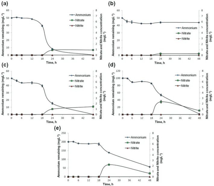
STB1 immobilized CA nanofibrous webs have shown efficient removal of ammonium at each concentration within 48 h, and the percentile ammonium removal capability of the web samples decreased as the initial ammonium concentrations increased (Fig. 4c, 4d and 4e). Bacteria-free CA webs displayed negligible decreases in ammonium concentrations (Fig. 4b), and the removal capability of STB1 immobilized CA nano-fibrous web samples was therefore attributed to bacterial nitro-gen metabolism. The ammonium removal capability of the STB1/CA nanofibrous web is very similar to that of the free bacteria sample at the initial ammonium concentration of 50 mg L−1(Fig. 4a and 4c), which shows that the STB1/CA nano-fibrous web can provide the same outcome as free-suspended bacteria at a defined w/v ratio without including any additional bacterial inocula into the aquatic system. The STB1/CA nano-fibrous web was capable of fully remediating an initial ammonium concentration of 50 mg L−1, and displayed 98.5% and 72% removal rates at initial ammonium concentrations of 100 mg L−1and 200 mg L−1, respectively. Increases in nitrate and nitrite concentrations were limited, and nitrite concen-trations in particular were below detectable limits. The increase in nitrate concentrations was likewise minimal, not exceeding 2 mg L−1 at the end of a 48 h period for each sample. As such, the concentrations of nitrite, nitrate and their sum were all below the legal limits for water quality man-agement,4 which suggests that the production of toxic meta-bolic by-products of ammonium is not a problematic issue for STB1/CA nanofibrous webs. Since nitrite and nitrate levels were minimal for STB1, we have tried to account for the remaining products of ammonium remediation in a previous study by performing total nitrogen (TN) analysis with an elemental analyzer.6The only nitrogen source in the bacterial growth medium was ammonium for STB1 cells; hence we were able to determine the percentage of ammonium incorporated into cellular biomass for the 100 mg L−1sample. Around 22% of the initial ammonium concentration was found to be intro-duced into the cell biomass, and a further 4% was initially incorporated into cell biomass and subsequently released to the supernatant. For this reason, we have concluded that STB1 cells converted most of the remaining ammonium into gaseous denitrification products.6A similar situation was pre-viously observed by Zhao and colleagues, who demonstrated that Acinetobacter calcoaceticus strain HNR could convert a considerable amount of ammonium into N2gas.5
3.3. Reusability and applicability of STB1 immobilized CA nanofibers
Ammonium removal capabilities of reused STB1 immobilized CA nanofibrous webs were tested for five cycles of reuse. Fig. 5
Fig. 3 SEM micrographs of bacteria-free electrospun CA nanofibers at (a) 2500× and (b) 200 000×; and STB1 immobilized nanofibers after 35 days of incubation at (c) 5000× and (d)10 000× magnification.
Fig. 2 General morphology ofAcinetobacter calcoaceticus STB1 under a Scan-ning Electron Microscope (SEM) at 5000× (a) and 15 000× (b) magnification.
Green Chemistry Paper
Published on 09 July 2013. Downloaded by Bilkent University on 16/05/2014 07:38:17.
shows the performance values of each cycle for the total of 5 cycles. 86% of the ammonium removal capacity was obtained for the final cycle (5th cycle) which suggests that STB1/CA nanofibrous webs can sustain their ammonium removal capacity under several cycles of reuse. This result is highly promising, and with a successful optimization, the STB1/CA nanofibrous web may be utilized repeatedly for ammonium remediation, constituting a reusable material for ammonium remediation from aquatic environments.
After the reusability experiments, CA webs were washed several times with PBS and fixed for SEM imaging. Fig. 6a and 6b show visible bacterial biofilms on nanofiber surfaces, suggesting that STB1 cells displayed stronger attachment to CA webs compared to the beginning of the reusability test. As such, washing and reuse of these webs did not lead to a decrease in the quantity of bacterial biofilms; in contrast, the
extent of biofilm formation increased and a stronger attach-ment to the nanofibrous matrix was observed.
The remediation of aquatic systems is an important issue and sustainable solutions, particularly novel and green approaches, for the removal of a wide host of pollutants have received considerable attention in recent times.29,30Since our bacterial isolate is not pathogenic or toxic6 and biological treatment methods are generally more sustainable and environ-mentally friendly than their physical and chemical equiva-lents,31 the use of our bacterial strain is advantageous for ammonium removal. In this study, we immobilized STB1 bac-terial strain on electrospun CA nanofibrous webs to analyze the efficiency of these bacteria in ammonium removal, which would be of considerable use in developing novel commercial removal techniques. We deem that this approach is successful since bacteria could attach strongly to nanofiber surfaces,
Fig. 4 Ammonium, nitrite and nitrate levels for: (a) free STB1 cells at the initial ammonium concentration of 50 mg L−1; (b) bacteria-free CA web at the initial ammonium concentration of 50 mg L−1; (c) STB1 immobilized CA web at the initial ammonium concentration of 50 mg L−1; (d) STB1 immobilized CA web at the initial ammonium concentration of 100 mg L−1; (e) STB1 immobilized CA web at the initial ammonium concentration of 200 mg L−1. Error bars represent the means of three independent replicates.
STB1 immobilized CA nanofibrous webs could remediate ammonium as effectively as freely floating STB1 bacterial inocula, and the biocomposite material could be used for several cycles for ammonium removal, as shown by reusability test results (Fig. 5). Similar approaches have been proposed in the literature. Eroglu et al. have immobilized microalgal cells on electrospun chitosan nanofiber mats for the removal of nitrate from liquid effluents.9In the present study, ammonium
was chosen as the target contaminant due to its high toxicity, and different concentration ranges of ammonium were tested to determine the efficiency of bacteria-immobilized nano-fibrous web samples for ammonium removal. The time required for complete removal of 100 mg L−1 ammonium by STB/CA nanofibrous webs was around 48 h. Zhao et al. reported an Acinetobacter calcoaceticus strain capable of completely removing 120 mg L−1 of the initial ammonium within 48 h, albeit without the immobilization of bacterial cells on a solid material, which is very similar to our results.5 The time required for complete removal can be reduced by further modi-fications, such as increasing the number of bacterial biofilms on nanofiber surfaces or performing optimization studies under heterotrophic conditions for enhanced nitrogen
metabolism. However, the presence of embedded bacterial cells within the biofilm complex prevents the estimation of total bacterial count on nanofiber surfaces, and it is therefore difficult to make a quantitative comparison between our results and previous reports in terms of the number of bacteria required for a defined ammonium removal rate.
We observed that the results of freely floating STB1 cells and STB1 immobilized CA nanofibrous web are quite close; however, using the STB1/CA nanofibrous web is more advan-tageous in terms of several points. While a small portion of the STB1 immobilized CA nanofibrous web is enough for ammonium bioremoval in a liquid environment, there should be a sufficient number of unimmobilized bacteria in its growth medium for the removal of ammonium. The STB1/CA nanofibrous web is more applicable and more cost-effective as well, since when we compare the required amount of STB1/CA nanofibrous web and free STB1 cells for removal of a defined concentration of ammonium, the weight of the required STB1/ CA nanofibrous web is much lighter in comparison to free STB1 cells (0.4 g of the STB1 immobilized CA web is equivalent to 1 L of bacteria containing liquid medium to show the same performance), which provides ease of application for large scale environments and lower transportation costs. In addition, since free STB1 cells are dispersed throughout the medium, it is much more difficult to isolate and reuse them in another ammonium-contaminated area. Finally, biofilm for-mation in the STB1/CA nanofibrous web brings some advan-tages over free STB1 cells such as higher resistance to environmental extremes and enhanced metabolic activity. Thus, although the results of the STB1/CA nanofibrous web and free STB1 cells are very close, the former one is more advantageous for ammonium removal due to the aforemen-tioned reasons.
In brief, ammonium removal by an STB1 immobilized CA nanofibrous web is very effective and easily applicable to be utilized in a wide variety of environments. The novel biocom-posite material described in the present study may therefore assist in the development of alternative green strategies for effective ammonium removal in a variety of freshwater and possibly marine environments.
4.
Conclusion
In this report, we demonstrate the production of a novel bio-composite material by immobilizing an efficient ammonium oxidizing bacterial strain on an electrospun CA nanofibrous web. SEM images of STB1 immobilized CA web samples show robust attachment of bacterial cells on nanofiber surfaces, even after 5 cycles of reuse and repeated washing steps. The ammonium removal capability of the STB1 immobilized CA nanofibrous web was observed to be very similar to that of sus-pended bacterial cells at the initial ammonium concentration of 50 mg L−1, and bacteria-free CA webs displayed a negligible ammonium removal capability compared to bacteria-immobi-lized webs. Ammonium removal was efficiently performed by
Fig. 5 Reusability test results of the STB1 immobilized CA web for 5 cycles of ammonium removal experiments at the initial ammonium concentration of 100 mg L−1. Error bars represent the means of three independent replicates.
Fig. 6 SEM micrographs of the STB1 immobilized CA web after the reusability tests, showing robust attachment of bacterial biofilms on nanofiber surfaces at (a) 2500× and (b) 10 000× magnification.
Green Chemistry Paper
Published on 09 July 2013. Downloaded by Bilkent University on 16/05/2014 07:38:17.
STB1 immobilized webs at each experimental concentration. Nitrite and nitrate, the principal by-products of ammonium, were at trace amounts and below the legal limits set by water quality management directives.4 Reusability test results demonstrate that bacteria-immobilized nanofibrous webs can be used in at least five consecutive removal experiments without significant losses in ammonium removal capacity, showing that the system can be used repeatedly for ammonium remediation with cost-effective properties com-pared to conventional chemical treatment methods. This bio-composite material may be easily applied by immersing into aquariums, ponds, ornamental pools and other aquatic environments, and is expected to be harmless for aquatic life during the ammonium removal process.
Acknowledgements
This work was supported by grants from the State Planning Organization of Turkey (DPT). Dr T. Uyar acknowledges EU FP7-Marie Curie-IRG for funding NANOWEB (PIRG06-GA-2009–256428). A. Celebioglu acknowledges TUBITAK-BIDEB for a National Ph.D. Scholarship. The authors thank Rabia Suluyayla, Yavuz Selim Dagdas and Yasin Tumtas for technical help and Alper Devrim Ozkan for his fruitful discussions.
References
1 J. A. Camargo and Á. Alonso, Environ. Int., 2006, 32, 831– 849.
2 WHO, Ammonia Health and Safety Guide, World Health Organization, Geneva, 1990.
3 US EPA, Draft 2009 Update of Aquatic Life Ambient Water Quality Criteria for Ammonia – Freshwater, United States Environmental Protection Agency, Washington, DC, 2009. 4 NJDEP, Facts; Nitrate and Nitrite in Drinking Water?
Depart-ment of Health and Senior Services, New Jersey, 1997. 5 B. Zhao, Y. L. He, J. Hughes and X. F. Zhang, Bioresour.
Technol., 2010, 101, 5194–5200.
6 O. F. Sarioglu, R. Suluyayla and T. Tekinay, Int. Biodeterior. Biodegrad., 2012, 71, 67–71.
7 S. M. Taylor, Y. He, B. Zhao and J. Huang, J. Environ. Sci., 2009, 21, 1336–1341.
8 S. Ramakrishna, K. Fujihara, W. Teo, T. Lim and Z. Ma, An Introduction to electrospinning and Nanofibers, World Scien-tific Publishing Company, Singapore, 2005.
9 J. H. Wendorff, S. Agarwal and A. Greiner, Electrospinning: Materials, Processing, and Applications, Wiley-VCH, Germany, 2012.
10 A. Greiner and J. Wendorff, Angew. Chem., Int. Ed., 2007, 46, 5670–5703.
11 S. Ramakrishna, K. Fujihara, W. E. Teo, T. Yong, Z. Ma and R. Ramaseshan, Mater. Today, 2006, 9(3), 40–50.
12 K. Yoon, B. Hsiao and B. Chu, J. Mater. Chem., 2008, 18, 5326–5334.
13 T. Uyar, R. Havelund, J. Hacaloglu, F. Besenbacher and P. Kingshott, ACS Nano, 2010, 4, 5121–5130.
14 E. Eroglu, V. Agarwal, M. Bradshaw, X. Chen, S. M. Smith, C. L. Raston and K. S. Iyera, Green Chem., 2012, 14, 2682– 2685.
15 L. Hall-Stoodley, J. W. Costerton and P. Stoodley, Nat. Rev. Microbiol., 2004, 2, 95–108.
16 Y. Liu, L. Gan, Z. Chen, M. Megharaj and R. Naidu, J. Hazard. Mater., 2012, 229–230, 419–425.
17 K. Y. Lee, L. Jeong, Y. O. Kang, S. J. Lee and W. H. Park, Adv. Drug Delivery Rev., 2009, 61, 1020–1032.
18 Y. Wang and Y. Hsieh, J. Polym. Sci., Part A: Polym. Chem., 2004, 42, 4289–4299.
19 S. Tungprapa, I. Jangchud and P. Supaphol, Polymer, 2007, 48, 5030–5041.
20 J. J. Stankus, D. O. Freytes, S. F. Badylak and W. R. Wagner, J. Biomater. Sci., Polym. Ed., 2008, 19, 635–652.
21 Z. Ma, M. Kotaki and S. Ramakrishna, J. Membr. Sci., 2005, 265(1–2), 115–123.
22 C. Huang, Y. Tang, X. Liu, A. Sutti, Q. Ke, X. Mo, X. Wang, Y. Morsi and T. Lin, Soft Matter, 2011, 7, 10812– 10817.
23 C. Huang, H. Niu, C. Wu, Q. Ke, X. Mo and T. Lin, J. Biomed. Mater. Res., Part A, 2013, 101, 115–122.
24 N. M. Bedford, M. Pelaez, C. Han, D. D. Dionysiouc and A. J. Steckl, J. Mater. Chem., 2012, 22, 12666–12674.
25 Z. Khatri, K. Wei, B.-S. Kim and I.-S. Kim, Carbohydr. Polym., 2012, 87, 2183–2188.
26 A. Celebioglu and T. Uyar, Mater. Lett., 2011, 65, 2291– 2294.
27 D. Greif, D. Wesner, J. Regtmeier and D. Anselmetti, Ultra-microscopy, 2010, 110, 1290–1296.
28 H. Liu and Y. Hsieh, J. Polym. Sci., Part B: Polym. Phys., 2002, 40, 2119–2129.
29 J. F. Brennecke, Green Chem., 2003, 5, G14–G15. 30 M. A. Keane, Green Chem., 2003, 5, 309–317.
31 US EPA, Process Design Manual for Nitrogen Control, United States Environmental Protection Agency, Washington, DC, 1993.