Results of a universal ultrasonographic hip screening
program at a single institution
Correspondence: Olcay Güler, MD. Medipol Üniversitesi Tıp Fakültesi,
Ortopedi ve Travmatoloji Anabilim Dalı, İstanbul, Turkey. Tel: +90 – 212 – 521 23 77 e-mail: [email protected]
Submitted: December 24, 2014 Accepted: May 28, 2015
©2016 Turkish Association of Orthopaedics and Traumatology
Available online at www.aott.org.tr doi: 10.3944/AOTT.2016.15.0024 QR (Quick Response) Code
doi: 10.3944/AOTT.2016.15.0024
Olcay Güler1, Ali Şeker1, Serhat Mutlu2, Mehmet Halis ÇerÇİ3, Baran köMür2, Mahir MAHİrOğullArı1 1Medipol University Faculty of Medicine, Department of Orthopedics and Traumatology, İstanbul, Turkey
2Kanuni Sultan Süleyman Training and Research Hospital, Department of Orthopedics and Traumatology, İstanbul, Turkey 3Nisa Hospital, Department of Orthopedics and Traumatology, İstanbul, Turkey
Objective: The aims of the present study were to determine the prevalence of developmental dysplasia
of the hip (DDH) in newborns screened by hip ultrasonography (US), to review outcome of follow-up and treatment of infants with DDH, and to evaluate the relationship between US-based diagnosis and risk factors.
Methods: A total of 9564 hips of 4782 newborns (2398 females, 50.1%) were evaluated with US. Risk
factors for DDH and accompanying deformities were also recorded. Graf classification type IIa(-), IIb, IIc, D, and III hips were treated with Pavlik harnesses.
Results: Abnormal US findings (type IIa, IIc, or D hips) were detected in 475 newborns (9.9%).
Risk factors and concomitant orthopedic deformities were similar in newborns with and without US abnormality and type I hips (p>0.05 for all). However, abnormal US findings were significantly more common in firstborn and female newborns. A total of 39 hips (5 bilateral, 20 left, 9 right) of 34 new-borns (31 females) were placed in Pavlik harnesses. Twelve newnew-borns had type IIc or D hips at initial evaluation, and 22 had type IIb or IIa(-) at follow-up. Mean recovery time was 12.5 (8–16) weeks for newborns with type IIc and D hips, 8.6 (8–12) weeks for those with type IIa(-) hips, and 11.2 (8–12) weeks for those with type IIb hips.
Conclusion: DDH is still common among newborns in Turkey. Hip US is recommended for detection
and follow-up of DDH in newborns.
Keywords: Developmental dysplasia of the hip; ultrasonography; Pavlik harness; risk factors.
Level of Evidence: Level III, Retrospective case control study.
Developmental dysplasia of the hip (DDH) is a devel-opmental disturbance generally defined as improperly formed hips, with a reported incidence of 2–76 per 1000 live births among various racial groups by geographical region.[1–4] Clinical presentation ranges from slight
ace-tabular dysplasia to severe hip dislocation with proximal femoral displacement.[5,6]
If not treated in time, DDH is associated with long-term morbidities, including gait abnormalities, chronic pain, and degenerative arthritis.[7] Therefore, early
de-tection of DDH is important in the prevention of hip deformities. However, incorrect diagnosis may lead to unnecessary treatment, which may be complicated by avascular necrosis.[6,8]
Although a number of studies have focused on the im-portance of early screening for DDH, no consensus has been reached regarding a standard time or method. Op-tions include clinical examination and ultrasonography (US) for all infants, and targeted US for infants at high
risk for DDH.[6,7] DDH screening programs in the UK
and the Netherlands recommend US screening of infants with one or more risk factors, including breech delivery, family history, and limited abduction.[8,9] However, the
late dislocation rate of 0.22–0.68 per 1000 live births in selective US screening programs should be considered.[6]
The number of studies comparing clinical examina-tion with US evaluaexamina-tion has been limited; further in-vestigation is needed in order to clarify the role of US in screening for DDH and determine whether all new-borns should be screened by hip US.
The present cross-sectional study was performed at a single institution to determine the prevalence of DDH in newborns screened by hip US, to report the outcome of follow-up and treatment of infants with DDH, and to investigate possible correlations between US-based DDH diagnosis, concomitant orthopedic deformities, and risk factors.
Patients and methods
The present study was approved by the institutional eth-ics committee and conducted in accordance with the lat-est revision of the Declaration of Helsinki. This was a cross-sectional prevalence study in which hospital files of 5439 newborns under routine clinical follow-up were reviewed between January 2010 and December 2012. A total of 9564 hips of 4782 newborns (2398 females, 50.1%; 2384 males, 49.9%) who had undergone hip US at 1 month of age were included. Thirty-four newborns were treated for DDH with a mean follow-up duration of 24.3 months (range, 18–42 months). Newborns who
did not undergo screening at 1 month or hip US exami-nation at the hospital in which the study was conducted, and those with neuromuscular diseases, neural tube de-fects, or genetic syndromes were excluded. Newborns on whom the Pavlik harness could not be used were also excluded.
Newborns included were initially evaluated by pe-diatricians at 1 month of age, a routine time for clinical follow-up at the hospital in which the study was con-ducted. Pediatricians performed physical examinations and determined presence of risk factors for DDH (e.g., breech presentation, family history, vaginal birth, female sex, first birth order, swaddling, and accompanying de-formities such as torticollis or foot deformity).
Newborns were screened for DDH with hip US, which was performed according to the Graf method, us-ing a 7.5-MHz linear array probe on both hips in the lat-eral decubitus position on a specially designed table.[10]
A minimum of 2 sonograms in the standard plane were obtained for each hip, and each sonogram was evaluated according to Graf classification (Table 1).[10,11]
Graf classification type I hips (mature) were exclud-ed from follow-up. Hips of other types were evaluatexclud-ed at orthopedic clinics. Type IIc, D, and III hips were im-mediately treated with Pavlik harnesses. A follow-up US examination at 4 weeks was performed for type IIa hips. Type IIa hips that progressed to type IIa(-) dur-ing follow-up were also treated with Pavlik harnesses. Type IIa(+) hips were followed at 4-week intervals, and Pavlik harnesses were used to treat hips progressing to type IIb. Correlations between risk factors, concomitant orthopedic pathologies, and Graf hip types were investi-gated. Some type IIa, immature, hips improved to type I during follow-up. An aim of the present study was to in-vestigate the improvement to type I by the third month of follow-up.
table 1. Graf classification of DDH.[10,11]
Classification Description Osseous roof Cartilaginous roof
(α angle) (β angle)
Type I Normal, fully mature hips ≥60 Ia: <55 (narrow)
Ib: ≥55 (wide based)
Type IIa IIa(+): Physiological immature–appropriate for age (infants <3 months)
IIa(–): Physiological immature–maturational deficit (infants <3 months) 50–59 55–77
Type IIb Physiological delay in ossification (infants >3 months) 50–59 55–77
Type IIc Abnormal, slightly dislocated hip (critical range) 43–49 ≤77
Type D On point of dislocation (decentric) 43–49 >77
Type III IIIa: Dislocated without structural alteration
IIIb: Dislocated with structural alteration <43 >77
Type IV Dislocated inferomedially <43 >77
The Pavlik harness is a dynamic abduction orthosis that places the hip in a position of flexion and modest abduc-tion.[12] As the well-established orthosis of choice for infants
with DDH,[6,8] Pavlik harnesses were applied by
orthope-dists for the treatment of type IIa(-) and more severe types of DDH after obtaining informed consent from families.
Patients in Pavlik harnesses were evaluated with US at 4-week intervals until mature (type I) hips had devel-oped; US was used to determine the end of Pavlik har-ness treatment. In addition to US, bilateral anteroposte-rior and frog-leg radiographs were taken at 6 months of Pavlik harness treatment.
Initial screening and follow-up were performed by the same radiologist and orthopedist.
Based on the 0.59% reported prevalence of DDH in Turkey,[13] sample size was calculated as 4780 subjects,
assuming an error rate of 0.0035, type I error of 0.05, and power of 80%. Post hoc analysis revealed a power of 99.99% with a DDH prevalence rate of 9.9% in 4782
newborns, compared to the study by Songür et al.[13]
Statistical analysis was performed using SPSS software for Windows (version 15.0; SPSS Inc., Chicago, IL, USA). Relationships between Graf classification and sex, breech presentation, family history, type of labor, history of swaddling, and presence of torticollis or foot deformity were evaluated using Pearson’s chi-square test. Fisher’s exact test was used for comparisons in which ex-pected cell count was less than 5. A p value less than 0.05 was considered statistically significant.
results
Type IIc or D DDH was detected in 12 (0.25%) of the 4782 newborns evaluated by US. Type IIa, immature,
hips were observed in 463 newborns (9.6%). A total of 8813 out of 9564 hips were type I (92.1%, 4307 new-borns), 737 were type IIa (7.7%, 463 newnew-borns), 12 were type IIc (0.1%, 11 newborns), and 2 were type D (0.02%, 1 newborn).
Data regarding risk factors and concomitant ortho-pedic deformities at initial evaluation could only be ob-tained from the hospital files of 475 newborns with type IIa (n=463), IIc (n=11), or D (n=1) hips and from the files of 1827 newborns with type I hips (1008 females, 819 males) (Table 2). Risk factors and concomitant or-thopedic deformities in newborns with type IIa, IIc, or D hips were similar to those observed in newborns with type I hips (vaginal birth, 63.4% vs 60.2%; family history, 22.5% vs 18.6%; breech presentation, 12.8% vs 10.6%; swaddling, 4.6% vs 5.5%; torticollis, 0.8% vs 1.9%; foot deformities, 1.3% vs 1.4%, respectively; p>0.05 for all). However, female gender and first birth order were more common among newborns with types IIa, IIc, or D hips and than among those with type I hips (female gender, 74.1% vs 55.2%, respectively; p<0.001; first birth order, 20.2% vs 7.8%, respectively; p<0.01).
Patients in Pavlik harnesses were examined with US at 4-week intervals, until mature (type I) hips were de-veloped; US was used to determine end of Pavlik har-ness treatment. Bilateral anteroposterior and frog-leg ra-diographs were taken at 6 months of harness treatment. Mean time to follow-up US evaluation was 36 (25– 44) days for newborns with type IIa (immature) hips. Pavlik harnesses were used to treat a total of 39 hips (5 bilateral, 20 left, 9 right) of 34 newborns (31 females). Fourteen of the 39 hips were type IIc or D at initial eval-uation and 16 were type IIa(-). Nine were type IIb at table 2. Risk factors and concomitant orthopedic deformities with respect to US Graf classification of newborn hips at initial evaluation.
Time of evalation Baseline
Type of Graf classification ı ııa ııc D ııa+ııc+D
Number of newborns 4307a 463 11 1b 475 pc Risk factors Firstborn child 142 (7.8) 91 (19.6) 4 (36.4) 1 (100) 96 (20.2) <0.01 Breech presentation 193 (10.6) 57 (12.3) 3 (27.3) 1 (100) 61 (12.8) 0.158 Family history 339 (18.6) 87 (18.8) 4 (36.4) 1 (100) 107 (22.5) 0.686 Vaginal birth 1099 (60.2) 291 (62.9) 9 (81.8) 1 (100) 301 (63.4) 0.201 Female gender 1008 (55.2) 342 (73.9) 2 (18.2) 1 (100) 352 (74.1) <0.001 Swaddling 101 (5.5) 22 (4.8) 0 (0.0) 0 (0.0) 22 (4.6) 0.439
Concomitant orthopedic deformities
Torticollis 34 (1.9) 4 (0.9) 0 (0.0) 0 (0.0) 4 (0.8) 0.121
Foot deformities 26 (1.4) 5 (1.1) 1 (9.1) 0 (0.0) 6 (1.3) 0.791
Data are presented as number of newborns (percentage to total newborns with corresponding Graf classification). aRisk factors and orthopedic deformities were
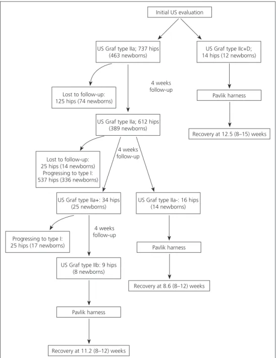
follow-up. Mean recovery time was 12.5 (8–16) weeks for type IIc and type D DDH, 8.6 (8–12) weeks for type IIa(-) DDH, and 11.2 (8–12) weeks for type IIb DDH (Figure 1). No patients experienced complications due to Pavlik harness. Three-month evaluation revealed that risk factors and concomitant orthopedic deformities were significantly more prevalent in newborns treated with Pavlik harness (Table 3). It was also observed that breech presentation, family history, and concomitant or-thopedic deformities were more prevalent in newborns with DDH that developed from type IIa (n=22) to type
IIb/IIa(-) than in those with DDH that developed from type IIa to type I (n=441) (Table 3).
Discussion
In the present cross-sectional study, 4782 newborns were screened for DDH using hip US at a single center in Istanbul. The center serves a population that has, for the most part, moved to the city from rural areas and has largely abandoned the practice of swaddling, which is still common in certain rural regions of Turkey. Al-though 10% of newborns were diagnosed with DDH Fig. 1. Follow-up and treatment of newborns diagnosed with DDH via hip US.
Initial US evaluation
US Graf type IIa; 737 hips (463 newborns)
US Graf type IIa; 612 hips (389 newborns)
US Graf type IIa+: 34 hips (25 newborns)
US Graf type IIb: 9 hips (8 newborns) Pavlik harness Pavlik harness Pavlik harness Recovery at 11.2 (8–12) weeks Recovery at 8.6 (8–12) weeks Recovery at 12.5 (8–15) weeks
US Graf type IIa-: 16 hips (14 newborns) Lost to follow-up: 125 hips (74 newborns) Lost to follow-up: 25 hips (14 newborns) Progressing to type I: 537 hips (336 newborns) Progressing to type I: 25 hips (17 newborns)
US Graf type IIc+D; 14 hips (12 newborns) 4 weeks follow-up 4 weeks follow-up 4 weeks follow-up
in the present study, no risk factors other than female sex and first birth order were significantly more preva-lent in infants with DDH. In addition to its significant role in diagnosing newborns with neither symptoms nor risk factors, hip US was effectively used during follow-up of DDH.
Studies from Turkey have revealed a wide range of DDH prevalence, 0.5–28.1%, depending on
screen-ing method, presence of risk factors, and region.[13–15]
Knowledge of DDH among primary care physicians in Turkey has been significantly improved by a hip screen-ing program in the recent years.[16] The aim of the
pres-ent study was to contribute to standards of screening for DDH.
Graf US was used to assess the relationship between the femoral head and acetabulum, and the classification of DDH. The Graf method, a quantitative classification system, is the most widely used, standardized technique
for DDH screening via US examination.[17]
Although US is currently the gold standard DDH diagnostic tool, no consensus exists regarding the use
of US for DDH screening.[18] Arti et al. recently used
US to screen 5800 newborns with risk factors or sus-picion upon clinical examination in Iran.[3] It was
deter-mined that 72% of hips included after US screening had been diagnosed as normal upon clinical examination, concluding that hip US is the gold standard method of evaluating DDH. In addition, Koşar et al. reported in a study that included 1321 male infants that up to 28% of DDH cases would have been missed by selective US screening protocol, concluding that all newborns should be assessed using hip US.[19] Dogruel et al. demonstrated
a low specificity for clinical findings and a need for US in diagnosing DDH, and Atalar et al. showed that low-grade dysplasia detected on US may go undetected on
radiography.[20,21] Furthermore, US has been reported
to have high interobserver reliability.[22] The findings of
the present study also demonstrate the effectiveness of hip US in the diagnosis of DDH. Prevalence of new-borns with abnormal hip US was approximately 10% (475/4782 newborns) at initial evaluation. Among these newborns, 12 were diagnosed with DDH (type IIc or D), and 463 had an immature hip, 22 of which devel-oped to types IIb or IIa(-) on follow-up.
Only gender and first birth order were found to cor-relate with increased risk of hip immaturity or DDH in the present study, a finding that sheds light on the im-portance of universal screening (Table 2). However, in 2 large trials that included 11925 and 15529 newborns who were randomly assigned to groups in which either all were screened (universal screening), only high-risk newborns were screened (selective screening), or none were screened, no statistically significant difference was determined regarding prevalence of late DDH among groups.[23,24] In addition, a recent Cochrane review
con-cluded that based on the available evidence, no clear recommendations for hip screening of newborn
in-fants can be given.[8] Currently, a combination of
uni-versal clinical screening and selective US screening is in practice.[14,25] During follow-ups, all risk factors and
concomitant deformities increased the risk of DDH (Table 3). However, Omeroglu et al. have reported that female newborns with type IIa are at greater risk for
DDH without spontaneous improvement.[26] In the
lit-erature, risk factors including breech presentation, fam-table 3. Risk factors and concomitant orthopedic deformities with respect to US Graf classification of newborn hips at third month.
Time of evalation third month
Type of Graf classification ı+ııa⇒ı ııa⇒ı ııa⇒ııb/ııa(-) ııa(-)+ııb+ııc+D
Number of newborns 1827+441a 441 22 34 Pb Pc Risk factors Firstborn child 222 (9.7) 80 (18.1) 11 (50.0) 16 (47.0) <0.001 <0.001 Breech presentation 241 (10.6) 48 (10.8) 9 (40.9) 13 (38.2) <0.001 <0.001 Family history 416 (18.3) 77 (17.4) 10 (45.4) 15 (44.1) <0.001 <0.001 Vaginal birth 1376 (60.6) 277 (62.8) 14 (63.6) 24 (70.5) 0.741 <0.05 Female gender 1331 (58.6) 323 (72.5) 19 (68.1) 22 (64.7) 0.142 <0.05 Swaddling 119 (5.24) 18 (4.08) 4 (18.1) 4 (11.7) <0.05 <0.05
Concomitant orthopedic deformities
Torticollis 37 (1.63) 3 (0.6) 1 (4.54) 1 (2.94) <0.05 <0.05
Foot deformities 30 (1.32) 4 (0.9) 1 (4.54) 2 (5.88) <0.05 <0.05
Data are presented as number of newborns (percentage to total newborns with corresponding Graf classification). aRisk factors and orthopedic deformities were
recorded for 2268 newborns at third month (1827 of the 4307 newborns with type I hips and 441 newborns with hips that developed from type IIa to type I);
ily history, and torticollis are shown to increase risk of DDH.[27–29]
As Pavlik harness has been proved superior to
ab-duction orthosis for primary management of DDH,[30]
harnesses were used as treatment in the present study. A recent review reported harness-related complications, including avascular necrosis and femoral nerve palsy.[31]
Outcome of treatment in the present study was success-ful in all cases without complication. In addition to its use as a screening tool, US has been used to monitor
re-duction during treatment.[25] US was also used to
moni-tor progress of DDH and response to harness treatment effectively in the present study.
Among subjects with abnormal US findings at ini-tial evaluation (n=475), most had immature (type IIa) DDH (n=463), the majority of whom were female (73.9%). Female gender is a known risk factor for DDH.
[20,32] In the present study, 22 newborns (21 girls)
diag-nosed as type IIb or IIa(-) were successfully treated with Pavlik harnesses.
In contrast to previous reports from Turkey demonstrating that swaddling is a risk factor for DDH,[20,26,30,32,33] swaddling rates among newborns with
and without DDH were similar in the present study. Re-cent literature supports the lack of association between
foot deformities such as clubfoot and DDH.[34]
Al-though the present findings run counter to widely held beliefs among many surgeons, they are similar to those of previous studies, which indicate a lack of association
between risk factors and DDH diagnosis.[19] Abnormal
findings concurrent with risk factors and concomitant orthopedic deformities occurred at similar rates in new-borns with and without DDH.
The main limitations of the present study were ret-rospective design and small sample size. Retret-rospective design led to the exclusion of newborns that had not been screened at the study center, inconsistent physical examination findings due to intraobserver variability, and examination of only a subset of patients for clini-cal risk factors, which may have introduced bias. Fur-thermore, several conclusions may have been influenced by the study of a population served by a single institu-tion. In addition, US evaluations were performed by the same radiologists throughout the study, which may also have introduced bias. Finally, prevalence of DDH in the present small sample may not reflect its prevalence in the larger Turkish population, precluding authors from forming conclusions regarding DDH risk factors. Nev-ertheless, the present findings contribute to DDH lit-erature, in which there is still no consensus on the use of US for screening and follow-up. Further large-scale and
prospective studies are needed to reach a definitive con-clusion regarding the necessity of universal US screen-ing, taking time and cost-effectiveness into account.
In conclusion, the findings of the present study sup-port US screening of firstborn and female newborns. Due to lack of statistical difference regarding risk factors and concomitant deformities among newborns with and without DDH, universal screening is recommended. Conflics of Interest: No conflicts declared.
references
1. Kokavec M, Bialik V. Developmental dysplasia of the hip. Prevention and real incidence. Bratisl Lek Listy 2007;108:251–4.
2. Loder RT, Skopelja EN. The epidemiology and demograph-ics of hip dysplasia. ISRN Orthop 2011;2011:238607. 3. Arti H, Mehdinasab SA1, Arti S2. Comparing results of
clinical versus ultrasonographic examination in develop-mental dysplasia of hip. J Res Med Sci 2013;18:1051–5. 4. Phelan N, Thoren J, Fox C, O’Daly BJ, O’Beirne J. De-velopmental dysplasia of the hip: incidence and treat-ment outcomes in the Southeast of Ireland. Ir J Med Sci 2015;184:411–5. CrossRef
5. Weinstein SL, Mubarak SJ, Wenger DR. Developmental hip dysplasia and dislocation: Part II. Instr Course Lect 2004;53:531–42.
6. Gulati V, Eseonu K, Sayani J, Ismail N, Uzoigwe C, Choudhury MZ, et al. Developmental dysplasia of the hip in the newborn: A systematic review. World J Orthop 2013;4:32–41. CrossRef
7. Shorter D, Hong T, Osborn DA. Cochrane Review: Screening programmes for developmental dysplasia of the hip in newborn infants. Evid Based Child Health 2013;8:11–54. CrossRef
8. van der Sluijs JA, De Gier L, Verbeke JI, Witbreuk MM, Pruys JE, van Royen BJ. Prolonged treatment with the Pavlik harness in infants with developmental dysplasia of the hip. J Bone Joint Surg Br 2009;91:1090–3. CrossRef
9. Gray A, Elbourne D, Dezateux C, King A, Quinn A, Gardner F. Economic evaluation of ultrasonography in the diagnosis and management of developmental hip dyspla-sia in the United Kingdom and Ireland. J Bone Joint Surg Am 2005;87:2472–9. CrossRef
10. Graf R. Hip sonography. Diagnosis and management of infant hip dysplasia, 2nd edn. Springer, Berlin 2006.
11. Graf R. Classification of hip joint dysplasia by means of sonography. Arch Orthop Trauma Surg 1984;102:248– 55. CrossRef
12. Pavlik A. Method of functional therapy with strap braces as a principle of conservative therapy of congenital disloca-tion of the hip in infants. [Article in German] Z Orthop
Ihre Grenzgeb 1957;89:341–52. [Abstract]
13. Songür M, Akel I, Karahan S, Kuzgun U, Tümer Y. Preva-lence of untreated hip dislocation in Turkish children aged 6 months to 14 years. Acta Orthop Traumatol Turc 2011;45:215–20. CrossRef
14. Omeroglu H, Koparal S, Bicimoglu A, Karademir A. The relationship between risk factors, clinical findings and ul-trasonographic findings in developmental hip dysplasia. Acta Orthop Traumatol Turc 1999;33:30–4.
15. Tümer Y, Omeroglu H. Prevention of developmental hip dysplasia in Turkey. Acta Ortop Traumatol Turc 1997;31:176–81.
16. Doğruel H, Atalar H, Yavuz Oy, Uraş İ, Günay C, Şayli U. An Evaluation of Developmental Hip Dysplasia Fre-quency and Screening Programs in Turkey: Review. Tur-kiye Klinikleri J Med Sci 2008;28:357–60.
17. Omeroğlu H. Use of ultrasonography in developmental dysplasia of the hip. J Child Orthop 2014;8:105–13. CrossRef
18. Scott-Jupp R. Ultrasound hip screening: why bother? Arch Dis Child 2013;98:985. CrossRef
19. Koşar P, Ergun E, Yiğit H, Gökharman FD, Kosar U. De-velopmental dysplasia in male infants: risk factors, insta-bility and ultrasound screening. Hip Int 2011;21:409– 14. CrossRef
20. Dogruel H, Atalar H, Yavuz OY, Sayli U. Clinical exami-nation versus ultrasonography in detecting developmental dysplasia of the hip. Int Orthop 2008;32:415–9. CrossRef
21. Atalar H, Dogruel H, Selek H, Tasbas BA, Bicimoglu A, Gunay C. A comparison of ultrasonography and ra-diography in the management of infants with suspected developmental dysplasia of the hip. Acta Orthop Belg 2013;79:524–9.
22. Copuroglu C, Ozcan M, Aykac B, Tuncer B, Saridogan K. Reliability of ultrasonographic measurements in sus-pected patients of developmental dysplasia of the hip and correlation with the acetabular index. Indian J Orthop 2011;45:553–7. CrossRef
23. Rosendahl K, Markestad T, Lie RT. Ultrasound screening for develop- mental dysplasia of the hip in the neonate: the effect on treatment rate and prevalence of late cases.
Pediatrics 1994;94:47–52.
24. Holen KJ, Tegnander A, Bredland T, Johansen OJ, Saeth-er OD, Eik-Nes SH, et al. UnivSaeth-ersal or selective screen-ing of the neonatal hip usscreen-ing ultrasound? A prospective, randomised trial of 15,529 newborn infants. J Bone Joint Surg Br 2002;84:886–90. CrossRef
25. Fitch RD. Ultrasound for screening and manage-ment of developmanage-mental dysplasia of the hip. N C Med J 2014;75:142–5. CrossRef
26. Omeroğlu H, Caylak R, Inan U, Köse N. Ultrasonograph-ic Graf type IIa hip needs more consideration in newborn girls. J Child Orthop 2013;7:95–8. CrossRef
27. Omeroğlu H, Koparal S. The role of clinical examination and risk factors in the diagnosis of developmental dyspla-sia of the hip: a prospective study in 188 referred young infants. Arch Orthop Trauma Surg. 2001;121:7–11. CrossRef
28. Tien YC, Su JY, Lin GT, Lin SY. Ultrasonographic study of the coexistence of muscular torticollis and dysplasia of the hip. J Pediatr Orthop 2001;21:343–7. CrossRef
29. Bache CE, Clegg J, Herron M. Risk factors for develop-mental dysplasia of the hip: ultrasonographic findings in the neonatal period. J Pediatr Orthop B 2002;11:212–8. 30. Sankar WN, Nduaguba A, Flynn JM. Ilfeld abduction
or-thosis is an effective second-line treatment after failure of Pavlik harness for infants with developmental dysplasia of the hip. J Bone Joint Surg Am 2015;97:292–7. CrossRef
31. Tibrewal S, Gulati V, Ramachandran M. The Pavlik meth-od: a systematic review of current concepts. J Pediatr Or-thop B 2013;22:516–20. CrossRef
32. Akman A, Korkmaz A, Aksoy MC, Yazici M, Yurdakök M, Tekinalp G. Evaluation of risk factors in developmen-tal dysplasia of the hip: results of infantile hip ultrasonog-raphy. Turk J Pediatr 2007;49:290–4.
33. Guner SI, Guner S, Peker E, Ceylan MF, Guler A, Turk-tas U, et al. Are consanguineous marriage and swaddling the risk factors of developmental dysplasia of the hip? J Membr Biol 2013;246:115–9. CrossRef
34. Chou DTs, Ramachandran M1. Prevalence of develop-mental dysplasia of the hip in children with clubfoot. J Child Orthop 2013;7:263–7. CrossRef
![table 1. Graf classification of DDH. [10,11]](https://thumb-eu.123doks.com/thumbv2/9libnet/5411233.102466/2.850.59.797.150.381/table-graf-classification-of-ddh.webp)