Mediterranean Journal of Hematology and Infectious Diseases
Scientific LetterDNA Repair Genes and Chronic Myeloid Leukemia: ERCC2 (751), XRCC1 (399),
XRCC4-Intron 3, XRCC4 (-1394) Gene Polymorphisms
Keywords: Polymorphisms; DNA repair genes; CML.
Published: March 1, 2021 Received: November 14, 2020 Accepted: February 12, 2021
Citation:Ozdilli K., Pehlivan M., Serin I., Savran F.O., Tomatir A.G., PehlivanS.DNA repair genes and Chronic Myeloid Leukemia: ERCC2 (751), XRCC1 (399), XRCC4-Intron 3, XRCC4 (-1394) gene polymorphisms. Mediterr J Hematol Infect Dis 2021, 13(1): e2021020, DOI:
http://dx.doi.org/10.4084/MJHID.2021.020
This is an Open Access article distributed under the terms of the Creative Commons Attribution License (https://creativecommons.org/licenses/by-nc/4.0), which permits unrestricted use, distribution, and reproduction in any medium, provided the original work is properly cited.
To the editor.
Chronic myeloid leukemia (CML), which is characterized by the overproduction of mature cells in the granulocytic series, is included in the group of
chronic myeloproliferative neoplasms.1 It is the first
disease ascertained as due to a specific chromosomal anomaly emerging from a reciprocal translocation between chromosomes 9 and 22. A chimeric gene denominated as the Philadelphia (Ph) chromosome is the product of the fusion of the Abelson oncogene (ABL) from chromosome 9q34 with the breakpoint cluster
region (BCR) on chromosome 22q11.2, t
(9;22)(q34;q11.2).1
New approaches are tried to be developed in evaluating the prognosis and treatment response. DNA repair mechanisms create a new study area for CML and constitute the subject of our study. There are more than 100 known DNA repair genes. Polymorphisms and/or functional gene variants occurring in these genes with environmental factors increase the cancer tendency by
disrupting the DNA repair mechanism.2
The ERCC2 (excision repair cross complementation group 2) gene acts on nucleotide excision repair (NER)
and is located in the 13.3rd district of the Q part in the
19th chromosome.5 Polymorphisms in the ERCC2 gene
provide information about DNA repair capacity and cancer risk. ERCC2 repair gene polymorphisms are
significantly associated with breast, colorectal,
pancreatic, bladder, lung, esophageal cancers and
hematological malignancies.3-5
The XRCC1 (X-ray repair cross-complementing group 1) gene is one of the BER genes and is located in
the 13.2 district of the q part in the 19th chromosome.
This gene has 17 exons required to synthesize DNA
proteins, including DNA polymerase.5 Polymorphisms
in the XRCC1 repair gene have been investigated, especially in colorectal, breast, pancreatic, head and neck, lung, prostate, and skin cancers.
The DNA repair protein XRCC4, also known as
X-ray repair cross-complementing protein 4, is a protein encoded in humans by the XRCC4 gene. XRCC4, an important non-homologous splice repair gene, acts as an essential scaffold protein between this complex and DNA Ligase IV in the DNA double-stranded break
repair pathway process.6
In our study, we aimed to examine the effect of ERCC2 (751), XRCC1 (399), XRCC4-Intron 3, and XRCC4 (-1394) gene polymorphism on CML, prognosis, and treatment response in patients.
Patients and Methods. Sixty-two (62) CML patients,
diagnosed and followed up in the Gaziantep University Hematology Clinic between January 2008 - January 2016, and a control group of 70 healthy people were included in the study. In addition to demographic data such as age and gender, initial Sokal risk scores, presence of splenomegaly, initial laboratory values
(hemoglobin, leukocytes, platelets), treatment
preferences (imatinib or interferon alfa), responses at 18 months according to European Leukemia Net (ELN) criteria, mortality, presence of any events, chromosome abnormalities, overall survival (OS) and event-free survival (EFS) durations (months) were recorded. The median age of all 62 patients included in the study was 41 (range: 20-74)
DNA isolation from peripheral blood leukocytes of CML patients and controls was performed using the
saline precipitation method (Miller et al.).7 ERCC2,
XRCC1, XRCC4-Intron 3, XRCC4 (-1394) gene polymorphism genotypes were analyzed by Polymerase chain reaction (PCR) and/or Polymerase chain reaction-restriction fragment length polymorphism (PCR-RLFP) method.
SPSS for Windows (version 13.0; SPSS, Chicago, IL) software was used for data analysis. Logistic regression analysis was used to determine the statistical significance of the differences between control groups and patients. The odds ratios (OR) and 95% confidence
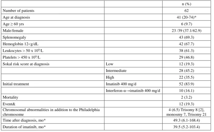
Table 1. Clinical Features of the Chronic Myeloid Leukemia in Chronic Phase Patients. n (%) Number of patients 62 Age at diagnosis 41 (20-74)* Age ≥ 60 yrs 6 (9.7) Male/female 23 /39 (37.1/62.9) Splenomegaly 43 (69.3) Hemoglobin 12<g/dL 42 (67.7) Leukocytes > 50 x 109/L 38 (61.3) Platelets > 450 x 109/L 29 (46.8)
Sokal risk score at diagnosis Low 12 (19.3)
Intermediate 28 (45.2)
High 22 (35.5)
Initial treatment Imatinib 400 mg/d 52 (83.9)
Interferon-α→imatinib 400 mg/d 10 (16.1)
Mortality 2 (3.2)
Event& 12 (19.3)
Chromosomal abnormalities in addition to the Philadelphia
chromosome
4 (6.5) Trisomy 8 [2], monosmy 7, Trisomy 21
Time after diagnosis, mo* 49.3 (6.1-168.4)
Duration of imatinib, mo* 39.5 (5.2-103.4)
ELN: European Leukemia Net, * median, mo: months, &death (2), progression to AP or blastic phase (2), loss of an MCyR (8).
intervals were used for this analysis. The X2 test was
used to compare the differences between the patient groups and the control group's DNA Repair Gene XRCC4 variable number tandem repeat (VNTR) at intron 3 and -1394), XRCC1, ERCC2 allele frequency. Fisher's test was used as needed. P values <0.05 were considered to indicate statistical significance. The Kaplan-Meier method was used to estimate the survival probabilities and the log-rank test to compare differences. The significance of risk factors was confirmed by applying The Cox stepwise regression analysis. In the multivariate analysis, the stepwise (backward) eliminated variables were used with a significance of less than 10%.
Results. Looking at the molecular responses of the
patients at 18 months, 42 were in the optimal (67.7%), 13 were in the warning (21%), and 7 (11.3%) were in the failure group. End-of-study mortality was 3.2% with two patients (Table 1).
Twelve (12) of the patients experienced any "event" (19.3%). Two of them were exitus (3.2%), 2 of them showed a progression to accelerated phase or blastic phase (3.2%), and 8 of them lost major molecular response (MMR) (12.9%). The median follow-up period was 49.3 months (6.1-168.4), and the median use of imatinib was 39.5 months (5.2-103.4) (Table 1).
When the genotype differences for ERCC2, XRCC1, and XRCC4 (-1394) between CML and healthy controls were analyzed, there was no statistically significant difference found between the two groups (p> 0.05).
When XRCC4-Intron 3 was examined, it was observed that there was a significant statistical difference in DD and II genotypes between CML and the control group (p = 0.018, p = 0.028). It was also observed that the DD genotype was 7.299 times protective factor for CML, and patients with II genotype have 2.379 times increased risk of CML (Table 2).
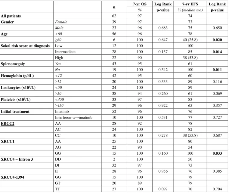
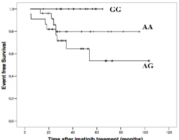
Four different factors were found to be statistically significant for EFS. Young age (<60) (p = 0.020), absence of splenomegaly (p = 0.011), presence of low Sokal risk score at initial diagnosis (p = 0.0148) and presence of XRCC1 GG genotype (p = 0.033) were statistically significant for better EFS (Table 3, Figure
1).
Discussion. The literature data about DNA repair
mechanisms in hematological malignancies is limited.
In a study conducted by Salimizand et al.,8 simultaneous
effects of polymorphism of three separate DNA repair genes were investigated on CML development. T allele of ABCB1 C3435T, T allele of XRCC1 Arg194Trp, and C allele of ABCG2 C421A polymorphisms were significantly higher CML patients compared to controls. TT genotype of ABCB1 and XRCC1 has been associated with a higher risk of developing CML.
In a meta-analysis, Wang et al.9 examined the
relationship between the Arg399Gln single nucleotide polymorphism (SNP) in the XRCC1 gene and the risk of leukemia. No association was found between XRCC1 and CML. Among the articles discussed in this meta-analysis, 2 of them were directly related to CML:
Table 2. Comparison of Frequencies of ERCC2, XRCC1, XRCC4 - Intron 3 and XRCC4 (-1394) gene Polymorphisms between Patients with
Chronic Myeloid Leukemia and Healthy Controls.
Genotype CML Healthy Control OR 95% CI p
n a (%) n b (%)
ERCC2 (751) AA 28 (45.2) 37 (52.9) 0.753* 0.264-2.146* 0.595*
AC 24 (38.7) 24 (34.3) 1.096* 0.362-3.317* 0.871*
CC 10 (16.1) 9 (12.8) 1.303& 0.492-3.451& 0.627&
XRCC1 (-399) AA 25 (40.3) 25 (35.7) 1.582* 0.650-3.851* 0.312*
AG 22 (35.5) 24 (34.3) 1.485* 0.600-3.677* 0.393*
GG 15 (24.2) 21 (30) 0.745& 0.343-1.615& 0.558&
XRCC4- Intron 3 DD 2 (3.2) 10 (14.3) 0.137* 0.027-0.708* 0.018*
DI 32 (51.6) 42 (60) 0.496* 0.232-1.058* 0.069*
II 28 (45.2) 18 (25.7) 2.379& 1.143-4.952& 0.028&
XRCC4-1394 GG 15 (24.2) 17 (24.3) 0.793* 0.321-1.956* 0.614*
GT 20 (32.3) 26 (37.1) 0.768* 0.344-1.715* 0.520*
TT 27 (43.5) 27 (38.6) 1.229& 0.613-2.463& 0.598&
an=62, bn=70, *:OR (95%CI) was adjusted by age and sex, &Fisher's Exact Test.
Table 3. Univariate analysis (Logrank test) of ERCC2, XRCC1, XRCC4 - Intron 3 and XRCC4 (-1394) Gene Polymorphisms in 62 Patients
with Chronic Phase -Chronic Myeloid leukemia.
n 7-yr OS Log Rank 7-yr EFS Log Rank
% p-value % (median mo) p-value
All patients 62 97 74
Gender Female 39 97 73
Male 23 96 0.683 75 0.650
Age <60 56 96 78
≥60 6 100 0.647 40 (25.8) 0.020
Sokal risk score at diagnosis Low 12 100 100
Intermediate 28 100 0.137 85 0.014 High 22 90 38 (53.8) Splenomegaly Yes 43 95 61 No 19 100 0.342 100 0.011 Hemoglobin (g/dL) <12 42 95 60 ≥12 20 100 0.333 89 0.116 Leukocytes (x109/L) <50 24 100 89 ≥50 38 94 0.260 61 0.069 Platelets (x109/L) <450 33 97 83 ≥450 29 96 0.922 65 0.357
Initial treatment Imatinib 52 96 76
Interferon-α→imatinib 10 100 0.531 77 0.727 ERCC2 AA 28 92 78 AC 24 100 82 CC 10 100 0.278 38 (53.8) 0.687 XRCC1 AA 25 100 80 AG 22 90 54 GG 15 100 0.160 100 0.033 XRCC4 – Intron 3 DD 2 100 50 DI 32 97 73 II 28 96 0.956 76 0.385 XRCC4-1394 GG 15 100 79 GT 20 89 79 TT 27 100 0.097 70 0.704
Figure 1. Kaplan-Meier Plots on Event-free Survival (EFS)
According to XRCC1 genotypes.
Deligezer et al.10 investigated the association of XRCC1
gene polymorphism Arg399Gln with CML and could not obtain a significant difference among patient groups. Similarly, in our study, no relationship was found between this polymorphism and CML. Annamaneni et
al.11 studied the XRCC1 effect on CML and
polymorphisms of XRCC1, codon 399, 280 and 194; similarly, no significant difference was detected.
Dhangar et al.12 investigated the correlation between
clinical response to therapy between CML and XRCC1
rs1799782, rs25487, and ERCC2 rs13181
polymorphisms; no significant relationship was found.
Banescu et al.13 also examined the relationship between
CML and XRCC1 Arg399Gln, Arg280His, Arg194Trp,
XRCC3 r241Met, and ERCC2 Lys751Gln
polymorphisms and showed that the ERCC2 Lys751Gln genotype increases the risk of CML.
Ozcan et al.14 investigated the place of ERCC2 and
XRCC1 gene polymorphisms in different hematological malignancies. In his study, he showed that a decrease in
the Gln / Gln genotype and the Gln allele in the ERCC2 codon 751 and XRCC1 codon 399 polymorphisms play a protective role in AML, and an increase in Lys/Lys genotype in acute leukemia was associated with early relapse.
Joshi et al.15 studied XRCC1 and ERCC2
polymorphisms in myelodysplastic syndrome (MDS), showing that the progression of MDS to AML be the result of the gradual accumulation of DNA mutations that create a defect in DNA repair. DNA repair gene XRCC1 (Arg280His) (p = 0.05) and ERCC2 (Lys751Gln) (p = 0.01). Polymorphisms were significantly higher in MDS patients compared to controls. There was a significant difference between RAEB I and XRCC1, being XRCC1 polymorphisms strongly associated with the advanced MDS subgroup.
In our study, different from the other two main studies, we also had the opportunity to evaluate XRCC4 and CML's relationship. When the genotype differences between CML and healthy control groups were statistically analyzed, no statistically significant difference could be found between them. However, when XRCC4-Intron 3 was examined, it was seen that there was a significant statistical difference in DD and II genotypes between CML and the control group. Additionally, it was observed that the DD genotype was 7.299 times protective factor for CML, and patients with II genotype have 2.379 times increased risk of CML.
The study also had some limitations. The most important limitation is the small patient population. It is thought that significant results can be obtained in terms of disease parameters and prognosis with the data in a larger patient group. Besides, only imatinib and interferon-related treatment results could be evaluated in our study. It would be more meaningful in terms of the
literature to conduct a study on 2nd generation tyrosine
kinase inhibitors (TKIs) throughout a broader period.
Kursat Ozdilli
1, Mustafa Pehlivan
2,3, Istemi Serin
2, Fatma Oguz Savran
4, Ayse Gaye Tomatir
5and Sacide
Pehlivan
4.
1
Medipol University, Faculty of Medicine, Department of Medical Biology.
2
University of Health Sciences, Istanbul Training and Research Hospital, Hematology Clinic.
3Gaziantep University, Faculty of Medicine, Department of Hematology.
4
Istanbul University, Istanbul Faculty of Medicine, Department of Medical Biology.
5Pamukkale University, Faculty of Medicine, Department of Medical Biology.
Competing interests: The authors declare no conflict of Interest.
Correspondence to: Istemi Serin, MD. University of Health Sciences, Istanbul Training and Research Hospital, Department of Hematology, Org.Nafiz GURMAN Cad, 34098, Fatih, Istanbul. Tel: 0090 532 3172393. E-mail: [email protected]
References:
1. Kurzrock R, Kantarjian HM, Druker BJ, Talpaz M. Philadelphia chromosome-positive leukemias: from basic mechanisms to molecular therapeutics. Ann Intern Med. 2003 May 20;138(10):819-30.
https://doi.org/10.7326/0003-4819-138-10-200305200-00010 PMid:12755554
acute myeloblastic leu-kemia. Hematol Oncol. 2000 Sep;18(3):99-110.
https://doi.org/10.1002/1099-1069(200009)18:3<99::AID-HON662>3.0.CO;2-Z
3. Taylor EM, Broughton BC, Botta E et al. Xeroderma pigmentosum and trichothiodystrophy are as-sociated with different mutations in the XPD (ERCC2) repair/transcription gene. Proc Natl Acad Sci U S A. 1997 Aug 5;94(16):8658-63.
https://doi.org/10.1073/pnas.94.16.8658 PMid:9238033 PMCid:PMC23065
4. Artac M, Bozcuk H, Pehlivan S et al (2010). The value of XPD and XRCC1 genotype polymor-phisms to predict clinical outcome in metastatic colorectal carcinoma patients with irinotecan-based regimens. J. Cancer Res. Clin. Oncol. 136: 803-809.
https://doi.org/10.1007/s00432-009-0720-3 PMid:19908066
5. Chacko P, Rajan B, Joseph T, Mathew BS, Pillai MR. Polymorphisms in DNA repair gene XRCC1 and increased genetic susceptibility to breast cancer. Breast Cancer Res Treat. 2005;89(1):15-21.
https://doi.org/10.1007/s10549-004-1004-x PMid:15666192
6. Lu J, Wang XZ, Zhang TQ et al. Prognostic significance of XRCC4 expression in hepatocellular carcinoma. Oncotarget. 2017 Sep 28;8(50):87955-87970.
https://doi.org/10.18632/oncotarget.21360 PMid:29152133 PMCid:PMC5675685
7. Miller SA, Dykes DD, Polesky HF. A simple salting out procedure for extracting DNA from human nucleated cells. Nucleic Acids Res. 1988 Feb 11;16(3):1215.
https://doi.org/10.1093/nar/16.3.1215 PMid:3344216 PMCid:PMC334765
8. Salimizand H, Amini S, Abdi M, Ghaderi B, Azadi NA. Concurrent effects of ABCB1 C3435T, ABCG2 C421A, and XRCC1 Arg194Trp genetic polymorphisms with risk of cancer, clinical out-put, and response to treatment with imatinib mesylate in patients with chronic myeloid leukemia. Tumour Biol. 2016 Jan;37(1):791-8.
https://doi.org/10.1007/s13277-015-3874-4
PMid:26250462
9. Wang F, Zhao Q, He HR et al. The association between XRCC1 Arg399Gln polymorphism and risk of leukemia in different populations: a meta-analysis of case-control studies. Onco Targets Ther. 2015 Nov 6;8:3277-87.
https://doi.org/10.2147/OTT.S92752 PMid:26609240 PMCid:PMC4644162
10. Deligezer U, Akisik EE, Dalay N. Lack of association of XRCC1 codon 399Gln polymorphism with chronic myelogenous leukemia. Anticancer Res. 2007 Jul-Aug;27(4B):2453-6.
11. Annamaneni S, Gorre M, Kagita S et al. Association of XRCC1 gene polymorphisms with chronic myeloid leukemia in the population of Andhra Pradesh, India. Hematology. 2013 May;18(3):163-8.
https://doi.org/10.1179/1607845412Y.0000000040 PMid:23320983
12. Dhangar S, Shanbhag V, Shanmukhaiah C et al. Lack of association between functional polymor-phism of DNA repair genes (XRCC1, XPD) and clinical response in Indian chronic myeloid leuke-mia patients. Mol Biol Rep. 2019 Oct;46(5):4997-5003.
https://doi.org/10.1007/s11033-019-04950-0 PMid:31286393
13. Bănescu C, Trifa AP, Demian S, Benedek Lazar E, Dima D, Duicu C, Dobreanu M. Polymorphism of XRCC1, XRCC3, and XPD genes and risk of chronic myeloid leukemia. Biomed Res Int. 2014;2014:213790.
https://doi.org/10.1155/2014/213790 PMid:24955348 PMCid:PMC4052066
14. Özcan, A, Pehlivan, M, Tomatir, A et al. (2011). Polymorphisms of the DNA repair gene XPD (751) and XRCC1 (399) correlates with risk of hematological malignancies in Turkish population. African Journal of Biotechnology. 10. 8860-8870. 10.5897/AJB10.1839.
https://doi.org/10.5897/AJB10.1839
15. Joshi D, Korgaonkar S, Shanmukhaiah C, Vundinti BR. Association of XPD (Lys751Gln) and XRCC1 (Arg280His) gene polymorphisms in myelodysplastic syndrome. Ann Hematol. 2016 Jan;95(1):79-85.
https://doi.org/10.1007/s00277-015-2528-3 PMid:26482462