APPLICATIONS OF BIOMATERIALS IN CANCER
DIAGNOSIS AND TREATMENT
A THESIS
SUBMITTED TO THE DEPARTMENT OF MOLECULAR BIOLOGY AND GENETICS
AND THE GRADUATE SCHOOL OF ENGINEERING AND SCIENCE OF BILKENT UNIVERSITY
IN PARTIAL FULFILLMENT OF THE REQUIREMENTS FOR THE DEGREE OF
MASTER OF SCIENCE
By
Damla Gözen
July, 2013
ii
I certify that I have read this thesis and that in my opinion it is fully adequate, in scope and in quality, as a thesis for the degree of Master of Science.
Assist. Prof. Dr. Kamil Can Akçalı (Advisor)
I certify that I have read this thesis and that in my opinion it is fully adequate, in scope and in quality, as a thesis for the degree of Master of Science.
Assoc. Prof. Dr. Ihsan Gürsel
I certify that I have read this thesis and that in my opinion it is fully adequate, in scope and in quality, as a thesis for the degree of Master of Science.
Prof. Dr. Gülay Bayramoğlu
Approved for the Graduate School of Engineering and Science
Prof. Dr. Levent Onural Director of the Graduate School
iii
ABSTRACT
APPLICATIONS OF BIOMATERIALS IN CANCER DIAGNOSIS AND TREATMENT
Damla Gözen
M.Sc. in Molecular Biology and Genetics Supervisor: Assist. Prof. Dr. Kamil Can Akçalı
July, 2013
Cancer remains to be a major burden of disease worldwide, despite the significant increase in the number of studies that focus on the development of novel diagnostic and treatment strategies. Recently, important part of these studies use biomaterials and their biomedical applications have been investigated extensively, due to their biocompatibility. Among these biomaterials two of them, carbon nanotubes (CNT) and polymer hydrogels have gained great importance due to their unique physical and chemical properties. The current study proposes new approaches that take advantage of these two biomaterials which could be used in the treatment and diagnosis of hepatocellular carcinoma (HCC). We first proposed the usage of CNTs as novel diagnostic tools for the determination of the aggressiveness of HCC. Two cell lines with different epithelial-to-mesenchymal (EMT) status, HUH7 and Snu182 were used and their attachment features on patterned CNT surfaces were compared. Our SEM images and MTT results revealed that the cells with epithelial phenotype (HUH7) attach and proliferate more on CNTs than the cells with mesenchymal phenotype (Snu182) which makes these surfaces promising diagnostic tools to differentiate HCC according to their aggressiveness. Secondly, polymer hydrogels with Dox release were suggested to be promising therapeutics to cure HCC. Our cell viability and cytotoxicity tests showed the inhibition of the proliferation of HCC line, SNU398 in the presence of drug-releasing hydrogels. This suggests the usage of hydrogels as drug delivery vehicles to have enhanced therapeutic efficacies in the HCC therapies.
iv ÖZET
BİYOMATERYALLERİN KANSER TEŞHİS VE TEDAVİSİNDEKİ KULLANIMI
Damla Gözen
Moleküler Biyoloji ve Genetik, Yüksek Lisans Tezi Tez Yöneticisi: Yard. Doç. Dr. Kamil Can Akçalı
Temmuz, 2013
Günümüzde birçok bilimsel çalışma kanser teşhis ve tedavisinde yeni ve daha etkili stratejiler geliştirmeye odaklanmış olsa da kanser hala dünyadaki başlıca hastalık nedenlerinden biridir. Bu çalışmalardan önemli bir kısmı biyomateryallerin biyolojik uyumlu özelliğinden dolayı biyomedikal alandaki uygulamalarını araştırmaya yönelmiştir. Bu biomateryaller arasında karbon nanotüpler (KNT) ve polimer hidrojeller eşsiz fiziksel ve kimyasal özellikleri sayesinde büyük önem kazanmaktadırlar. Bu çalışma bu iki biyomateryalin özelliklerinden yaralanarak karaciğer kanseri teşhis ve tedavisi için yeni yaklaşımlar getirmeyi amaçlamaktadır. İlk olarak, karaciğer kanserinin agresifliğini belirlemek için teşhis esnasında KNTlerin kullanımı test edildi. Değişik epitel ve mezenkimal fenotipteki iki hücre hattının, HUH7 ve Snu182, KNT yüzeyleri üzerindeki yapışma özellikleri karşılaştırıldı. Taramalı elektron mikroskobunda elde edilen görüntüler ve MTT sonuçları epitel fenotipteki HUH7 hücrelerinin KNT yüzeyler üzerindeki yapışma ve büyümesinin mezenkimal fenotipteki Snu182 hücrelerine göre daha fazla olduğunu göstermiştir. Bu sonuç KNT yüzeylerinin karaciğer kanserinin agresifliğini belirlemek için kullanılabileceğini göstermektedir. Bu çalışmadaki ikinci hedefimiz, Dox içeren polimer hidrojellerin karaciğer kanseri tedavisinde etkili olarak kullanılabileceğini göstermekti. Yapılan hücre sayım ve sitotoksisite testleri ile karaciğer kanseri hücre hattı Snu398in çoğalmasının ilaç içeren hidrojellerin varlığında engellendiği gösterilmiştir. Bu sonuçlar, hidrojellerin ilaç taşıyıcısı olarak
kullanıldığında kaaciğer kanseri tedavisinin tesirini arttırabileceğini öne sürmektedir.
v
vi
Acknowledgements
I would like to express my gratitude to my supervisor Assist. Prof. Dr. Can Akçalı for giving me the opportunity to work in his lab, and providing his personal and academic guidance throughout my studies. Working with him was a great honor for me.
I would like to thank the members of Akçalı Group, Verda Bitirim, Merve Aydın, Ece Akhan and Şahika Cıngır for being very friendly, patient, helpful and encouraging. I could not have accomplished this without their tremendous help during my research. I am very grateful to Assist. Prof. Dr. Erman Bengü for his supervision and to the members of Bengü group, especially Gökçe Küçükayan for her tremendous help during the chemistry studies.
I would like to thank Prof. Dr. Yakup Arıca and Prof. Dr. Gülay Bayramoğlu from Gazi University, for providing the hydrogels for my studies.
I would like to offer my special thanks to the MBG faculty, for providing me with full support and academic knowledge that I will need throughout my future life. I would like to especially thank Assoc. Prof. Dr. İhsan Gürsel for his contribution.
I would like to thank Derya, Merve Ç. and Hande, for being more than friends and for their endless support and encouragement.
I am indebted to all the past and present members of MBG family, especially to Şahika, Mehmet, Azer, Pelin, Ceyhan and the members of Öztürk group for their friendship and support.
vii
I would like to express my gratitude to my family, especially to my dearest mother, for her unconditional and endless love and support not only in my academic life but also in all the decisions I make throughout my whole life.
viii
Table of Contents
1 Introduction ... 1
1.1 Hepatocellular Carcinoma ... 2
1.2 The use of VA-CNTs in Cancer Diagnosis ... 3
1.2.1 Epithelial-to-Mesenchymal Transition ... 3
1.2.2 The use of CNTs in Cancer Diagnosis... 5
1.3 The use of Hydrogels in Cancer Treatment ... 5
1.3.1 Doxorubucin ... 6
1.3.3 Hydrogels and Cancer Treatment ... 7
1.4 Subject Outline of the Thesis ... 9
1.4.1 The use of VA-CNTs in Cancer Diagnosis ... 9
1.4.2 The use of Hydrogels in Cancer Treatment ... 9
2 Materials and Methods ... 10
2.1 Materials ... 10
2.1.1 Synthesis of VA-CNTs ... 10
2.1.2 Preparation of Dox-releasing p(HPMA/PEG–MEMA) hydrogel films formulation ... 11
2.1.3 Cell Culture ... 12
2.1.4 Standard Buffers and Solutions ... 12
2.1 Methods ... 12
2.2.1 Total RNA Isolation... 12
ix
2.2.3 Reverse Transcriptase Polymerase Chain Reaction (RT-PCR) ... 13
2.2.4 Agarose Gel Electrophoresis ... 14
2.2.5 Imaging of the Cells on VA-CNTs ... 15
2.2.6 Determination of the Proliferation of Cells on VA-CNTs ... 15
2.2.7 Antitumor activity of Dox loaded hydrogel Formulations ... 15
2.2.8 Cytotoxicity of Biomaterials on Cells ... 16
2.2.9 Controlled release studies of Dox from the hydrogel film formulations ……… ... 16
2.2.10 Statistical Analysis ... 17
3 Results ... 18
3.1 The use of VA-CNTs in Cancer Diagnosis ... 18
3.1.1 Preparation of VA-CNTs ... 18
3.1.2 EMT Marker Expression of HCC ... 19
3.1.3 Proliferation and Attachment of HCCs on Patterned CNTs ... 20
3.2 The Use of Hydrogels in the Treatmentof Cancer ... 25
3.2.1 The Characteristics of Hydrogels ... 25
3.2.2 In vitro Dox-relese studies ... 27
3.2.3 The effects of drug releasing hydrogels on the proliferation features of cancer cells ... 30
4 Discussion ... 34
5 Future Perspectives ... 39
6 References ... 41
7 Appendicex ... 48
x
List of Figures
Figure 1.1. Mechanisms for Hepatocellular carcinogenesis ... 2
Figure 1.2. The Epithelial-to-Mesenchymal Transition. . ... 4
Figure 1.3. Contribution of EMT to cancer progression. ... 5
Figure 1.4. The structural formula of Doxorubucin. ... 6
Figure 3.1. Side and top view SEM images of VA-CNTs and the schematic representation of CNT surfaces after patterning and collagen coating.. ... 19
Figure 3.2. EMT marker expression patterns ofHUH7 and Snu182 cell lines at mRNA level . ... 20
Figure 3.3. Low vacuum SEM images of HUH7 and SNU182 cancer cells after seeded and cultured for 3 days on collagen coated and non-coated patterned CNT surfaces ... 22
Figure 3.4. High vacuum SEM image of a collagen coated patterned CNT surface... .... 23
Figure 3.5. MTT assay of cancer cells on non-coated and collagen coated CNT surfaces... ... 24
Figure 3.6. LDH assay of cancer cells on non-coated and collagen coated CNT surfaces ... 25
Figure 3.7.The effect of PEG chains length of the macromonomer on the Dox release kinetics ... 28
Figure 3.8. The effect of the cross-linker density on the Dox release kinetics ... 29
Figure 3.9. The cell numbers of Snu398 cells in the presence of different formulation of Dox releasing hydrogels ... 31
Figure 3.10. LDH assay of Snu398 cells in the presence of different formulation of Dox releasing hydrogels... . 33
xi
List of Tables
Table 2.1. Primers used in RT-PCR. ... 13 Table 2.2. The contents of the reaction mixture... 14 Table 2.3. The steps of the RT-PCR. ... 14 Table 3.1. Formulation of hydrogels and their density, water content and permeability to Dox.. ... 27
xii
Abbreviations
5-FU: 5-Fluorouracil
cDNA: complementary DNA CNT: Carbon Nanotubes ddH2O: double distilled water DOX: Doxorubucin
DMEM: Dulbecco’s Modified Eagle Medium DNA: deoxyribonucleic acid
DNAse: Deoxyribonuclease ECM: Extracellular matrix
EMT: Epithelial to Mesenchymal Transition HC: High control HBV: Hepatitis B Virus HCC: Hepatocellular Carcinoma HCV: Hepatitis C Virus Iv: Intravenous LC: Low Control LDH: Lactose dehydrogenase
xiii
MET: Mesenchymal to Epithelial Transition
L: Microliter
mL: Milliliter
MTT: 3-(4,5-dimethylthiazol-2-yl)-2,5-diphenyltetrazolium bromide, a yellow tetrazole nM: nanoMolar
PD: Poorly differentiated RNA: ribonucleic acid
ROS: Reactive Oxygen Species rpm: revolution per minute
RPMI: Roswell Park Memorial Institute medium RT: Room Temperature
RT-PCR: Reverse Transcriptase Polymerase Chain Reaction WD: Well differentiated
1
Chapter 1
Introduction
Cancer is a major public health problem worldwide. The number of patients diagnosed with cancer has been increasing each year [1]. Recently, many studies have been performed to find novel diagnostic strategies for early and easier recognition of the disease and determination of its stage. There is also a huge interest in the studies focusing on the development of new therapies with increased efficacy and decreased side effects.
Along with other fields, biomaterial and nanotechnology research gain great importance in biomedical applications; especially in cancer diagnosis and treatment; due to their biocompatible characteristics. They have been studied for targeted drug delivery, gene delivery, chemotherapy and tumor imaging as well as many other uses. The studies of application of biomaterials to develop safer medicines with increased treatment efficacy have promising results. [2–4] Two of these biomaterials; Carbon nanotubes (CNT) and polymer hydrogels have shown considerable promise both in cancer diagnosis and therapy. [5,6]
2
The current study proposes new approaches that take advantage of two biomaterials, CNTs and hydrogels which could be used in the treatment and diagnosis of Hepatocellular carcinoma. CNTs are proposed as novel diagnostic tools for the determination of the aggressiveness of Hepatocellular Carcinoma (HCC); while polymer hydrogels with Dox release are suggested to be promising therapeutics to cure HCC. In order to understand the application of these biomaterials in treatment and diagnosis of HCC, the first part of the introduction of this thesis includes some information about HCC. Then, the use of VA-CNTs in HCC diagnosis and the use of hydrogels in the treatment of HCC would be explained in further parts.
1.1
Hepatocellular Carcinoma
Hepatocellular carcinoma (HCC) is an epithelial cancer with one of the worst prognosis among the other cancers. There are many causes of HCC development such as HBV and HCV infections, alcohol and tobacco consumption, aflotixin B1, obesity, etc [7] (Figure 1.1). HCC can occur after inflammation and fibrosis resulting in cirrhosis. It is among the most aggressive cancers [7] and early diagnosis is very important in the effective treatment of it [8].
3
HCC has four stages including well differentiated, moderately differentiated, poorly differentiated and undifferentiated tumors. The poorly differentiated cells gain mesenchymal phenotype through Epithelial-to-Mesenchymal Transition (EMT) to become more independent from the underlying tissue, with increased capacity to invade and metastase [10].
HCC is the 5th common cause of cancer related death. Many treatment strategies including surgical resection, liver transplantation and radiation are used for cure; but they are only efficient when the tumor is in the early stage. Most of the patients have HCC in advanced stage due to the late diagnosis; and in that case the commonly used treatment strategy is chemotherapy; to which HCC is generally resistant. This leads to decreased treatment efficacy.
1.2
The Use of VA-CNTs in HCC Diagnosis
Cancer is a devastating disease and responsible for high mortality rates [11]. The aggressiveness of a cancer cell depends on its interaction with neighboring cellular structure and the metastasis of these cells to distant organs is one of the main causes of cancer mortality [12,13]. Hence, the factors that regulate the attachment of cancer cells are one of the heavily studied areas of biology. Epithelial-to-Mesenchymal Transition (EMT) is one of the processes that has important roles in the prevention of the adhesion of cancer cells; leading to an increase in invasiveness and metastatic potentials [14]. Hence, the determination of the EMT status of a cancer cell during diagnosis is an important step for the efficiency of the treatment.
1.2.1 Epithelial-to-Mesenchymal Transition (EMT)
During development, EMT is a normal physiological process that has critical roles in embryogenesis. Epithelial and mesenchymal cells are two different cell types with distinct cell morphologies and functions [14]. The cells with epithelial characteristic are polarized and immobile; which are attached to each other and interact with basement membrane. On the other hand, cells in mesenchymal phenotype lose their polarity to
4
become more migratory, invasive and resistant to apoptosis with distinct morphology, protein expression and gene signatures.
EMT is the process which is initiated by several steps including; activation of transcription factors such as Twist and Snail, expression of specific proteins and microRNAs, reorganization of cytoskeleton and production of certain enzymes as a result of which an epithelial cell gain mesenchymal characteristic (Figure 1.2). EMT and the reverse process MET are required for several steps of embryogenesis including mesoderman and endodermal mesenchyme formation. They are also essential for tissue regeneration and organ fibrosis. [15,16]
Figure 1.2 [15]: Epithelial-to-Mesenchymal Transition.
The cells undergoing EMT typically lose the expression of epithelial cell markers such as E-cadherin, and gain mesenchymal marker expressions, like α-SMA, N-Cadherin, vimentin and fibronectin [16]. (Figure 1.2)
Many in vivo studies have demonstrated that tumor cells can gain mesenchymal phenotype by the activation of EMT-inducing transcription factors such as Sail, Slug, ZEB1 and Twist. The tumors in early stage undergoing EMT process become aggressive malignancies with the prevention of adhesion; leading to increased invasiveness, metastasis and survival abilities [17,18] (Figure 1.3)
5
Figure 1.3 [15]: Contribution of EMT to cancer progression.
1.2.2 Use of CNTs in Cancer Diagnosis
Based on their cellular responses, the nature and type of cancer cells can be characterized. Novel strategies have been developed to classify and characterize the stage of cancer cells. The nanostructured materials such as nanowires [19], metal nanoparticles [20,21] and quantum dots [22] have been widely used both for cancer treatment and diagnostic applications. Among these nanostructures, the recent advances in the field of biomaterials have opened a new era for the use of CNTs to figure out the cellular interactions [23].
CNTs are single or multilayered cylindrical structures which consist of carbon allotropes. They are widely used in biomaterial studies because of their unique electronic and mechanical properties; which make them suitable tools for tumor imaging, gene and drug delivery [2]. There are many studies reporting the use of CNTs as nanofibrous scaffold for living cells [24–29]. Furthermore, the unique electronic, mechanical and chemical properties bring CNTs forward out of other nanostructured materials as a promising tool for the detection of different types of cancer cells (oral, prostate and lung cancers) not only by taking advantage of functionalizing and binding specific markers on CNTs [30–35] but also by deriving benefit from their rigid surface properties for entrapping cancer cells [36,37].
1.3
The Use of Hydrogels in Cancer Treatment
Since chemotherapy and radiotherapy give damage to healthy tissues and cells, novel treatment strategies are necessary to cure HCC more effectively with the aim of
6
destroying only tumor cells without damaging the body tissues. [38–41] Recently, various combinations of chemotherapeutic agents have been studied and some promising results have been taken from phase II studies of the combinational use of Doxorubicin, 5-fluorouracil, cisplatin and interferon Alfa. [42,43]
1.3.1 Doxorubicin
Doxorubicin (Dox) is isolated from Streptomyces peucetius var. Caesius cultures. As it is mentioned in the drug description, it is an anthracycline antibiotic which is cytotoxic to cells. It is used as a compound with hydrochloride and the chemical compound is 5,12-Naphthacenedione, 10-[(3-amino-2,3,6-trideoxy-α-L-lyxo-hexopyranosyl)oxy]-7,8,9,10-tetrahydro-6,8,11-trihydroxy-8-(hydroxylacetyl)-1-methoxy-, hydrochloride (8S-cis)-. The structural formula is presented in Figure 1.4.
Figure 1.4: The structural formula of Doxorubucin.
Dox interacts with nucleic acids and binds to cell membranes as well as plasma proteins. Its cytotoxic effects are due to this nucleotide base intercalation, which inhibits DNA replication and RNA transcription by preventing DNA and RNA polymerase activity [44]. The binding of Dox to the cell membrane also has some effects on enzymatic activity of the cells by generating free radical formation. This characteristic of Dox make it cardiotoxic due to Cu(II) and Fe(III) reduction [45]. Due to these interactions, Dox treatment results in morphological changes in cells, leading to apoptosis.
7
(http://dailymed.nlm.nih.gov/dailymed/lookup.cfm?setid=1fd148fb-0fbc-4b6f-b790-23546fb46a71#nlm34089-3)
Although it is a promising drug for HCC treatment, Dox has many side effects, since it is also toxic to normal tissues; which limits its clinical use. There are many studies to overcome the side effects, including finding new delivery systems to directly target the tumor cells. Once the toxicity of the drug to the normal cells is prevented, it is a promising agent for HCC treatment due to its strong antitumor activity. The animal studies also showed the effects of Dox activity on tumors, by inducing toxic effects [44]. Dox hydrochloride compound is approved by FDA as a safe and effective drug. The nanoencapsulated form of the compound cannot be absorbed from the gastrointestinal tract and is not stable in gastric acid, so iv administration was found to be the best way for cancer treatment, for now[39] (http:// pubchem.ncbi.nlm.nih.gov /summary/summary.cgi? cid=31703&loc=ec_rcs).
1.3.2 Hydrogels and Cancer Treatment
Although many drugs are being used in the treatment of cancer, they have short half-lives and therefore the efficacy of the therapy largely depends on the way that the drug is administered. One approach to increase the duration of the in vivo effects of the drug is the use of a delivery vehicle to load the drug. Hydrogel is such a biomaterial that allows continuous drug release, increasing treatment efficacy. [46]
Hydrogels are insoluble, hydrophilic polymers that are able to absorb water in very large amounts. They swell as they retain water and this property of hydrogels come from the hydrophilic functional groups, network elasticity, porous structures that they have and the cross-link density. [47] Recently, they have been investigated extensively; because of their potential use in biomedical applications. Their similarity to soft tissues makes them promising tools for drug delivery, clinical and plastic surgery, interventional therapy, etc. [48,49]
8
Fully swollen hydrogels have very similar properties to living tissues; therefore they have reduced irritation to the surrounding tissues. The hydrogel surface and body fluid have low interfacial tension; which decreases protein adsorption and cell adhesion to the surface, decreasing the probability of the stimulation of an immune response. In addition, they can be modified by the addition of multiple drug molecules; which can be administered for controlled release of drug; making them effective drug delivery vehicles. Coupling anticancer drugs to hydrogels is a promising strategy due to their good chemical stability and high biocompatibility. The toxic effects of free drug can be reduced with the controlled release of it by hydrogel in the target area. The characteristic of polymers provide enhanced drug residence time and tissue permeability, increasing the treatment efficacy. In this system, the amount of drug needed is also decreased, lowering the cost. [49,50]
Recently, several hydrogel-drug conjugates with the controlled drug release have been developed. One of these conjugates that is being investigated extensively is the hydrogels loaded with Dox. The highly toxic property of this drug damage normal tissue and to prevent the possible side effects a controlled release of the drugs could be provided by this hydrogel conjugate.
9
1.4 Subject and outline of the Thesis
1.4.1 The Use of VA-CNTs in HCC Diagnosis
HCC is a common cancer with high mortality rates, worldwide. Many treatment strategies in HCC therapy are only efficient when the tumor is in the early stage. However, most of the patients have HCC in advanced stage when diagnosed; due to the late recognition; which leads to decreased treatment efficacy. [38–40] Thus, novel strategies for diagnosis are necessary.
In this thesis, firstly, we have focused on a new approach to detect the invasiveness and metastatic degree of cancer cells based on their EMT status, by using patterned VA-CNTs as a potential diagnostic tool without any further surface functionalization. By using this tool; EMT status of the tumor cells could be determined; which is an indicator of the stage and aggressiveness of the tumor. This result could be assessed during diagnosis to determine the most suitable therapy strategy according to the stage of the tumor.
1.4.2 The Use of Hydrogels in Cancer Treatment
Recently, many studies have been performed with several classes of chemotherapeutics and the efficacy of their use as single or combinational regimens as cancer therapy has been assessed in great detail.However, due to high occurrence rates of many tumors and their low response rates to the current treatments, novel strategies with increased treatment efficacy remains to be determined. HCC is one of the most common cancer type for which more efficient treatment strategies are needed to overcome the limitations and side effects of the existing ones. [1,43,51]
In the thesis, secondly, we tested the antitumor effects of different formulations of polymer hydrogels with continuous release of Dox on Snu398 human HCC line. The results were evaluated by the comparison with the antitumor activity of drug free counterparts of these hydrogels in the same conditions.
10
Chapter 2
Materials and Methods
2.1
Materials
2.1.1 Synthesis of VA-CNTs
VA-CNTs were prepared with Bilkent University, Department of Chemistry, Assist. Prof. Dr. Erman Bengü’s group. They were grown by alcohol catalyzed chemical vapor deposition (ACCVD) method on oxidized Si (100) surfaces as described before in a previous study [26]. Sandwich catalyst layers (Al/Co/Al) were prepared for the growth of VA-CNTs by using the electron beam and thermal evaporation techniques [26]. Si substrates with the aforementioned layers were introduced into the ACCVD furnace for the growth of VA-CNTs through reduction and reaction steps using ethanol as a carbon source at temperature of 625°C under flowing H2 and Ar gases (20 sccm and 100 sccm,
respectively). After the growth, patterning was induced to the VA- CNTs by using a dropper filled with deionized water. Following this step, 1µg/µl sterilized collagen solution was added by a micropipette onto some of the patterned CNTs for every cm2 (approximately 10:1 weight ratio of collagen to CNT), resulting in two separate groups of patterned CNT arrays; one non-coated and the other collagen coated.
11
2.1.2 Preparation of the Dox-releasing p(HPMA/PEG–MEMA) hydrogel films formulation
The Dox-releasing hydrogels were prepared in Gazi University, Department of Chemistry, by Prof. Dr. Gülay Bayramoğlu. Here, the protocol used to prepare the different formulations is explained briefly. The Poly(Hydroxypropyl methacrylate/ polyethylene glycole-methylether methacrylate) (p(HPMA/PEG–MEMA)) hydrogel formulations in the film form were prepared using UV-induced redox polymerization process. To test the effects of PEG chains length on the drug entrapment efficiency and release rates, PEG-MEMA macromonomers with three different averages Mn (i.e., 300, 475, and 950) were used in the initial polymerization mixture, and here after the formulations were referred to p(HPMA/PEG-300)X1, p(HPMA/PEG–475)X1, and p(HPMA/PEG–950)X1, respectively. The monomer (hydroxypropyl methacrylate; 0.75 mL), macro-monomer (polyethylene glycole-methylether; methacrylate, 0.75 mL) cross-linker (i.e., N,N-methylenebisacrylamide; 10 mg, it was donated by X), amonium persulphate (5.0 mg), TEMED (10L from 10% solution w/v), absolute ethanol (0.5 mL) were transferred in phosphate buffer (1.0 mL, 50 mM, pH 7.0). To test the effects of the cross-linker density on the film properties and drug-release efficiency in the initial polymerization mixture, the amount of added BisAA were varied between 10 and 40 mg, and the same amount macromonomer PEG–MEMA-300 was used as described above. Here after, the formulations were referred as p(HPMA/PEG–300)X1 p(HPMA/PEG– 300)X2 p(HPMA/PEG–300)X3. These resulting mixtures were equilibrated at 4°C for 30 min in a thermostatic water bath. Each polymerization mixture was transferred in a flat glass mould (internal diameter: 6.5 cm). After sealing, a nitrogen atmosphere was created by purging nitrogen gas for 5 min, it was then exposed to UV radiation (TUW series, Philips; UV-C, 254-nm short wave, 24 W, Ankara, Turkey) for 30 min. The hydrogels obtained were cut into disks by means of a perforator (0.75 cm in diameter and about 600 m in thickness), and stored at 4°C in the same buffer until use. For the preparation of drug-loaded formulations, Dox-HCl was supplied from Fresenius Ltd.,
12
UK and it was added to the polymerization mixture in two different concentrations (0.25 or 0.5 mg/mL).
2.1.3 Cell Culture
We used two human HCC lines (Snu182 and HUH7) in the first experimental part: The use of VA-CNTs for HCC diagnosis. Snu182 is poorly differentiated and HUH7 is well differentiated hepatocellular cancer cell line. For the second part; the use of hydrogels in cancer treatment; we used Snu398 HCC line to test the effects of DOX-releasing hydrogels. Snu182 and SNU398 cells were cultured in RPMI medium (LONZA). HUH7 cells were cultured in DMEM medium (LONZA). Both mediums were supplemented with 10% fetal bovine serum, 1% non-essential amino acids and 1% Penicillin/Streptomycin antibiotic.
2.1.4 Standard Buffers and Solutions
Ingredients of the standard solutions and buffers mentioned throughout the Materials and Methods section is given in the Appendix Section.
2.2
Methods
2.2.1 Total RNA isolation
When the cells were 80-90% confluent, they were trypsinated at 37°C for 3-5 minutes. After the inhibition of the trypsin with the appropriate media, the cells were centrifuged at 1500 rpm for 3 minutes. Total RNAs were extracted from the precipitate according to the manufacturer’s protocol of NucleoSpin RNA II Kit (MN Macherey-Nagel, Duren, Germany). NanoDrop ND-1000 (NanoDrop Technologies, USA) was used to measure the concentrations and the OD260 and OD280 values of the RNA samples.
13
2.2.2 Synthesis of cDNAs
ProtoScript M-MuLV First Strand cDNA Synthesis Kit (New England Biolabs) was used to prepare cDNAs from the isolated total RNA samples according to the manufacturer’s protocol. 2 μg RNA was used for each reaction with a total volume of 40 μl.
2.2.3 Reverse Transcriptase Polymerase Chain Reaction (RT-PCR)
Primers were designed by using the online Primer3 v.0.4.0 (http://frodo.wi.mit.edu /primer3/) and NCBI primer BLAST tools (http://www.ncbi.nlm.nih.gov/tools/primer-blast/). The amplifications were performed using Taq DNA Polymerase with Standart Taq (Mg-free) Buffer kit (New England, BioLabs) according to the manufacturer’s protocol. The primers and product sizes were listed in Table 2.1.
Table 2.1: Primers used in RT-PCR
Gene Name Product
Size
Forward Primer (5’-3’) Reverse Primer (5’-3’) TM
°C
E-Cadherin 119 GACTCGTAACGACGTTGCAC GGTCAGTATCAGCCGCTTTC 60
Vimentin 110 GCAGGAGGAGATGCTTCAGA ATTCCACTTTGCGTTCAAGG 60
Fibronectin 108 AATATCTCGGTGCCATTTGC CAGTAGTGCCTTCGGGACTG 60
-SMA 165 TATCAGGGGGCACCACTATG GCTGGAAGGTGGACAGAGAG 60
N-Cadherin 139 TCCAGACCCCAATTCAATTAATATTAC AAAATCACCATTAAGCCGAGTGA 60
Slug 159 CTTTTTCTTGCCCTCACTGC AGCAGCCAGATTCCTCATGT 60
Twist1 201 GGAGTCCGCAGTCTTACGAG TCTGGAGGACCTGGTAGAGG 60
SIP1 132 TGTAGATGGTCCAGAAGAAATGAA TTGGCAAAGTATTCCTCAAAATCT 60
GAPDH 119 GGCTGAGAACGGGAAGCTTGTCAT CAGCCTTCTCCATGGTGGTGAAGA 60
14
Table 2.2: The contents of the reaction mixture
Ingredients Volume (l)
cDNA 1
10x Taq Reaction Buffer 2.5
10 mM dNTP 0.5 50 mM MgCl2 1.5 Forward Primer 1 Reverse Primer 1 Taq Polymerase 0.2 ddH2O 17.3 Total 25
The conditions for the RT-PCR was common for all of the primers and it is shown in Table 2.3.
Table 2.3: The steps of the RT-PCR
Step Temperature °C Duration Cycles
Initial Denaturation 94 30 sec.
Denaturation 94 30 sec.
Annealing 60 30 sec.
Extension 68 30 sec.
Final Extension 68 5 min.
2.2.4 Agarose Gel Electrophoresis
1.5% agarose gel was prepared in 1XTAE Buffer and ethidium bromide (30ng/mL) was added into the solution. 6X Agarose Gel Loading Dye was used to prepare the samples with the final concentration of 1X and they were loaded on gel to be ran at 100 V for 30
15
minutes. 100 bp Gene Ruler DNA Ladder Mix was used as marker. Transiluminator was used for visualization and the gel photos were taken with ChemiCap software.
2.2.5 Imaging of the Cells on VA-CNTSs
The VA-CNTs were placed in 6-well plates. 3x10⁵ cells of each cell line were culture on each well in 3 ml medium. A negative control group with VA-CNT excluding any cultured cells was also prepared. At the 3rd day of cell culture, the cells were washed with PBS and prepared for imaging by scanning electron microscope (SEM) at low vacuum mode (40 bar) and accelerating voltage of 10 kV.
2.2.6 Determination of Cell Proliferation Rates on VA-CNTs
The VA-CNTs were placed in 96-well plates in triplicates. 104 cancer cells of each type were cultured on each well in 100 l medium. Roche® Cell Proliferation Kit I (MTT) was used to perform the assay and all the steps were done according to the manufacturer’s protocol. At the 3rd day of culturing, 10 l/well MTT Labeling Reagent was added. After a 4-hour-incubation in a humidified incubator, 100 l/well Solubilization Solution was added and the samples were left for incubation in humidified incubator, O/N. The readings were done by using an ELISA reader at the wavelength value of 551 nm.
For the calculations, the average value of background group readings was subtracted from all of the sample readings. The average value of control group readings were taken as 100% cell proliferation and the percentage of cell proliferation of each cell on the VA-CNTs were calculated by direct proportion.
2.2.7 Antitumor activity of Dox loaded hydrogel formulations
The different Dox-relasing hydrogel formulations and their counterparts without drug release were placed in 24-well plates in triplicates. 3x10⁴ SNU398 cells were cultured on each well in 1.0 mL medium. Control group with 3x10⁴ cells cultured directly without
16
any hydrogel was also included. The medium was changed at day 3 and cell counting was performed at day 6 with a hemocytometer after trypsinization.
2.2.8 Cytotoxicity of biomolecules on cells
The VA-CNTs and polymer hydrogels were placed in 96-well plates in triplicates. 2x10⁴ cancer cells of each type were cultured on wells in 200 l medium. LDH Cytotoxicity Detection Kit (Clontech®) was used to measure the cytotoxicity according to the manufacturer’s protocol. Two control groups, high control (HC) and low control (LC) were also included, in which the cells were cultured directly on bare plate surface. In the LC group, 200 l medium was added onto the cells in order to test the minimum cytotoxicity, while in the HC group 1% Triton-X was added into the medium to test the maximum cytotoxicity value for each cell line. The readings were performed by using an ELISA reader at the wavelength value of 490 nm. For the calculations, the average value of background group readings was subtracted from all of the sample readings.
2.2.9 Controlled release studies of Dox from the hydrogel film formulations
The in vitro release studies were conducted in Gazi University, Department of Chemistry, by Prof. Dr. Gülay Bayramoğlu, in a continuous flow system with constant stirring. Here, the protocol is explained briefly. Dox-loaded five different hydrogel formulations (0.5 g) were placed in the continuous flow drug-release cell, and a physiological buffer solution (pH 7.4) was introduced through the bottom inlet port of the flow cell at a flow rate of 10 mL/h at 37°C with a peristaltic pump (model IPC, Ismatec, Düsseldorf, Germany). The effluent was collected at predetermined time intervals, and the released Dox (at a maximum wavelength of 490 nm) was determined spectrophotometrically (model 1601, Shimadzu). The analyzed solution was added back to the collected dissolution media to maintain a constant volume. To study the effect of the Dox loading on the release rate, similar procedures were followed for 0.5, and 1.0 mg/mL drug-loaded samples. All experiments were repeated three times to minimize the
17
error variation. Therefore, the data presented in the graphs show the average values of three experiments. The model drug concentration in the collected dissolution medium was calculated with a calibration curve obtained from samples of known concentrations.
2.2.10 Statistical Analysis
All the results were analyzed with ANOVA. Minitab® 15 Statistical Software was used to make multiple comparisons of control to experimental groups. All the comparisons were performed using Fisher’s test with 95% confidential interval. The graphs were drawn by the usage of GraphPad® program.
18
Chapter 3
Results
3.1
The Use of CNTs in Cancer Diagnosis
3.1.1 Preparation of VA-CNTs
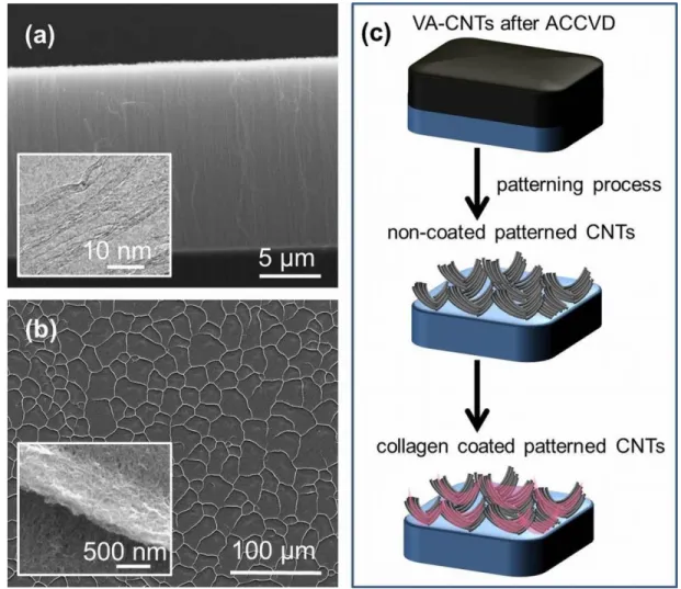
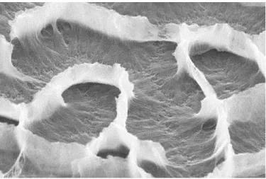
VA-CNTs were synthesized through ACCVD method on Si substrates using sandwich catalyst design (Al/Co/Al) as reported in a previous study [26]. They were observed under SEM and the side view of the images revealed that the height of the CNTs were 10 µm whereas the average diameters of the CNTs were around 10 nm, which was shown by TEM analysis (Figure 3.1a). Before the cell culturing step on CNTs, we created asperities on VA-CNTS for the growth. This patterning step was induced by dropping deionized water on these surfaces. As a result of water contact, some of the aligned CNTs were collapsed which were surrounded by standing CNTs; forming cavities (Figure 3.1b). The average width of these cavities was measured as 10 µm. The mechanism behind the formation of self-assembled patterning is based on the elasto-capillary effect and hydrophobic property of aligned CNTs [26,52]. Following aforementioned patterning process, patterned CNTs were either treated with collagen or
19
not. The experimental design of patterning was shown in Figure 3.1c. We generated two surfaces designated as patterned non-coated and collagen coated CNT surfaces.
Figure 3.1: (a) Side view SEM images of VA-CNTs. Inset shows TEM image of CNTs.
(b) Top view SEM image of patterned CNT surface with the inset showing a high magnified image from the side of pockets. (c) Schematic representation of CNT surfaces after patterning and collagen coating.
3.1.2 EMT Marker Expressions of HCC
The EMT marker expression profiles of two HCC lines; HUH7 and Snu182 were monitored at mRNA level (Figure 3.2). Our RT-PCR results showed that SNU182 cell line was positive for the expression of all of the mesenchymal markers tested; except
20
SIP1 (Vimentin, Fibronectin, a-Sma, N-Cad, Slug, Twist1). In contrary to SNU182, only two of the tested mesenchymal markers (Vimentin and N-Cad) were expressed in HUH7 cells. Hence the expression profiles of these cell lines confirmed that SNU182 cells are poorly differentiated whereas HUH7 cells are well differentiated HCC cell line [10]. In parallel to previous reports, these results also indicated that HUH7 had a more epithelial characteristic compared to SNU182 cells [10].
Figure 3.2: EMT expressions of HUH7 and SNU182 cell lines at m-RNA level.
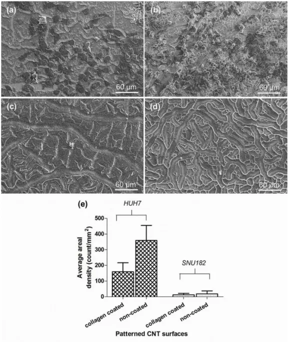
3.1.3 Proliferation and attachment of HCCs on patterned CNTs
After the CNT surfaces were modified and sterilized by UV for one hour, poor (SNU182) and well (HUH7) differentiated HCC s were cultured on both non-coated and collagen coated patterned CNT surfaces. The cells were incubated for 3 days on the
21
patterned CNT surfaces and at the 3rd day of culture, low vacuum SEM imaging was performed to assess the relationship between their differentiation levels and attachment ability both on collagen coated and non-coated patterned CNT surfaces (figure 3.3). In order to compare the number of attached cells on patterned CNTs, we counted the attached cells under SEM and calculated the average areal cell density by dividing the total cell number on the surface to total surface area. The density plot given in Figure 3.3e indicates that the average attached cell number for well differentiated HUH7 cells were higher than the number of attached poor differentiated Snu182 cells on CNT surfaces. The difference between the areal density of well and poor differentiated cells was statistically significant. This suggests that patterned CNT surfaces could be used as tools for the definition of the differentiation level and EMT status of HCCs by taking advantage of the difference in the attachment rates.
22
Figure 3.3: Low vacuum SEM images of HUH7 and SNU182 cancer cells after seeded
and cultured for 3 days on (a,c) collagen coated and (b,d) non-coated patterned CNT surfaces, respectively.
In many studies, collagen is used as a coat in cell culture systems [53]. In our study, collagen was used as a coating on patterned CNTs instead of composite usage where a significant effect was observed. The number of well differentiated cells on non-coated
23
patterned CNTs was comparably higher than the ones on collagen coated surfaces. Contrarily, there was no significant effect of collagen coating for poor differentiated cells (Figure 3.3e). It is already well known that surface topography has great effects on the attachment abilities of cells [54]. Since collagen coating would smoothen the patterned CNT surfaces by infiltrating and filling the voids between each CNT, the addition of collagen results in decreased surface area as it was shown in Figure 3.4. Thereby, less surface area that is available for cell attachment would result in less number of cell attachment and growth on collagen coated surfaces. Our results suggest that the difference in the attachment ability of poor and well differentiated HCC lines on non-coated CNT surfaces could be used for evaluating the differentiation level and thus aggressiveness of HCC lines.
Figure 3.4: High vacuum SEM image of a collagen coated patterned CNT surface.
In addition to the imaging of the attached cells on patterned CNT surfaces, we performed MTT assay to measure the viability of the two cell lines on both non-coated and collagen coated patterned CNT surfaces (Figure 3.5). It is important to note that the reading of control group was obtained from cells on 96-well culture plates excluding CNT surfaces. Although cell viabilities for both cell lines decrease when compared to the control, our results showed that the viability of HUH7 cells were more on non-coated patterned CNTs when compared to the viability of SNU182 (Figure 3.5) and the
24
difference was statistically significant. Thus, our MTT data, together with the SEM images and the areal density data revealed that the proliferation and attachment rate of HUH7 is more than Snu182 on patterned CNT surfaces, making CNT a promising tool for the evaluation of the differentiation level of the tumor.
Figure 3.5: MTT assay of cancer cells on non-coated and collagen coated CNT surfaces.
Control group represent the case where there is no CNT surface.
Toxicity of CNTs has been a controversial issue for researchers since there are conflicting reports on their toxicity [55–57]. Therefore, in order to investigate the cytotoxic effect of patterned CNT surfaces on HCC lines we performed LDH cytotoxicity analysis both on collagen coated and non-coated patterned CNTs (Figure 3.6).In LDH test, two control groups were used; low control (LC) and high control (HC) in which HCCs were seeded directly on bare plate surfaces. LC group shows the minimum LDH release while HC represents the maximum release. The closer the absorbance readings of the samples to HC, the more cytotoxic the conditions are for that sample. Our LDH results clearly showed that HUH7 and SNU182 cell lines have minimum LDH release when cultured both on the non-coated and coated patterned
25
CNTs. This result indicates that CNTs are not cytotoxic for these HCC lines when used as surfaces for the attachment of the cells. This data also suggests that the difference in the attachment rates of the two cell lines are due to difference in cells’ interaction characteristics with CNT surfaces; not because of the cytotoxic effects of the CNT surfaces. Therefore, the WD HCC line, HUH7 has greater tendency to attach on CNT surface when compared to the PD HCC line Snu182.
Figure 3.6: LDH assay of cancer cells on non-coated and collagen coated CNT surfaces.
Low control (LC) and high control (HC) groups represent the case where there is no CNT surface.
3.2
The Use of Hydrogels in Treatment of Cancer
3.2.1 The characteristics of the Dox-releasing hydrogels
We used Dox-releasing and Dox-free hydrogel networks consisting of poly(hydroxypropyl methacrylate, and polyethylene glycole-methylether methacrylate) with five different PEG chain length and crosslinker amount combinations to investigate the effects of the drug release on the proliferation of Snu398 human HCC line. Two different concentrations (I1: 1.0 mg/ml and I2: 0.5 mg/ml) of drug were used to load the
26
hydrogels. The preparation and characterization of them were done by Prof. Dr. Gülay Bayramoğlu, as mentioned in Materials and Methods section. For the characterization of the hydrogels FTIR, SEM, swelling, permeability and contact angle studies were performed. In these studies, five formulations; p(HPMA/PEG-300)X1, p(HPMA/PEG– 475)X1, p(HPMA/PEG–950)X1, p(HPMA/PEG-300)X2, and p(HPMA/PEG–300)X3 were used as the most effective drug release systems.
The entrapment efficiency (%) of a drug depends on the type of matrix material, entrapment method of the drug and the polymer networks preparation conditions. In this study, it was shown that the drug-loading efficiency of the hydrogel film formulations depend on the PEG chain length and crosslinker density of the formulations (Table 3.1). The permeability to Dox was found to be increased as the PEG chain lengths in the hydrogel formulations increased due to the higher hydrophilicity of the hydrogel. As seen in Table 3.1 the p(HPMA/PEG-950)X1 formulation had the highest PEG chain length and therefore had a substantially higher water content and a permeability to Dox; while this permeability decreases as the length of PEG chain decrease to 300 from 950. Additionally, as the crosslinker density increased from X1 to X3, Dox-loading efficiency of the hydrogel formulations increased and the drug release rate decreased.
In the same study, by the protein adsorption tests, it was shown that the increase in the PEG chain lengths create pulsive force against the negatively charged blood proteins resulting in the reduction in the adsorbed protein amounts. This characteristic of the hydrogel formulations make them suitable tools to deliver drugs with minimum protein adsorption.
27
Table 3.1: Formulation of hydrogels and their density, water content, permeability to Dox and drug loading efficiencies.
Hydrogel formulations Monomer ratio HPMA/PEG-MEMA (mmoL/mmoL) Cross- linker (moL) Density (g/cm3) Water content (%) Permeability to doxorubicin (cm2/s x10-9) Drug loading efficiency (%) p(HPMA/PEG-950)X1 5.55/0.80 6.5 1.036 579 91.36 75 p(HPMA/PEG-475)X1 5.55/1.71 6.5 1.089 433 79.64 81 p(HPMA/PEG-300)X1 5.55/2.63 6.5 1.103 171 32.83 84 p(HPMA/PEG-300)X2 5.55/2.63 12.7 1.158 107 19.81 86 p(HPMA/PEG-300)X3 5.55/2.63 19.2 1.176 77 12.35 89
3.2.2 In vitro Dox-release studies
Drug release from hydrogel matrices is affected by many factors, such as their swelling properties, network porosity, and the erosion of the hydrogel matrix, as well as drug– polymer interactions (REF). In this study, the effects of the PEG chain length of the macromonomer (i.e., PEG–MEMA), cross-linker density and the amount of loaded drug on the Dox release were studied in a continuous drug release system. The kinetics of drug release from p(HPMA/PEG–MEMA) formulations were also analyzed. The effect of the PEG chain length of the macromonomer on the Dox release kinetics for the p(HPMA/PEG-300)X1, p(HPMA/PEG-475)X1, and p(HPMA/PEG-950)X1 formulations are presented in Figure 3.7.The release profiles indicate that the amount of Dox released and the rate of the release increased when the PEG chain length of the
28
hydrogel increased. The p(HPMA/PEG-300)X1 hydrogel films showed 60.3% release, whereas the p(HPMA/PEG-475)X1 and p(HPMA/PEG-950)X1 hydrogel films showed 85.4 and 98.3% release, respectively at 37°C and 480 min with 1.0 mg/mL Dox loading (Figure 3.7B). These results should be attributed to the more hydrophilic nature of the p(HPMA/PEG-950)X1 hydrogel formulations (Table 3.1).
Figure 3.7: The effect of the PEG chains length of the macromonomer on the Dox
release kinetics for the p(HPMA/PEG-300)X1, p(HPMA/PEG-475)X1, and p(HPMA/PEG-950)X1 at 0.5 mg/mL and (B) 1.0 mg/mL drug loading
The release profiles of Dox in p(HPMA/PEG-300)X1, p(HPMA/PEG-300)X2, and p(HPMA/PEG–300)X3 hydrogel formulations, cross-linked with different ratios of BisAA, are shown in Figure 3.8. It showed that the release rate of Dox for the p(HPMA/PEG-300)X1 hydrogel formulation was higher and faster than those for the p(HPMA/PEG-300)X2 and p(HPMA/PEG–300)X3 formulations. This was due to the lower cross-linking density and higher swelling ratio of p(HPMA/PEG-300)X1. Figure 3.8 shows the cumulative Dox release profiles for the hydrogel formulations. The p(HPMA/PEG-300)X1 hydrogel films showed 60.3% release, whereas the
B) A)
29
p(HPMA/PEG-300)X2 and p(HPMA/PEG-300)X3 hydrogel films showed 53.2 and 43.1% release, respectively, at 37°C and 480 min with 1.0 mg/mL Dox loading. The initial release rate of the drug from the hydrogels was rapid, and this initial fast release may be attributed to the rapid diffusion of Dox that was loaded close to the surface of the hydrogels. Later on, the drug was released more slowly from the hydrogels.
Figure 3.8: The effect of the cross-linker density on the Dox release kinetics for the
p(HPMA/PEG-300)X1, p(HPMA/PEG-300)X2, and p(HPMA/PEG-300)X3 at 0.5 mg/mL and (B) 1.0 mg/mL drug loading.
The hydrogel formulations had similar release profiles, despite different Dox contents. This indicates that at a lower drug concentration, most of the drug molecules remained bound to the polymeric chains of the hydrogels, whereas at a higher drug concentration, the percentage of unbound drug molecules increased in the polymer networks. The maximum Dox release for the p(HPMA/PEG-950)X1 hydrogel formulation with different drug amounts loaded (0.5 and 1.0 mg/mL) was determined to be 61.3 and 98.3% after 480 min, respectively. Dox is freely soluble in a physiological phosphate
30
buffer (pH 7.4); as a result, the rate of depletion of the drug in the polymer at a high concentration was faster than at a low concentration.
3.2.3 The effects of drug releasing hydrogels on the proliferation features of cancer cells
After the hydrogel formulations were sterilized by UV for 1 hour, Snu398 cells were seeded on 24-well plates and all formulations of hydrogels, including counterparts without drug release were added into the wells. The Snu398 cells were trypsinized and counted at the 6th day of culture by hemocytometer. Graphs showing the cell numbers were drawn to assess the relationship between the drug release and proliferation features of the HCC in the absence and presence of Dox (Figure 3.9).
Our results showed that the number of Snu398 cells was decreased, significantly, when they were cultured in the presence of all five Dox-releasing hydrogel formulations (Figure 3.9). We did not see any significant difference in the cell numbers in the presence of the two different concentrations of Dox (I1 and I2). The hydrogels alone were found to have no effects on the cell proliferations when there is no drug release. It is important to note that at Day 0, 3x10⁴ cells were seeded on each well and the control group was excluding hydrogels. At the 6th date of the culture, the increase in the cell number of the control group was 30 fold higher. Similar increase in the cell numbers were detected when the counterparts of the formulations without drug release were present. In addition, Dox release decreased the proliferation of Snu398 cells up to 90 fold for all formulations. These results indicate that Dox-releasing hydrogels prevent the proliferation of Snu398 cells, in vitro and can be used as a promising strategy to treat HCC.
31
Figure 3.9:The cell numbers of Snu398 cells in the presence of different formulation of Dox releasing hydrogels, counted at the 6th day of culture. D0 represents the initial number of cells seeded at the 1st day and the control group represents the cell numbers at Day 6 where hydrogels were excluded.
32
In order to investigate the cytotoxic effects of the drug-releasing hydrogels and their counterparts without drug release on the HCC line, we performed LDH cytotoxicity analysis (Figure 3.10).In LDH test, two control groups were used as it was explained in the first part; LC and HC in which HCCs were seeded on bare plate surfaces. The closer the absorbance readings of the samples to HC, the more cytotoxic the conditions are for that sample. Our LDH results clearly showed that, the drug-free counterparts of all hydrogel formulations were not cytotoxic to Snu398 cells since the absorbance values were very similar to the LC. In addition, the cytotoxicity of most of the formulations were increased when they were loaded with Dox and 1 mg/ml (I1) Dox-loaded p(HPMA/PEG-300)X3 formulation was found to be the most cytotoxic hydrogel to the cells. This data, together with the cell number analysis show that the usage of Dox-releasing hydrogels has cytotoxic effects on Snu398 cells as a result of which cell proliferation is inhibited.
33
Figure 3.10: LDH assay of Snu398 cells in the presence of different formulation of Dox
releasing hydrogels. Low control (LC) and high control (HC) groups represent the case where there is no hydrogel.
34
Chapter 4
Discussion
Biomaterials have been studied extensively for their biomedical applications including cancer research. The biocompatibility and safety of these materials make them promising tools for the cancer therapy and diagnosis. Previous studies showed the usage of different biomaterials in targeted drug delivery[58], gene delivery[59] and tumor imaging[60]; resulting in increased treatment and diagnostic efficacies. Among these biomaterials, CNTs and polymer hydrogels have gained great importance due to their unique physical, electrical and chemical properties [2–4].
CNTs have gained considerable attention in biomaterial studies for their applications in tumor imaging, gene and drug delivery[2]. In many studies CNTs were used as nanofiber scaffolds for living cells by mimicking the collagen network in connective tissues and designing them with similar structure to native extracellular matrix (ECM)24–
29,54
. By binding specific markers on CNTs, researchers were able to detect various types of cancer cells such as lung and prostate cancer[30–35]. Furthermore, in a previous study, CNTs were used as arrays to differentially entrap colon cancer cells with two different metastatic grades by taking advantage of their rigid surface properties. It was
35
reported that higher metastatic cancer cells entrapped more compared to the lower metastatic ones and this was explained by the morphology differences of cells at different grades [36]. Moreover, by the same group, it was shown that biosensors made of VA-CNTs could be used for the detection of cancer by entrapping them on VA-CNTs [37]. However, in these studies, only the difference in deformability properties of cancer cells with different metastatic grades was taken into consideration. Until the current study, there was not any study that examined the differential attachment features of cancer cells with different EMT status on CNTs.
In this study, firstly, we examined the difference in attachment of HCC lines with different EMT status on patterned CNT surfaces to find out a facile way to differentiate the cells according to their aggressiveness. Two cell lines HUH7 and Snu182 were used. The EMT marker expression results revealed that HUH7 had a more epithelial characteristic compared to SNU182 cells, which were in mesenchymal phenotype. Regarding to our SEM and MTT results well differentiated HUH7 cells attached more both on collagen coated and non-coated patterned CNT surfaces than the poor differentiated SNU182 cells. This suggest that the difference in the attachment ability of poor and well differentiated HCC lines on patterned CNT surfaces could be used for evaluating the differentiation level and thus aggressiveness of HCC lines making them promising diagnostic tools.
There are many conflicting results on the toxic effects of CNTs to cells[62]. Many studies state that they do not have any cytotoxic effects[25,56]. Yet, relatively few studies report the health hazards that might be related to these biomaterials[55,63]. According to these studies, CNTs cause cell damage, genotoxicity and carcinogenicity due to their effects on cell signaling, membrane perturbation and the production of cytokines, chemokines and reactive oxygen species (ROS), hence, decreasing cell viability[64]. These cytotoxic effects were found to depend on the presence of large amounts of catalysts such as Co and Fe, which can generate ROS in cellular environments [63,64]. Nevertheless, CNTs can be in variety of forms with difference in
36
their surface chemistries, processing methods, lengths, diameters, contaminants, and more and all of these variations affect the cytotoxicity of these materials[63].
In the current study, in order to test the cytotoxic effect of patterned CNT surfaces on HCC lines we performed LDH cytotoxicity analysis. LDH is normally a cytoplasmic enzyme and it is not secreted outside of the cells. However, upon damage to the cell membrane, LDH leaks out. By using the LDH cytotoxicity analysis, it is possible to measure the release of LDH from cells based on a colorimetric quantitation. According to our results, CNTs were found to have no cytotoxic effects on the attached cells. Therefore, it can be concluded that CNTs are not cytotoxic to the cells when used as surfaces for the attachment of the cancer cells as explained in this study. Moreover, the difference in the attachment features of the WD and PD HCC lines are not due to any possible cytotoxic effect of CNTs, preventing the attachment of cells. It was the result of the difference in the cells own interaction characteristics with CNTs and this makes CNTs promising tools to differentiate HCC according to their EMT status by taking advantage of the difference in their attachment features.
Recently, there are many potent drugs which are used in the treatment of HCC. However, the efficacies of them are constrained by their side effects, limited cellular entry and drug resistance. Most of the drugs also have very short half-lives under physiological conditions. Because of these reasons the efficacy of a cancer therapy largely depends on the way that the drug is administered and it is important to develop efficient delivery systems to enhance cellular uptake and decrease any possible harm given to normal body tissues[46].
One of the novel approaches to increase the duration of the in vivo effects of the drug is to use vehicles for the drug delivery. Polymer hydrogel is such a biomaterial that allows continuous drug release in the target area, increasing treatment efficacy and decreasing side effects[46]. Recently, research on hydrogels have gain great importance; because of their potential use in biomedical applications, especially in drug delivery, clinical and plastic surgery, interventional therapy, and more[48,49]. They are reported to have very
37
similar properties to living tissues and this prevents the recognition of the material as foreign by body, reducing irritation to the normal tissues. In addition, they are designed to keep the protein adsorption and cell adhesion in minimum level, which inhibits any possible harm given to the normal cells. Because of their high biocompatibility and stability, they could be coupled with anticancer drugs to use them as drug delivery vehicles. With this system, many studies showed that toxicity of free drug can be reduced because of the controlled release of it in the target area.[49,50]
Numerous hydrogel-drug formulations have been developed for controlled release of active ingredient over time. Poly(hydroxypropyl methacrylate), p(HPMA) and its copolymer hydrogels have found extensive applications in the biomedical field because of their good chemical stability and high biocompatibility. Poly(ethylene glycol) (PEG) is a water-soluble, nontoxic, and non-immunogenic polymer. Hydrogels containing PEG are interesting biomaterials because they exhibit low degrees of protein adsorption and cell adhesion.
In this study, secondly, we used different formulations of p(HPMA/PEG-MEMA) hydrogels with Dox release and tested the effects of the drug on the proliferation of HCC to propose the usage of this biomaterial as novel way in drug administration. The amino groups of Dox are positively charged at pH 7.4 and the Lewis acid groups (γ
-) of the PEG molecules have partially negative charge. Thus, in this hydrogel system, polymer and drug can interact electrostatically at pH 7.4. Snu398 was used as the HCC line. Our results clearly show that release of Dox significantly decreased the proliferation of HCC when compared to the cell proliferations in the presence of hydrogels without drug-release. These results suggest the usage of hydrogels in HCC treatment. Further, by LDH cytotoxicity assay, the cytotoxic effects of the different formulations of Dox releasing hydrogels and their counterparts without drug release on Snu398 cells were tested. Our results showed that the hydrogels without any drug release were not cytotoxic to HCC, as it was expected. However, the cytotoxicity of the formulations were increased significantly when they were loaded with Dox and
p(HPMA/PEG-38
300)X3 was found to be the most cytotoxic hydrogel. Overall, these results indicate that Dox- releasing p(HPMA/PEG-300)X3 formulation was found to be the most effective formulation among the five formulations due to their significant cytotoxic effects and the inhibition of the proliferation of cells.
The proposed novel administrative strategy is promising for the treatment of HCC due to the controlled drug release, which will increase the drug residence time leading to more efficient therapy. The amount of drug needed for the therapy is decreased and this lowers the cost. It is possible to place these hydrogels near tumor cells which will decrease the damage given to the normal body tissues, inhibiting the possibility of side effects.
Overall, in this study, it was shown that patterned CNT surfaces are promising diagnostic tools for the determination of the EMT status and therefore the aggressiveness of tumors, which may be useful for the early detection and characterization of tumors. It was also shown that Dox-loaded hydrogels could be used in the treatment of HCC, with increased efficacy and decreased side effects.
39
Chapter 5
Future Perspective
In order to better understand the difference in the attachment features of tumor cells according to their aggressiveness on CNTs, first of all, usage of more HCC lines with different epithelial and mesenchymal phenotypes could be very informative to see whether the difference in the attachment rates of WD and PD cells is also true for other cell lines. Once it is tested, primary tumor cells acquired from patients could be examined for their attachment behaviors on CNTs, which could be an important data since it would represent a better experimental model for tumor behavior.
For the examination of the effects of drug-releasing hydrogels on HCC, the usage of additional cell lines and primary tumor cells could also be necessary. Further, in vivo protocols should be used. HCC mouse models would be suitable for this step. By placing the hydrogel in the tumor area, the effects of drug release on the tumor size and the overall survival of the organism could be tested which would give a better idea about the efficacy of this administration method in the cancer treatment.
Once the efficiencies of each biomaterial in cancer diagnosis and treatment of HCC is further proved by these experimental steps, they could also be modified for other cancer
40
types. These modifications could include preparation of different patterns on CNTs with increased capability to differentiate the tumor cells according to EMT status, the trial of different formulations of hydrogels to optimize the effects according to the tissue type and the usage of other drug conjugates.
Therefore, the final application of these biomaterials would be as follows: During the diagnosis of a HCC patient, pathological sample would be taken from the tumor by biopsy. Some of these tumor cells would be cultured on CNTs which would be designed as chips to determine the EMT status and aggressiveness of the tumor cells. By evaluating the attachment features of the tumor cells on these chips, the aggressiveness of the tumor would be determined. This information would be used to choose the most appropriate therapy strategy for increased treatment efficacy. During the treatment of a HCC patient, Dox-releasing hydrogels would be placed directly to the tumor area. Dox release would kill the cancer cells in that target area, which would decrease the side effects of the drug and increase the treatment efficacy.
![Figure 1.1 [9]. Mechanisms for Hepatocellular carcinogenesis](https://thumb-eu.123doks.com/thumbv2/9libnet/5569769.108819/15.918.229.781.690.995/figure-mechanisms-for-hepatocellular-carcinogenesis.webp)
![Figure 1.2 [15]: Epithelial-to-Mesenchymal Transition.](https://thumb-eu.123doks.com/thumbv2/9libnet/5569769.108819/17.918.182.833.465.674/figure-epithelial-to-mesenchymal-transition.webp)
![Figure 1.3 [15]: Contribution of EMT to cancer progression.](https://thumb-eu.123doks.com/thumbv2/9libnet/5569769.108819/18.918.206.859.152.276/figure-contribution-emt-cancer-progression.webp)