cium gluconate and glucagon are considered as possible adjuvant therapy for CCB toxicity (3, 6, 7). According to currently available data these therapies should be considered for the refractory cases (7). Owing to high protein binding and extensive tissue distribution of CCBs hemofiltration or dialysis are not useful in overdose cases (4).
Physicians should be aware that patients may not be telling the truth every time. Especially in some psychiatric disorders such as factitious disorder (Münchausen syndrome); patient may act as if he/ she has an illness by deliberately producing or exaggerating symp-toms by taking drugs with overdoses (8). Factitious disorders are not rare psychiatric disorders and many clinicians may encounter them during their career. Our patient can be considered as malingering, because he had false physical symptoms intentionally produced and motivation of the behavior involved external incentives. Whereas factitious disorder has no other incentive than to be a patient and experience the sick role (8, 9).
Conclusion
This report underlines that the reliability of anamnesis should be questioned in complicated, suspicious shock cases with unknown ori-gin. Drug overdoses and psychiatric disorders should be kept in mind in such suspicious patients.
References
1. Vincent JL, De Backer D. Circulatory shock. N Engl J Med 2014; 370: 583. 2. DeWitt CR, Walksman JC. Pharmacology, pathophysiology and manage-ment of calcium channel blocker and β-blocker toxicity. Toxicol Rev 2004; 23: 223-38. [CrossRef]
3. Saravu K, Balasubramanian R. Near-fatal amlodipine poisoning. J Assoc Physicians India 2004; 52: 156-7.
4. Ghosh S, Sircar M. Calcium channel blocker overdose: Experience with amlodipine. Indian J Crit Care Med 2008; 12: 190-3. [CrossRef]
5. Humbert VH Jr, Munn NJ, Hawkins RF. Noncardiogenic pulmonary edema com-plicating massive diltiazem overdose. Chest 1991; 99: 258-9. [CrossRef]
6. Upreti V, Ratheesh VR, Dhull P, Handa A. Shock due to amlodipine over-dose. Indian J Crit Care Med 2013; 17: 375-7. [CrossRef]
7. Patel NP, Pugh ME, Goldberg S, Eiger G. Hyperinsulinemic euglycemia therapy for verapamil poisoning: a review. Am J Crit Care 2007; 16: 498-503.
8. Füessl HS. Factitious disorders: doctors be aware. MMW Fortschr Med 2013; 155: 33-6. [CrossRef]
9. Folks DG. Munchausen's syndrome and other factitious disorders. Neurol Clin 1995; 13: 267-81.
Address for Correspondence: Dr. Cihan Altın,
6471/5 Sokak, No:7, Yalı Mahallesi, Bostanlı, Karşıyaka, İzmir-Türkiye Phone: +90 232 241 10 00
E-mail: [email protected] Available Online Date: 25.06.2014
©Copyright 2014 by Turkish Society of Cardiology - Available online at www.anakarder.com DOI:10.5152/akd.2014.5581
Successful elimination of a Mahaim
pathway using an 8 mm tip cryoablation
catheter in a child
Neslihan Kıplapınar, Celal Akdeniz, Yakup Ergül, Volkan Tuzcu* Departments of Pediatric Cardiology/Electrophysiology and *Pediatric Arrhythmia Center, Mehmet Akif Ersoy Cardiovascular Research and Training Hospital; İstanbul-Turkey
Introduction
Mahaim fibers typically demonstrate decremental conduction properties and constitute approximately 3% of preexcitation syndromes (1). Mahaim pathways are usually right-sided, however several left-sided cases have been reported (2). Conventionally Mahaim pathway ablation is performed with radiofrequency ablation (RFA). We report a patient who presented with a wide QRS tachycardia with left bundle branch block (LBBB) pattern. The electrophysiology study demonstrat-ed Mahaim tachycardia and the patient was successfully treatdemonstrat-ed with cryoablation following a failed attempt with RFA.
Case Report
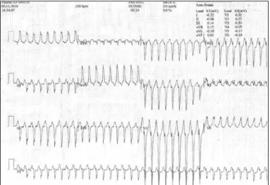
An 8-year-old girl was referred to our clinic with the preliminary diagnosis of ventricular tachycardia. Her palpitations started at the age of 2. Metoprolol was started 2 months prior to the presentation, how-ever tachycardia episodes continued. A resting 12-lead electrocardio-gram showed no abnormalities. A 24-hour Holter monitoring demon-strated wide QRS tachycardia with leftward superior axis deviation and LBBB morphology. During the exercise test tachycardia was induced and the test had to be terminated due to sustained wide QRS tachycar-dia (Fig. 1).
An electrophysiology study was carried out under general anesthe-sia, following the parents` consent for the procedure. Two quadripolar catheters and a decapolar catheter (St. Jude Medical Inc., St. Paul, MN) were positioned at the high right atrium, coronary sinus and right ventricle. The Ensite Velocity system (St. Jude Medical Inc., St. Paul, MN) was used for mapping and navigation of the catheters. A wide QRS tachycardia with LBBB and superior axis started spontaneously. Intravenous adenosine was administered as 200 mcg/kg, resulting in sudden termination of the tachycardia. However, following a few ven-tricular escape complexes, sinus rhythm returned with pre-excitation with the same QRS morphology similar to that of during the tachycardia. With programmed ventricular stimulation, earliest ventriculoatrial con-duction was observed in the His region with decremental properties. When the diagnostic catheter was at the His position, there was no His signal preceding the ventricular signal during the wide QRS tachycar-dia. All of these electrophysiological features were strongly suggestive Figure 1. The patient’s initial chest X-ray was normal (A). But on the
chest X-ray taken next day; there was perihilar consolidations, increased width of vascular pedicle and peribronchial cuffing due to the newly occurring pulmonary edema (B).
A B
Case Reports Anadolu Kardiyol Derg 2014; 14: 549-57
of a Mahaim pathway. Mahaim potential could not be identified around the tricuspid valve annulus. Therefore, ventricular pace mapping was done around the tricuspid annulus to locate the insertion site of the Mahaim pathway. Consequently, we induced the tachycardia with similar morphology in the right posterolateral area where pace map-ping QRS morphology was very similar to that of during the tachycardia. Radiofrequency application was performed with a power of 48-55 W and temperature of 47°C for 30-60 sec at each successful ablation site. Radiofrequency applications were only temporarily effective despite good catheter stability. Therefore, we suspected a deeply seated epi-cardial pathway. After that an 8-mm tip cryocatheter (FreezorMax, Cryocath Technologies Inc, Kirkland, Canada) was used (Fig. 2). During cryoablation tachycardia terminated within 13 seconds (Fig. 3). Cryoablation was carried out 4 times for 4 minutes and once for 6
min-utes. Cryoablation was applied for total of 1320 seconds. There was no further conduction via the accessory pathway and tachycardia was not inducible by stimulation and metaproterenol infusion. The procedure time was 180 minutes and fluoroscopy time was 2.1 minutes, at a dose of 5.2 mGy. She was discharged in normal sinus rhythm on the following day.
Discussion
To our knowledge, this is the first report of successful cryoablation of a Mahaim pathway using an 8-mm tip cryocatheter following a failed attempt with RFA.
Mahaim potential can be mapped at the lateral tricuspid annulus in the majority of patients with Mahaim tachycardia and RFA at that site is associated with a high success rate (3, 4). In our case, Mahaim poten-tial could not be recorded during the procedure, however due to the electrophysiological properties, Mahaim pathway was suspected and ablation was attempted. RFA failed to eliminate the Mahaim pathway in our patient. Parvez et al. (5) showed that lesion size created with an 8 mm tip cryocatheter is larger than lesion of open irrigated radiofre-quency catheter. Potential advantages of 8 mm tip catheter are stability and effectiveness for deeper targets. Short-term results in patients with atrioventricular nodal reentry tachycardia showed that the use of 8 mm tip cryocatheter reduced recurrences (6). Due to mentioned properties above we used 8-mm tip cryocatheter and we achieved the elimination of Mahaim pathway.
Conclusion
Conventionally Mahaim pathway ablation is performed with RFA. However, in patients with failed radiofrequency attempts, cryoablation with an 8-mm tip catheter can be an alternative approach.
References
1. Mahaim I. Kent’s fibers and the A-V paraspecific conduction through the upper connection of the bundle of His-Tawara. Am Heart J 1947; 33: 651-3. [CrossRef]
2. Johnson CT, Brooks C, Jaramillo J, Mickelsen S, Kusumoto FM. A left free-wall, decrementally conducting, atrioventricular (Mahaim) fiber: diagno-sis at electrophysiological study and radiofrequency catheter ablation guided by direct recording of a Mahaim potential. Pacing Clin Electrophysiol 1997; 20: 2486-8. [CrossRef]
Figure 2. The cryoablation location for the Mahaim pathway is shown in anterio-posterior view in this Figure
Figure 3. The ablation site (right posteriolateral area) and catheter is shown in Ensite-NavX electroanatomic mapping system
Figure 1. Electrocardiogram tracing showed wide QRS tachycardia with leftward-superior axis and LBBB pattern during pretest period in Bruce protocol exercise test
Case Reports
3. McClelland JH, Wang X, Beckman KJ, Hazlitt HA, Prior MI, Nakagawa H, et al. Radiofrequency catheter ablation of right atriofascicular (Mahaim) accessory pathways guided by accessory pathway activation potentials. Circulation 1994; 89: 2655-66. [CrossRef]
4. Bohora S, Dora SK, Namboodiri N, Valaparambil A, Tharakan J. Electrophysiology study and radiofrequency catheter ablation of atriofas-cicular tracts with decremental properties (Mahaim fibre) at the tricuspid annulus: Europace 2008; 10: 1428-33. [CrossRef]
5. Parvez B, Pathak V, Schubert CM, Wood M. Comparison of lesion sizes produced by cryoablation and open irrigation radiofrequency ablation catheters: J Cardiovasc Electrophysiol 2008; 19: 528-34. [CrossRef]
6. Silver ES, Silva JN, Ceresnak SR, Chiesa NA, Rhee EK, Dubin AM, et al. Cryoablation with an 8-mm tip catheter for pediatric atrioventricular nodal reentrant tachycardia is safe and efficacious with a low incidence of recurrence: Pacing Clin Electrophysiol 2010; 33: 681-6. [CrossRef]
Address for Correspondence: Dr. Volkan Tuzcu,
İstanbul Medipol Üniversitesi Hastanesi, Pediyatrik Kardiyoloji Bölümü, Pediyatrik ve Genetik Aritmi Merkezi,
İstanbul-Türkiye Phone: +90 212 460 77 82 Fax: +90 212 471 94 94 E-mail: [email protected] Available Online Date: 25.06.2014
©Copyright 2014 by Turkish Society of Cardiology - Available online at www.anakarder.com DOI:10.5152/akd.2014.5291
Near zero fluoroscopy radiation exposure
during successful catheter ablation of
atrial tachycardia from the non-coronary
aortic cusp using 3-dimentional
electroanatomic mapping system
Ekrem Güler, Oğuz Karaca, Filiz Kızılırmak, Gamze Babür Güler, Fethi Kılıçaslan
Department of Cardiology, Faculty of Medicine, İstanbul Medipol University; İstanbul-Turkey
Introduction
Atrial tachycardia (AT) rarely originates from the paraHisian region (1). Catheter ablation of paraHisian AT carries a substantial risk of atrio-ventricular (AV) block. Cryoablation offers a safer ablation strategy for these patients (2). There is a small risk of AV block with cryoablation. Ablation of paraHisian AT from the non-coronary aortic cusp (NCC) is an option in these patients. We report a patient with paraHisian AT that was successfully ablated from the NCC by 3-Dimentional (3-D) mapping system with using near zero fluoroscopy (NZF).
Case Report
A 62-year-old woman, was referred to our institution due to symp-tomatic supraventricular tachycardia. She had undergone paraHisian AT ablation from right atrium at another center one year ago. She had recur-rence of AT after 3 months. At the time of her admission, electrocardio-gram (ECG) during tachycardia revealed AT. Echocardioelectrocardio-gram showed mild mitral regurgitation and normal left ventricular function. An
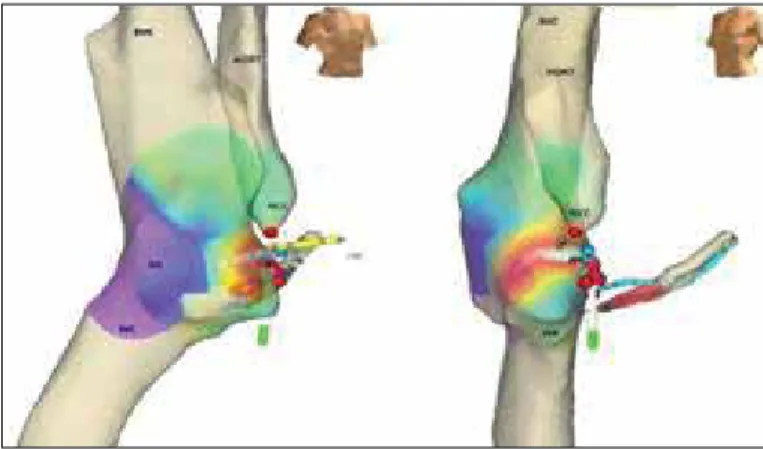
electro-physiological (EP) study was undertaken. Multi-electrode catheters were positioned into His bundle region and coronary sinus and an electro-anatomic mapping system was used (EnSite NavX® system, St. Jude Medical, Inc., St. Paul, MN, USA). His region was tagged on the 3-D anatomy of the right atrium (RA). Supraventricular tachycardia was induced by programmed atrial stimulation. AT was diagnosed by using the EP maneuvers. The tachycardia was mapped by using the En-site system and the earliest activation was found at the His region. Cryoablation cath-eter was advanced to the His region. Then, cryoablation could not be performed due to transient AV block during cryomapping of the tachycar-dia at the point of earliest activation. There was AV block even during manipulation of the cryoablation catheter. Then, 3-D anatomy of the aorta and aortic cusps were constructed (Fig. 1). The earliest local activation was detected within the NCC. Here, local atrial activity preceded onset of the p wave by 53 msec (Fig. 2). Location of the radiofrequency (RF) cath-eter was confirmed using fluoroscopy and electro mapping system. A single application of RF energy (power: 30 Watts, maximum temperature 55 degree) completely terminated the tachycardia. Total fluoroscopy time was 1 min 38 seconds. The patient was asymptomatic, ECG and rhythm Holter recordings showed no recurrence of AT.
Discussion
Focal ATs may originate in the right atrium around crista terminalis, coronary sinus, paraHisian region, tricuspid annulus and right atrial appendage. In left atrium, they primarily originate from pulmonary veins, mitral annulus, left atrial appendage and left septum (3). ATs originating Figure 1. 3-D geometry of aorta was constructed by En-Site system HB - his bundle; IVC - inferior vena cava; NCC - non-coronary cusp; RA - right atrium; SVC - superior vena cava
Figure 2. Local atrial activity preceded onset of the P wave by 53 msec
Case Reports Anadolu Kardiyol Derg 2014; 14: 549-57