20
ycosis fungoides (MF) is the most common type of cutaneous T-cell lymphoma and characterized by proliferation of small to medium T lymphocytes with cerebriform nuclei in most cases. However, the diagnosis of MF may be very difficult in certain cases, par-ticularly in those variants of MF such assyringotropic MF, granulomatous MF and folliculotropic MF.1Folliculotropic MF is a rare variant, which
histopathologically is characterized by pronounced folliculotropism of neo-plastic T cells, with or without follicular mucinosis, and clinically by an
im-Folliculotropic Mycosis Fungoides Associated
with Alopecia in a Case
AABBSSTTRRAACCTT Mycosis fungoides (MF) is the most common type of cutaneous lymphoma and char-acterized by proliferation of small to medium T lymphocytes with cerebriform nuclei in most cases. However, the diagnosis of MF may be very difficult in certain cases, particularly in those variants of MF such assyringotropic MF, granulomatous MF and folliculotropic MF. The classic histopatho-logic feature is the presence of atypical T-cells with a tropism to hair follicle epithelium. The clin-ical presentation of folliculotropic MF often differs from the patches and plaques of classic MF and may be associated with decreased clinical suspicion for folliculotropic MF. The average time inter-val from onset of lesions till diagnosis of folliculotropic MF was 2 years. Folliculotropic MF dis-plays resistance to standard treatment modalities, has an unfavourable course and diversity in the histological spectrum. Here we reported a rare case who presented to our dermatology polyclinic with a complaint of hair loss on his back firstly. On the follow-up, the case was diagnosed as fol-liculotropic MF and treated with interferon-α2a and PUVA successfully.
KKeeyywwoorrddss:: Mycosis fungoides; alopecia areata; skin neoplasms Ö
ÖZZEETT Mikozis fungoides (MF); birçok olguda, küçük ve orta boyuttaki serebriform çekirdekçiklere sahip T lenfositlerinin çoğalmasıyla karakterize en sık olarak görülen kutanöz lenfoma tipidir. Bu-nunla birlikte; özellikle asiringotropik, granülomatöz ve follikülotropik MF gibi olgularda tanı koy-mak güç olabilmektedir. Folikülotropik MF’de klasik histopatolojik görünüm; atipik T hücrelerinin folliküler epitele tropizmi ile karakterizedir. Folikülotropik MF’in klinik görünümü özellikle kla-sik MF’de görülen yama ve plaklardan farklılık arz etmektedir ve bu durum da klinik olarak foli-külotropik MF öntanısını güçleştirmektedir. Hastalığın başladığı dönemle tanı arasındaki süre ortalama 2 yıldır. Folikülotropik MF, standart tedavi seçeneklerine dirençlidir, istenmeyen klinik gidişata sahiptir ve histopatolojik olarak farklılıklar arz etmektedir. Biz burada; sırtta kıl dökülen alanlar şikayeti ile polikliniğimize başvuran, takiplerinde Folikülotropik MF gelişen ve PUVA ve in-terferon- α2a ile başarıyla tedavi edilen oldukça nadir görülen bir olguyu sunduk.
AAnnaahhttaarr KKeelliimmeelleerr:: Mikozis fungoides; alopesi areata; deri neoplazileri
Ali BALEVİ,a
Mustafa ÖZDEMİR,a
Pelin Doğa ÜSTÜNER,a
Hatice TOYb
aDepartment of Dermatology, İstanbul Medipol University Faculty of Medicine, İstanbul bDepartment of Pathology, Konya Necmettin Erbakan University Meram Faculty of Medicine, Konya Ge liş Ta ri hi/Re ce i ved: 03.03.2016 Ka bul Ta ri hi/Ac cep ted: 04.11.2016 Ya zış ma Ad re si/Cor res pon den ce: Ali BALEVİ
İstanbul Medipol University Faculty of Medicine,
Department of Dermatology, İstanbul, TURKEY/TÜRKİYE
Cop yright © 2017 by Tür ki ye Kli nik le ri
OLGU SUNUMU DOI: 10.5336/dermato.2016-51209
Ali BALEVİ et al. Turkiye Klinikleri J Dermatol 2017;27(1):20-3
21 paired prognosis compared to classic MF. The clin-ical presentation of folliculotropic MF often differs from the patches and plaques of classic MF and may be associated with decreased clinical suspicion for MF.2Here we reported a rare folliculotropic MF
case and treated with interferon (IFN)-α2a and psoralen and ultraviolet A (PUVA) successfully.
CASE REPORT
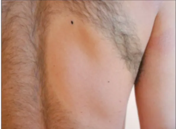
In 2011, a 30 year-old-male presented to our terti-ary dermatology unit with a 1-week history of alopesic patch of the right arm and right side of lower scapular region. When the alopesic lesion first appeared, the surface of the lesion was smooth and slightly red skin color without any skin alter-ations like atrophic, scaling and follicular changes. The patient was diagnosed as having alopecia areata (AA). Treatment with topical/intralesional steroid was attempted for 3 months but without success. Then slightly erythematous atrophic scale ap-peared within alopesic sites and we performed skin punch biopsy from the lower scapular region (Figure 1). The histopathologic examination re-vealed a marked and predominant involvement of hair follicles, mostly consisting on perifollicular and intrafollicular infiltration by small and medium atypical T lymphocytes (Figures 2, 3). The histopathological findings were consistent with folliculotropic MF. Within a few weeks, new alopesic patches and MF lesions apperared on body and extremities. Our patient was administered both with PUVA therapy and IFN- α2a 3x 10⁶ units
thrice every week for 1 year. After the clinical and histopathological improvement were achieved at 12thmonth of treatment, IFN-α2a was stopped and
PUVA therapy was then tapered gradually a period of 1 year. There was no recurrence in follow-up for 1 year. Neverthless, no hair regrowth was observed within folliculotropic MF plaques during treatment and follow-up period.
DISCUSSION
Folliculotropic MF has a broad clinical spectrum with acneiform lesions, comedones, plaques with follicular papules, alopecia, cysts and nodules, fre-quently on head and neck areas. Other important
observations in folliculotropic MF are the occa-sional presence of peculiar lesions such as excori-ated nodules, xanthomatous changes, or pustules which are very uncommonly found in conven-tional MF forms. The extent of lesions can be quite
FIGURE 1: Erythematous slightly atrophic scale within alopesic site and
ery-thematous atrophic macule on the right arm.
FIGURE 2: Atypical T lymphocytes in upper dermis and squamous epithelium
(HE, x40).
FIGURE 3: Perifollicular and intrafollicular infiltration by small and medium
Ali BALEVİ et al. Turkiye Klinikleri J Dermatol 2017;27(1):20-3
22 variable.3 Folliculotropic MF occurs mostly in
adults with a male predominance, but it has also been reported in children and adolescents.4
The diverse cutaneous involvement delays correct diagnosis in folliculotropic MF.5The
aver-age time interval from onset of lesion stil diagnosis of folliculotropic MF is 2 years.6In our patient, the
time interval was 3 months. Among the diverse cu-taneous involvements, alopecia is tended to occur after the onset folliculotropic MF symptoms.7In
the literature, there is only 1 case report include folliculotropic MF mimicking alopecia areata.8
Contrary to limited alopesic patch in the latter case, alopesic patches and atrophic erythematous plaques were more and spreaded to other body areas very quickly in our case. In a previous study, 1550 MF patients were reviewed and only 5 pa-tients had documented alopecia before or within 1 year of onset of skin symtoms. And additionally, it is stated that alopecia within MF lesions mostly de-velop more than 1 year after the onset of skin symptoms.7It may be difficult at least initially to
distinguish alopecia areata, follicotropic MF, as in our case. Whether or not our patient first had alopecia areata and then developed folliculotropic MF later or whether this was initially undiagnosed folliculotropic MF is uncertain. It is proposed that folliculotropic MF should be kept in mind espe-cially for patients who do not respond to alopecia area treatment.6
PUVA, local radiotherapy, bexarotene, total skin electronbeam, topical steroids, surgery, pred-nisone, chlorambucil, IFN-α2a are used in the treatment of folliculotropic MF. When IFN-α2a is used in combination with PUVA, both response
and response duration are reported to be improved, with recent studies reporting overall response and complete response rates of 98% and 84%, respec-tively.9Our patient responded to PUVA and
IFN-α2a treatment. Folliculotropic MF lesions were cleared and no recurrence were seen in the follow-up period. Nevertheless, no hair regrowth was ob-served within folliculotropic MF plaques during treatment and follow-up period.
In conclusion; this type of folliculotropic MF onset led to late diagnosis in regards of atypical lo-calisation. The lesson to draw from this case is that folliculotropic MF is a great mimicker and may ini-tially resemble alopecia areata. If AA resist to con-ventional treatments and erythematous papules and/or atrophy appear within alopesic lesions, fol-liculotropic MF should be kept in mind.
C
Coonnfflliicctt ooff IInntteerreesstt
Authors declared no conflict of interest or financial support.
A
Auutthhoorrsshhiipp CCoonnttrriibbuuttiioonnss
I
Iddeeaa//CCoonncceepptt:: CCoonnssttrruuccttiinngg tthhee hhyyppootthheessiiss oorr iiddeeaa ooff rreesseeaarrcchh a
anndd//oorr aarrttiiccllee:: Ali Balevi; DDeessiiggnn:: PPllaannnniinngg mmeetthhooddoollooggyy ttoo r
reeaacchh tthhee ccoonncclluussiioonnss:: Ali Balevi, Pelin Üstüner; CCoonnttrrooll//SSuu--p
peerrvviissiioonn:: OOrrggaanniizziinngg,, ssuuppeerrvviissiinngg tthhee ccoouurrssee ooff pprrooggrreessss aanndd t
taakkiinngg tthhee rreessppoonnssiibbiilliittyy ooff tthhee rreesseeaarrcchh//ssttuuddyy:: Mustafa Özdemir; DDaattaa CCoolllleeccttiioonn aanndd//oorr PPrroocceessssiinngg:: TTaakkiinngg rreessppoonnssii--b
biilliittyy iinn ppaattiieenntt ffoollllooww uupp,, ccoolllleeccttiioonn ooff rreelleevvaanntt bbiioollooggiiccaall m maa--t
teerriiaallss,, ddaattaa mmaannaaggeemmeenntt aanndd rreeppoorrttiinngg,, eexxeeccuuttiioonn ooff tthhee e
exxppeerriimmeennttss:: Ali Balevi, Pelin Üstüner, Hatice Toy; AAnnaallyyssiiss a
anndd//oorr IInntteerrpprreettaattiioonn:: TTaakkiinngg rreessppoonnssiibbiilliittyy iinn llooggiiccaall iinntteerr--p
prreettaattiioonn aanndd ccoonncclluussiioonn ooff tthhee rreessuullttss:: Ali Balevi; LLiitteerraattuurree R
Reevviieeww:: TTaakkiinngg rreessppoonnssiibbiilliittyy iinn nneecceessssaarryy lliitteerraattuurree rreevviieeww ffoorr t
thhee ssttuuddyy:: Ali Balevi; WWrriittiinngg tthhee AArrttiiccllee:: TTaakkiinngg rreessppoonnssiibbiill--i
ittyy iinn tthhee wwrriittiinngg ooff tthhee wwhhoollee oorr iimmppoorrttaanntt ppaarrttss ooff tthhee ssttuuddyy:: Ali Balevi, Pelin Üstüner, Mustafa Özdemir, Hatice Toy.
Ali BALEVİ et al. Turkiye Klinikleri J Dermatol 2017;27(1):20-3
23
1. Wang L, Wang G, Gao T. Granulomatous sy-ringotropic mycosis fungoides with two lesions having reactive B-cell proliferation. J Cutan Pathol 2014;41(3):400-6.
2. Lehman JS, Cook-Norris RH, Weed BR, Weenig RH, Gibson LE, Weaver AL, et al. Fol-liculotropic mycosis fungoides: single-center study and systematic review. Arch Dermatol 2010;146(6):607-13.
3. Cerroni L, Fink-Puches R, Bäck B, Kerl H. Follicular mucinosis: a critical reappraisal of clinicopathologic features and association with mycosis fungoides and Sézary syndrome. Arch Dermatol 2002;138(2):182-9.
4. Ahn CS, ALSayyah A, Sangüeza OP. Mycosis fungoides: an updated review of clinicopatho-logic variants. Am J Dermatopathol 2014;36(12):933-48.
5. Marschalkó M, Erős N, Kontár O, Hidvégi B, Telek J, Hársing J, et al. Folliculotropic myco-sis fungoides: clinicopathological analymyco-sis of 17 patients. J Eur Acad Dermatol Venereol 2015;29(5):964-72.
6. Bi MY, Curry JL, Christiano AM, Hordinsky MK, Norris DA, Price VH, et al. The spectrum of hair loss in patients with mycosis fungoides and Sézary syndrome. J Am Acad Dermatol 2011;64(1):53-63.
7. Iorizzo M, El Shabrawi CL, Vincenzi C, Mis-ciali C, Tosti A. Folliculotropic mycosis fun-goides masquerading as alopecia areata. J Am Acad Dermatol 2010;63(2):50-2. 8. de Masson A, Battistella M,
Vignon-Penna-men MD, Cavelier-Balloy B, Mouly F, Rybojad M, et al. Syringotropic mycosis fungoides: clin-ical and histologic features, response to treat-ment, and outcome in 19 patients. J Am Acad Dermatol 2014;71(5):926-34.
9. Lehman JS, Cook-Norris RH, Weed BR, Weenig RH, Gibson LE, Weaver AL, et al. Fol-liculotropic mycosis fungoides: single-center study and systematic review. Arch Dermatol 2010;146(6):607-13.