O R I G I N A L A R T I C L E
H. Y. Mahmoud Uslu Æ A. B. Erkek Æ A. Cakmak U. Sozener Æ L. Soylu Æ A. G. Turkcapar
E. Kuterdem
Incisional hernia treatment with polypropylene graft:
results of 10 years
Received: 19 December 2005 / Accepted: 26 May 2006 / Published online: 14 July 2006 Ó Springer-Verlag 2006
Abstract Purpose: To report herein our results of ten-sion-free repair of large incisional hernia with polypro-pylene mesh using a modification of the method that was described by Usher. Method: Two hundred ninety-one patients who were operated on between January 1994 and December 2004 were studied. Two hundred thirty-two patients were female (79.7%), and 59 were male (20.3%). The average follow-up period was 55 months. The patients were evaluated for infection, recurrences, hematoma and seroma formation, sinuses and entero-cutaneous fistula formation. Results: Infection was ob-served in eight patients (2.7%). Graft removal due to infection was encountered only in two patients (0.6%). Recurrence was observed in six patients (2.1%). Two patients (0.6%) developed hematoma while another two developed seroma. No patient developed enterocutane-ous fistula. Conclusion: By using our modified technique we can decrease the expected complications after ten-sion-free repair of large incisional hernias.
Keywords Incisional hernia Æ Polypropylene mesh Æ Tension-free repair
Introduction
In spite of the progress that has been made in preoper-ative and postoperpreoper-ative care as well as in suture
materials and operative techniques, incisional hernias remain a common problem that general surgeons face after major abdominal surgery. The incidence ranges between 2 and 20% [1–4]. Incisional hernias can also be seen after laparoscopic interventions at large trocar (10 mm) sites in adults and even at small trocar sites in children; these are termed trocar-insertion-site hernias [5–7]. Although small incisional hernias less than 5 cm in size can be treated successfully by primary repair, this is not true for large hernias. Different studies have shown different recurrence rates of 20–54% after primary conventional closure of large incisional hernias [8–10]. Many efforts have been made to improve treatment re-sults. First, steel mesh was tried as a prosthetic material to reinforce the fascia in the hope of decreasing recur-rences rate in 1940s [2]. In 1958, Usher reported his first successful plastic prosthetic-graft-mesh usage in hernia repair [11]. After the introduction of prosthetic grafts for hernia repair, the incidence of recurrence decreased to between 0 and 10% [12,13].
We have modified the technique that was described by Usher involving onlay mesh on approximated tissue and employed it for our postoperative hernia patients hoping to obtain better results. We have previously published our results with polypropylene mesh usage in incisional hernia repair of 45 patients using the same modified technique described below and our results were found to be promising [13]. Acknowledging the group of 45 patients as a small number, we have increased the number of our patients to 291 and the follow up to 10 years because the highest rate of incisional hernia was obtained within the first 5 years after laparotomy, while the highest rate of recurrence was observed within the first 3 years after repair [15].
Patients and method
A total of 291 patients out of 319 who underwent an incisional hernia repair with polypropylene mesh using the Modified Usher Technique (described below)
The authors of the manuscript state that this manuscript has not been submitted for publication elsewhere.
H. Y. Mahmoud Uslu (&) Department of General Surgery,
Ufuk University Medical School, Ankara, Turkey E-mail: [email protected]
Tel.: +90-536-5776695 Fax: +90-312-4283292
A. B. Erkek Æ A. Cakmak Æ U. Sozener Æ L. Soylu A. G. Turkcapar Æ E. Kuterdem
Department of Surgery,
Ankara University Medical School, Ankara, Turkey
between January 1994 and December 2004 were taken into the study. Patients with an incisional hernia of less then 5 cm in length were treated by primary closure without graft insertion. Due to their small fascial de-fects, those patients were not included in the study. Only patients with incisions more than 5 cm in length treated with onlay mesh placement were included. The defect sizes were 6–25 cm in length (average length 9 cm) and 3–8 cm in width (average width 4 cm). In the presence of multiple defects, we transformed them into one large defect, when possible, and proceeded with our tech-nique. When the transformation was not possible, we treated each defect separately and if the size of the de-fects was less than 5 cm, we did not apply any graft, thus excluding the patients from the study.
The patients were reviewed at the first month, the sixth month and finally in 2005. They were asked whe-ther they had pain, redness, leakage of fluid or bulging at the insertion point. Then they were physically examined. The findings were recorded. One hundred eighty-four patients (63.2%) had midline, 56 patients (19.2%) had transverse, 16 patients (5.5%) had paramedian and 21 patients (7.2%) had sub-costal incisions. Three patients had Rocky-Davis and 12 patients had lumbar incisions.
Operative techniques
All the operations were performed under general anes-thesia. A first generation cephalosporin (1–2 g) was administered intravenously at the time of induction of anesthesia. Skin preparation was with Povidone iodine solution. The skin was incised and subcutaneous tissue was dissected until it was 5–7 cm away from the fascia margins. Then the hernia sac was opened and the adhesions beneath the fascia margins were dissected when necessary. The two fascia margins were approxi-mated to each other by non-absorbable 0/0 polypro-pylene suture using far–near near–far technique. Each bite was taken 1–1.5 cm away from one fascia margin 0.5 cm away from the opposite fascia margin, then 0.5 cm from the first margin and 1–1.5 cm away from the opposite fascia margin. The stitches were repeated within 1.5 cm from each other along the defect length.
Then the fascia was evaluated for the presence of tension. When no tension was observed, polypropylene mesh (PROLENE polypropylene, Ethicon, Summer-ville, NY, USA) was placed on the fascia leaving a margin of 5 cm from the suture line on each side (176 patient). Then the mesh was fixed to the fascia by interrupted 2/0 polypropylene sutures placed 1 cm from the suture line on each side and 2 cm distant from each other vertically along the graft length. Second and third fixing-stitch rows were placed 3 and 5 cm away from the first one.
When tension was present after closure, relaxation incisions were applied on the fascia anterior leaf (Fig.1). We did not have any objective method to determine tension, but decided whether there was tension based on
our experience. Afterwards the graft was fixed in the same manner as described above. Seventy-five patients needed relaxation incisions.
In cases of very large hernias (40 patients), the hernia sac was dissected and incised at the appropriate site dividing the sac into two leaves. One leaf was fixed to the opposite fascia margin just beneath the other leaf. The other leaf was fixed to the opposite fascia margin on top of the first leaf with 2/0 vicryl suture in a continuous pattern. The graft was placed on the fixed hernia sac and on the fascia with the graft margin 5 cm away from the fascia margin. The first fascia graft-fix-ing stitches were placed along the fascia margins with interrupted 2/0 propylene. Then second and third su-ture lines were applied as described before (Fig. 2). No stitch was applied on the hernia sac while fixing the graft. After hemostasis, two suction vacuum drains were placed on top of the repaired fascia on each side. The subcutaneous tissue was approximated, and the skin was closed with interrupted 3/0 non-absorbable suture material. The drains were kept in place until daily drainage was below 20 cc.
Results
Two hundred thirty-two patients were female (79.7%), and 59 patients were male (20.3%). There was no selection of patients. We tried to monitor all our pa-tients. The youngest patient was 20 years old while the oldest was 87. The average age was 59±12.6 years. Ninety-one percent of the patients were followed. The patients were followed for 1 month to 10 years, the average follow-up period being 55 months. They were evaluated for recurrence, infection, hematoma, seroma, sinus, and enterocutaneous fistula formation. All the patients were examined clinically. Recurrence was present in six patients (2.1%). Four of them had prior recurrences while two experienced a recurrence for the first time. All recurrences were observed within the
Fig. 1 Prolene graft (A) was placed over the approximated fascial wall (B). Relaxation incisions (C) were applied to the anterior rectus sheath to reduce tension
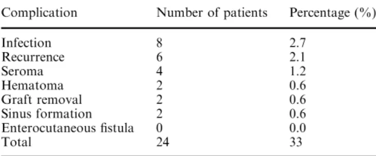
first year after treatment. Two patients had hematoma formation; seroma was present in four patients. These complications were noticed at early postoperative days. While hematoma showed regression, seroma needed intermittent repeated aspirations. Infection was encountered in eight patients (2.7%), six of whom were within the first 10 days after operation and treated with antibiotics without the need for graft excision. The other two had serious infection within the first month after treatment, necessitating graft removal with open wound treatment in addition to antibiotic administra-tion. Sinus formation was seen in two patients who were treated by curettage. No case of enterocutaneous fistula formation was seen. The results are summarized in Table 1.
Discussion
Incisional hernia is a common complication that can be encountered after major abdominal surgery. Its fre-quency ranges between 2 and 20% [1–3]. It can also be observed after relatively minor interventions like lapa-roscopy. This is known as trocar-site hernia, and the frequency has been found to be about 1% [5, 6]. Treatment of large incisional hernias by primary repair, i.e., conventional suturing technique, has been unsatis-factory [8–10]. Patients’ age, body weight beyond nor-mal, and associated diseases are among patient-related factors that predispose to hernia development or to its recurrence after primary repair. Some other factors including the type of incision, suture material used during abdominal closure or hernia repair and wound infections are also considered to be predisposing factors for hernia development or its recurrence [16–19].
High recurrence rates after primary repair could be due to extensive tension on the suture line in addition to the predisposing factors mentioned above [3, 18, 25]. The size of the fascial defect has been reported to be in
direct proportion to the recurrence rate after primary hernia repair [17, 18, 20–22]. This may be due to extensive tension that appears after primary closure of large fascial defects, leading to the abdominal corsage effect, which will ultimately lead to tissue ischemia, su-ture deficiency and finally fascia tear. This effect is de-fined as restriction of abdominal wall motility with feeling of tightness [16]. We have overcome this effect by applying relaxation incisions on the anterior leaf of the rectus fascia, using the hernia sac as fascia in cases of extensive tension when the fascia ends were approxi-mated.
The high rate of recurrence after primary repair has led us to use polypropylene mesh-reinforced type hernia repair. Ideal synthetic graft material should be biolog-ically inert, strong, stable in the presence of infections, non-carcinogenic and non-allergenic [23]. We think all these characteristics are present in the graft (polypro-pylene), we have used. Infection was observed in eight patients (2.7%); only in two patients (0.6%) was graft removal needed to control the infection. In our previ-ous study infection was observed in three patients (6.6%) [13]. In another study, where a second-genera-tion cephalosporin was used, 7% infecsecond-genera-tion rate and 2.3% graft removal rate were reported [16]. In Koren-kov’s study, although third-generation cephalosporin was used as a prophylactic antibiotic, the infection rate was 8.5% [24]. In our the study the infection rate and graft removal rate were far lower compared to other studies [1, 2, 24, 25]. We think there is no need for prophylactic purposes to use an antibiotic with a spectrum wider than that of first-generation cephalo-sporin.
There is no doubt tension-free mesh repairs for large hernias have reduced the incidence of recurrence, but the rate is still considered high [9, 23, 26]. It was found to range between 5 and 20% [2,16, 23,25]. Wound infec-tion, and defect size are among the factors most asso-ciated with recurrence [1, 17, 23]. In the body, polypropylene mesh shrinks by 30% from its original size [1]. This explains why the graft used should be 4– 6 cm larger than the defect size. In our study prophy-lactic antibiotic usage, relaxation-incision application when tension was present, and suitable mesh size usage were among the factors that led to low recurrence rate. Few studies have revealed as low a recurrence rate as
Fig. 2 The fascial defect was covered with peritoneum (A), and the tension-free prolene graft (B) was employed on it. Sutures were applied to the margins of the fascial defect
Table 1 Complications encountered after hernia repair with mesh Complication Number of patients Percentage (%)
Infection 8 2.7 Recurrence 6 2.1 Seroma 4 1.2 Hematoma 2 0.6 Graft removal 2 0.6 Sinus formation 2 0.6 Enterocutaneous fistula 0 0.0 Total 24 33
ours [1,4,14–16]. But in these studies polypropylene was inserted retro muscular. This is disadvantageous when compared to ours because its application and removal are technically difficult. We should not forget that, al-though it is rare, sometimes mesh removal is needed for one reason or another.
Our average patient follow up was 55 months. Studies have shown that most recurrences appear within the first 3 years after repair [27,28]. We think the follow-up period was long enough and the patient number was large enough to give us statistically meaningful results. Sometimes because of short follow-up time, a falsely low recurrence rate has been recorded.
Hematoma and seroma formation are not serious complications though they are disturbing ones. Patients developing hematoma were treated by tied dressing and observation. Re-operations were not needed. Seroma formation can be prevented by avoiding unnecessary wide fascial dissection and the use of a good drainage system. Patients with seroma formation were treated by repeated aspirations.
Enterocutaneous fistula (E.C. fistula) formation after incisional hernia repair with mesh is a rare complication [26,29]. Leber et al. reported a 3.5% incidence of E.C fistula formation [25,29]. We did not encounter any E.C fistula. We think if the propylene mesh is kept away from direct contact with intestine no E.C fistula for-mation will be observed, while others have reported in-tra-abdominal use of meshes to be safe [21, 29].
Conclusion
Covering the herniation site by suturing the hernia sac to the opposite fascia and applying mesh graft on top of it allows the achievement of a tension-free repair. This also prevents the adhesion of the omentum and intestines to the mesh graft, increasing the development of ileus and E.C. fistulas. In our technique, hernia sac and graft act as a dual mesh graft. This combination is not only cheaper but also more physiologically sound than actual dual mesh graft.
Mesh application in treatment of abdominal hernias has revealed good results when compared to primary suture. But some complications are still bothersome. Our modified technique with polypropylene mesh seemed to decrease all the expected complications after incisional hernia repair. The patient number and the follow-up period in this study are almost the largest and longest, respectively, when compared to other studies in the literature. Our modified operative tech-nique revealed a recurrence rate of 2.1%, a serious infection rate of only 0.6%, no enterocutaneous fistula formation, and insignificant incidence rates of hema-toma and seroma formation. We think tension and infection prevention, wide spectrum of mesh usage and usage of large enough mesh are the factors that allowed these good results.
References
1. Paajanen H, Hermunen H (2004) Long-term pain and recur-rence after repair of ventral incisional hernias by open mesh: clinical and MRI study. Langenbecks Arch Surg 389(5):366– 370
2. Leber GE, Garb JL, Alexander AI, Reed WP (1998) Long-term complications associated with prosthetic repair of incisional hernias. Arch Surg 133(4):378–382
3. Johnson D, Harrison DH (1999) A technique for repairing massive ventral incisional hernias without the use of a mesh. Br J Plast Surg 52(5):399–403
4. Bucknall TE, Cox PJ, Ellis H (1982) Burst abdomen and inci-sional hernia: a prospective study of 1,129 major laparotomies. Br Med J (Clin Res Ed) 284(6320):931–933
5. Sanz-Lopez R, Martinez-Ramos C, Nunez-Pena JR, Ruiz de Gopegui M, Pastor-Sirera L, Tamames-Escobar S (1999) In-cisional hernias after laparoscopic vs open cholecystectomy. Surg Endosc 13(9):922–924
6. Tonouchi H, Ohmori Y, Kobayashi M, Kusunoki M (2004) Trocar site hernia. Arch Surg 139(11):1248–1256
7. Bowrey DJ, Blom D, Crookes PF, Bremner CG, Johansson JL, Lord RV, Hagen JA, DeMeester SR, DeMeester TR, Peters JH (2001) Risk factors and the prevalence of trocar site herniation after laparoscopic fundoplication. Surg Endosc 15(7):663–666 8. Clark JL (2001) Ventral incisional hernia recurrence. J Surg
Res 99(1):33–39
9. Langer S, Christiansen J (1985) Long-term results after inci-sional hernia repair. Acta Chir Scand 151(3):217–219 10. Gecim IE, Kocak S, Ersoz S, Bumin C, Aribal D (1996)
Recurrence after incisional hernia repair: results and risk fac-tors. Surg Today 26(8):607–609
11. Usher FC, Ochsner J, Tuttle LL Jr (1958) Use of marlex mesh in the repair of incisional hernias. Am Surg 24(12):969–974 12. Usher FC (1970) The repair of incisional and inguinal hernias.
Surg Gynecol Obstet 131(3):524–530
13. Turkcapar AG, Yerdel MA, Aydinuraz K, Bayar S, Kuterdem E (1998) Repair of midline incisional hernias using polypro-pylene grafts. Surg Today 28(1):59–63
14. Schumpelick V, Conze J, Klinge U (1996) Preperitoneal mesh-plasty in incisional hernia repair. A comparative retrospective study of 272 operated incisional hernias. Chirurg 67(10):1028– 1035
15. Mudge M, Hughes LE (1985) Incisional hernia: a 10 year prospective study of incidence and attitudes. Br J Surg 72(1):70–71
16. Machairas A, Misiakos EP, Liakakos T, Karatzas G (2004) Incisional hernioplasty with extraperitoneal onlay polyester mesh. Am Surg 70(8):726–729
17. Petersen S, Henke G, Freitag M, Faulhaber A, Ludwig K (2001) Deep prosthesis infection in incisional hernia repair: predictive factors and clinical outcome. Eur J Surg 167(6):453– 457
18. Houck JP, Rypins EB, Sarfeh IJ, Juler GL, Shimoda KJ (1989) Repair of incisional hernia. Surg Gynecol Obstet 169(5):397– 399
19. Condon RE (1995) Incisional hernia. In: Nyhus LM, Condon RE (eds) Hernia, 4th edn. Lippincott, Philadelphia, pp 319–328 20. Larson GM, Vandertoll DJ (1984) Approaches to repair of ventral hernia and full-thickness losses of the abdominal wall. Surg Clin N Am 64(2):335–249
21. Shukla VK, Gupta A, Singh H, Pandey M, Gautam A (1998) Cardiff repair of incisional hernia: a university hospital expe-rience. Eur J Surg 164(4):271–274
22. Hesselink VJ, Luijendijk RW, de Wilt JH, Heide R, Jeekel J (1993) An evaluation of risk factors in incisional hernia recurrence. Surg Gynecol Obstet 176(3):228–234
23. Morris-Stiff GJ, Hughes LE (1998) The outcomes of nonab-sorbable mesh placed within the abdominal cavity: literature review and clinical experience. J Am Coll Surg 186(3):352–367
24. Korenkov M, Sauerland S, Arndt M, Bgrad L, Neugebauer EA, Troidl H (2002) Randomized clinical trial of suture repair, polypropylene mesh or autodermal hernioplasty for incisional hernia. Br J Surg 89(1):50–56
25. Trombetta F, Scamuzzi M, Moscato R, Mussa B, Goss M (2002) Surgical treatment for incisional hernias. Panminerva Med 44(2):141–144
26. McLanahan D, King LT, Weems C, Novotney M, Gibson K (1997) Retrorectus prosthetic mesh repair of midline abdominal hernia. Am J Surg 173(5):445–449
27. San Pio JR, Damsgaard TE, Momsen O, Villadsen I, Larsen J (2003) Repair of giant incisional hernias with polypropylene mesh: a retrospective study. Scand J Plast Reconstr Surg Hand Surg 37(2):102–106
28. Langer S, Christiansen J (1985) Long-term results after inci-sional hernia repair. Acta Chir Scand 151(3):217–219 29. Burger JW, Luijendijk RW, Hop WC, Halm JA, Verdaasdonk
EG, Jeekel J (2004) Long-term follow-up of a randomized controlled trial of suture versus mesh repair of incisional her-nia. Ann Surg 240(4):578–583; discussion 583–585