The triangle between the anterior and posterior
cruciate ligaments: an arthroscopic anatomy study
Correspondence: Alper Kaya, MD. Acibadem University, Department of Orthopaedics and Traumatology,
Acibadem Kadikoy Hospital, İstanbul, Turkey. Tel: +90532 - 654 77 34 e-mail: [email protected]
Submitted: November 10, 2014 Accepted: February 15, 2015
©2015 Turkish Association of Orthopaedics and Traumatology
Available online at www.aott.org.tr doi: 10.3944/AOTT.2015.14.0402 QR (Quick Response) Code
doi: 10.3944/AOTT.2015.14.0402
Alper KAyA1, Murat KöKen2, Burak AKAn3, Doğaç KArAgüven3, Berk güçlü3
1Acıbadem University Faculty of Medicine, Department of Orthopedics and Traumatology, İstanbul, Turkey 2Samsun Gazi State Hospital, Department of Orthopaedics and Traumatology, Samsun, Turkey 3Ufuk University Faculty of Medicine, Department of Orthopaedics and Traumatology, Ankara, Turkey
Objective: The goal of anterior cruciate ligament (ACL) reconstruction is to place the graft in closest
proximity to the native ACL anatomy. This study aims to examine the angular relation between intact anterior and posterior cruciate ligaments (PCL) from an arthroscopic perspective.
Methods: Forty patients (20 male, 20 female) with a mean age of 35.12 (range: 18–40) years that
un-derwent knee arthroscopy for reasons other than ACL rupture were included in the study. Following diagnostic examination and repair of the primary pathology, the triangle between ACL and PCL was seen at different flexion degrees of the knee joint (120, 90, 60, and 30°) through standard anterolateral (AL) and anteromedial (AM) portals. The narrow top angle of the triangle between the long intersect-ing axes of ACL and PCL was measured usintersect-ing recorded images by 3 blind observers.
Results: The average ACL-PCL angle was 61°, (standard deviation±2°) at 90°of knee flexion. The
angles were narrower when viewed through the AM portal. The degree of the angles was not affected by age, sex, body mass index (BMI), or the side (right or left) on which the procedure was performed. There was good-to-excellent intra- and interobserver reliability.
Conclusion: The angular relation between intact ACL and PCL has the potential to provide a better
view of the anatomy during arthroscopic ACL surgery. To perform better anatomic reconstructions, it is important to create a 60° angle between the ACL graft and PCL (as viewed through AL portal) at 90°of knee flexion.
Keywords: Anatomy; anterior cruciate ligament; arthroscopy; posterior cruciate ligament;
reconstruc-tion; triangle.
Level of Evidence: Level IV Therapeutic Study
Previous studies on anterior cruciate ligament (ACL) have mainly focused on its anatomy and function, in an attempt to develop better reconstruction techniques. The anatomy of the ACL has been detailed in cadaveric, bio-mechanical, and embryological studies,[1–10] which have
provided useful data for the identification of femoral and
tibial tunnel locations for anatomic ACL reconstruction. Following arthroscopic ACL reconstruction, a trian-gular space should be visible at the apex of the notch with the knee in 90° of flexion.[11] The borders of the triangle
consist of the roof of the notch, the anteromedially direct-ed fibers of the posterior cruciate ligament (PCL), and
the posterolaterally directed fibers of the ACL graft.[12]
The aim of this study was to demonstrate the angular relationship between ACL and PCL using arthroscopy. Since tunnels are established when the knee is in a flexed position during ACL reconstruction, the relationship between ACL and PCL at various flexion angles of the knee could provide helpful information about the recon-structed ligament’s conformity to native ACL.
Patients and methods
Patients who were admitted to our hospital and under-went knee arthroscopy for conditions other than ACL rupture were included in this observational study. Pa-tients <18 or >40 years and paPa-tients with a history of ACL injury, osteoarthritis, or findings of instability on physical examination were excluded. Twenty male and 20 female patients were studied, with 10 left and 10 right knees in each gender group. Demographical data including age, sex, site of involvement, height, and body mass index (BMI) were recorded. Mean age and BMI of patients were 35.12 (range: 18–40) years and 26.70 (range: 20–32) kg/m2, respectively. The study protocol
was approved by the ethics committee of university (ap-proval number: 8088), and informed consent was ob-tained from all patients prior to study entry. The study was conducted according to the principles of the Decla-ration of Helsinki.
Two surgeons performed the arthroscopic opera-tions in the study. Diagnostic knee arthroscopy using standard anterolateral (AL) and anteromedial (AM) portals was performed under spinal anesthesia with 4 mm, 30° arthroscope. The AL portal is located at the center of the soft spot area between the lateral border of patellar tendon, lateral femoral condyle, and the proxi-mal tibia. For optiproxi-mal insertion point, the AM portal was found with the help of a spinal needle, located just proximal to the meniscal rim, 1 cm distal and medial to the inferior pole of patella and halfway between the pa-tellar tendon and anterior fibers of the medial collateral ligament. Attention was paid to determinate the portal locations relative to each other as much as possible with a standardized method as described above by marking these anatomical landmarks in every patient. Patients who were morbidly obese or in whom these landmarks were not clearly visible were excluded from the study. After diagnostic arthroscopy was performed, the follow-ing primary pathologies were treated: medial meniscus tears (n=14), lateral meniscus tears (n=7), medial pa-tellar plicae (n=5), loose body (n=4), chondral lesions (n=6; 5 on medial and 1 on lateral femoral condyle), and patellofemoral problems (n=1). The ACL was
exam-ined by arthroscopy probe, and patients were excluded from the study if ACL was elongated or if there was any doubt regarding the function of the ACL. Debridement, including excision of ligamentum mucosum and limited excision of synovial tissue between ACL and PCL, was performed in patients in whom the intercondylar notch was not clearly visible. The triangle between ACL and PCL was visualized at different flexion degrees of the knee joint (120, 90, 60, and 30° of flexion angle was determined with a sterile goniometer) through AL and then AM portals, respectively. The scope position was standardized as much possible; the camera was held vertical to the floor and the light source was located inferomedially and directed to the intercondylar femo-ral notch while viewed from the medial portal, located inferolaterally and directed to the intercondylar notch while viewed from the lateral portal. Prior to recording the images, the surgeon confirmed that 1/3 of the lateral femoral condyle, cartilage border of the medial femoral condyle, PCL, and ACL were included in the image. The scope was held posterior to the intermeniscal ligament while recording the images. From the 2 portals and at the 4 different knee flexion degrees, a total of 8 photo-graphs were obtained for each patient.
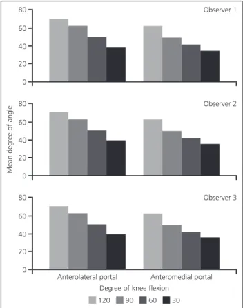
Triangles formed between ACL and PCL at each flexion degrees and as viewed through AL or AM por-tals were examined using the recorded images. This is a 3-dimensional angle, but it can be measured 2-dimen-sionally. The narrow angle of the triangle between the long intersecting axes of the medial border of the AM bundle of ACL and lateral border of PCL was measured (Figure 1a). In some cases, the direction of ACL was not straight (curved over PCL); thus, points were marked on the medial border of the ACL at the intersection area with PCL and connected to form a line. Samples of the measurements in a left knee at 90, 60, and 30° of knee flexion are shown in Figures 1b–d. Measurements were performed using image processing software (Ado-be Photoshop CS5, San Jose, CA, USA) and numeri-cal values were recorded. Three independent observers measured and recorded the narrow angle of the triangles twice at different times (Figure 2).
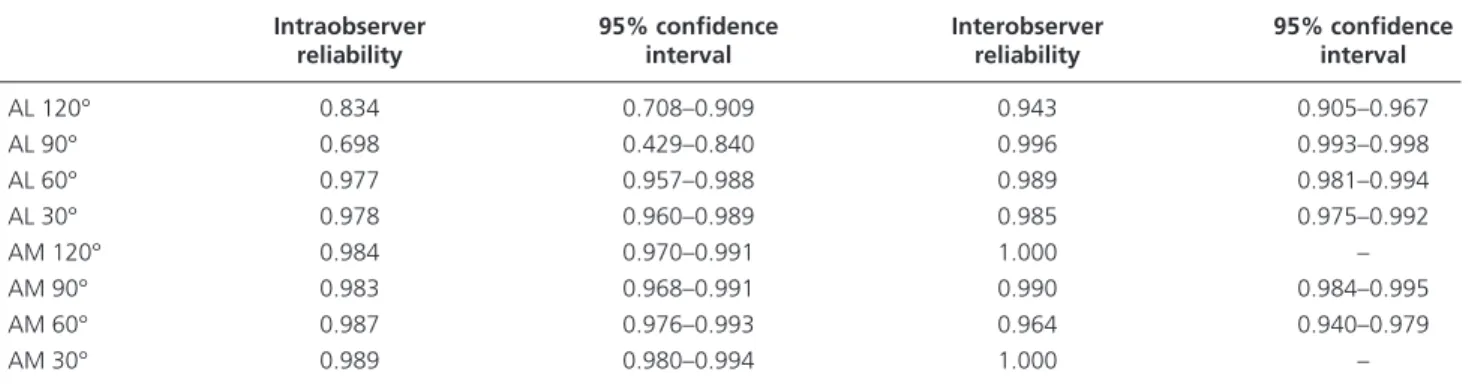
Descriptive statistics including frequencies, mean, range, and standard deviation were recorded from all different measured variables. The mean measurement of all 3 observers was used for further statistical analysis. Independent t-test was used to determine if there was a difference in ACL-PCL angle between male and female subjects. The alpha level for statistical significance was set at 0.05. Intra- and interobserver reliability was cal-culated using the intraclass correlation coefficient (ICC).
results
Results of the demographics and measurements for each flexion angle are displayed in Table 1. Intra- and interob-server reliability is demonstrated in Table 2.
Discussion
This was an observational study on young patients with normal ACL and PCL, and results of the study showed that there is approximately 60° of angle between ACL and PCL at 90–120° flexion of the knee joint while look-ing from the AL portal with scope. To the best of our knowledge, no such study has been previously performed.
Successful ACL reconstruction depends on various factors, including graft fixation, graft tensioning, as well as femoral and tibial tunnel positioning.[10,13,14] Detailed
anatomy, including the tibial and femoral attachments
of the human ACL, have been comprehensively de-scribed dede-scribed in previous studies.[1,2,4–8,10,13–15]
Ac-curate measurements of the insertion points are critical when determining placement of bone tunnels during ACL reconstruction. More anatomic ACL reconstruc-tions and improvement of knee mechanics were the aims of these studies. Patients in this study were in an age group in which ACL injuries are most frequent and most arthroscopic ACL reconstructions are performed, and the angular relation was documented from an
ar-Fig. 1. (a) The triangle formed between ACL and PCL. (b) A 61O angle between ACL and PCL, as viewed through the AL portal when the left knee
joint is at 90O flexion. (c) A 50O angle between ACL and PCL as viewed through the AL portal when the left knee joint is at 60O flexion. (d)
A 36O angle between ACL and PCL as viewed through the AL portal when the left knee is at 30O flexion. (ACL: Anterior cruciate ligament;
PCL: Posterior cruciate ligament; LFC: Lateral femoral condyle; AM: Anteromedial bundle of ACL; PL: Posterolateral bundle of ACL.) [Color figures can be viewed in the online issue, which is available at www.aott.org.tr]
(a) (b) (c) (d)
Fig. 2. Mean measurements of angles by 3 different observers.
80 60 40 20 0 Mean deg ree of ang le Observer 1 80 80 60 60 40 40 20 20 0 0 Observer 2 Anterolateral portal
Degree of knee flexion
Anteromedial portal Observer 3
120 90 60 30
Table 1. No significant difference was found in AC-PCL angle
between men and women for any of the measured portal or knee flexion angles.
Frequency Mean range SD
Sex 20:20 Age 35.12 18–40 6.354 Height 167.85 151–196 8.279 Weight 75.20 49–98 10.737 Side 20:20 BMI 26.70 20–32 3.360 AL 120° 69 68–72 1.00 AL 90° 62 59–63 1.00 AL 60° 52 50-–55 1.90 AL 30° 40 36–45 1.70 AM 120° 62 59–65 1.75 AM 90° 53 50–56 2.00 AM 60° 42 40–45 2.10 AM 30° 31 30–35 1.80
throscopic point of view, the method routinely used for ACL repair.[5,6]
This study showed the angles of a triangle between the longitudinal axes of the ACL and PCL and the roof of the notch. The angle between the longitudinal axis of ACL and PCL changes with knee motion. The measure-ments of the observers were consistent, suggesting that these angles can be used after the suture or graft passage during arthroscopic ACL reconstructions to provide additional guidance to the surgeon. Our results demon-strated that age, sex, height, and BMI do not have any significant relation with ACL-PCL intersection angle. The angles may change according to the portal locations and position of light source; however, in this study we aimed to observe the angular relation of ACL and PCL with standard AL and AM portals, viewed with the light source directed to the intercondylar notch. Accordingly, the values obtained from this study can better inform surgeons on the angular relation between ACL and PCL.
Selecting the localization of the femoral tunnel in particular presents a greater challenge than selecting that of of the tibial tunnel. Due to the double-bundle morphology of the ACL, its femoral footprint is larger than the diameter of the ACL body. Femoral insertion site anatomy changes with varying degrees of knee flex-ion. The placement of traditional ACL grafts in a high and proximal position at the femoral attachment and PCL grafts at the tibial attachment results in vertical graft orientation.[16,17] This graft placement results in a
limited ability to provide rotational stability and is infe-rior when compared to an anatomic reconstruction. Tra-ditional 2-dimensional evaluations such as the subjective clockwise method did not prove sufficient to adequately define the position of the femoral tunnel, as the knee is flexed to 90° and the horizontal plane cannot be well-matched with the clock face.[10] In a cadaveric study,
Mo-chizuki et al. showed that the center of the AM bundle
of ACL was at a clock position of 1:40 in the left knee.
[5] Siebold et al. showed that the ACL is aligned
hori-zontally when the femoral shaft axis was lifted 12° from the horizontal plane,[6] which can be obtained in a knee
flexion angle of 102°. With this flexion angle, centers of the AM and PL bundles of the ACL align horizontally, allowing for use of the clock method. Other techniques such as the AM portal technique may be used in de-termining the insertion points of grafts.[18] Our study
supports the horizontal or low tunnel position to form a broad triangle. If the femoral tunnel is drilled via the transtibial portal, the surgeon may use these angles with the guide pin in order to determine the tunnel location. Transtibial drilling guides the wire to a more vertical direction, resulting in a highly-positioned femoral tun-nel. This approach results in a narrow angle between ACL and PCL and may cause roof impingement. Stud-ies show that horizontal graft placement provides better results.[19,20] Zantop et al. showed that non-anatomical
double-bundle reconstruction may fail to demonstrate any clinical superiority to single-bundle reconstruction.
[14] These results may indicate that the primary critical
factor is the anatomic placement of the graft rather than the choice to use single- or double-bundle procedure.
This study provides additional information on the dy-namic anatomy of ACL from an arthroscopic perspective. For more anatomic reconstructions, it is important to create a triangle of approximately 60° between ACL and PCL, as viewed through the AL portal at 90–120° de-grees of knee flexion. The angle between ACL and PCL was not affected by sex in this study. In addition, age, sex, BMI, and side (right or left) on which the procedure was performed did not affect mean degree of the angle. Acknowledgement
We greatly thank to Prof. Freddie H. Fu, M.D., and Dr. Carola F. Van Eck, M.D. for their kindly contribution and statistical help for our study.
Table 2. The reliability was good-to-excellent for all measured dimensions.
Intraobserver 95% confidence Interobserver 95% confidence
reliability interval reliability interval
AL 120° 0.834 0.708–0.909 0.943 0.905–0.967 AL 90° 0.698 0.429–0.840 0.996 0.993–0.998 AL 60° 0.977 0.957–0.988 0.989 0.981–0.994 AL 30° 0.978 0.960–0.989 0.985 0.975–0.992 AM 120° 0.984 0.970–0.991 1.000 – AM 90° 0.983 0.968–0.991 0.990 0.984–0.995 AM 60° 0.987 0.976–0.993 0.964 0.940–0.979 AM 30° 0.989 0.980–0.994 1.000 –
Conflics of Interest: No conflicts declared.
references
1. Colombet P, Robinson J, Christel P, Franceschi JP, Djian P, Bellier G, et al. Morphology of anterior cruciate liga-ment attachliga-ments for anatomic reconstruction: a cadav-eric dissection and radiographic study. Arthroscopy 2006;22:984–92.
2. Edwards A, Bull AM, Amis AA. The attachments of the anteromedial and posterolateral fibre bundles of the ante-rior cruciate ligament. Part 2: femoral attachment. Knee Surg Sports Traumatol Arthrosc 2008;16:29–36.
3. Ferretti M, Levicoff EA, Macpherson TA, Moreland MS, Cohen M, Fu FH. The fetal anterior cruciate liga-ment: an anatomic and histologic study. Arthroscopy 2007;23:278–83.
4. Luites JW, Wymenga AB, Blankevoort L, Kooloos JG. De-scription of the attachment geometry of the anteromedial and posterolateral bundles of the ACL from arthroscopic perspective for anatomical tunnel placement. Knee Surg Sports Traumatol Arthrosc 2007;15:1422–31.
5. Mochizuki T, Muneta T, Nagase T, Shirasawa S, Akita KI, Sekiya I. Cadaveric knee observation study for de-scribing anatomic femoral tunnel placement for two-bun-dle anterior cruciate ligament reconstruction. Arthroscopy 2006;22:356–61.
6. Siebold R, Ellert T, Metz S, Metz J. Femoral insertions of the anteromedial and posterolateral bundles of the an-terior cruciate ligament: morphometry and arthroscopic orientation models for double-bundle bone tunnel place-ment--a cadaver study. Arthroscopy 2008;24:585–92. 7. Siebold R, Ellert T, Metz S, Metz J. Tibial insertions of
the anteromedial and posterolateral bundles of the ante-rior cruciate ligament: morphometry, arthroscopic land-marks, and orientation model for bone tunnel placement. Arthroscopy 2008;24:154–61.
8. Takahashi M, Doi M, Abe M, Suzuki D, Nagano A. Ana-tomical study of the femoral and tibial insertions of the anteromedial and posterolateral bundles of human ante-rior cruciate ligament. Am J Sports Med 2006;34:787–92. 9. Tansatit T, Saowaprut S, Kanchanatawan W, Chom-kerd T. Pattern of angular change of the anterior cruciate ligament across the range of knee flexion and the related anatomical dimensions. J Med Assoc Thai 2005;88 Suppl 4:95–102.
10. Tsukada H, Ishibashi Y, Tsuda E, Fukuda A, Toh S. Ana-tomical analysis of the anterior cruciate ligament femoral and tibial footprints. J Orthop Sci 2008;13:122–9. 11. Lawhorn K, Howell SM. Avoiding ACL graft
impinge-ment: principles for tunnel placement using the transtibial tunnel technique. In Jackson DW, editor. Master Tech-niques in orthopaedic surgery:reconstructive knee surgery. Philadelphia:Lippincott Williams & Williams 2008. 12. Longino D, Clerck N, Fowler PJ, Giffin JR. Technique in
ACL reconstruction: hamstring reconstruction. In Bonnin M et al. editors. The Knee Joint. Surgical techniques and strategies. Paris: Springer-Verlag 2012;17:195–202. 13. Musahl V, Plakseychuk A, VanScyoc A, Sasaki T, Debski
RE, McMahon PJ, et al. Varying femoral tunnels between the anatomical footprint and isometric positions: effect on kinematics of the anterior cruciate ligament-reconstructed knee. Am J Sports Med 2005;33:712–8.
14. Zantop T, Diermann N, Schumacher T, Schanz S, Fu FH, Petersen W. Anatomical and nonanatomical double-bundle anterior cruciate ligament reconstruction: impor-tance of femoral tunnel location on knee kinematics. Am J Sports Med 2008;36:678–85.
15. Harner CD, Baek GH, Vogrin TM, Carlin GJ, Kashiwa-guchi S, Woo SL. Quantitative analysis of human cruciate ligament insertions. Arthroscopy 1999;15:741–9. 16. Heming JF, Rand J, Steiner ME. Anatomical limitations
of transtibial drilling in anterior cruciate ligament recon-struction. Am J Sports Med 2007;35:1708–15.
17. Noyes FR. The function of the human anterior cruciate ligament and analysis of single- and double-bundle graft reconstructions. Sports Health 2009;1:66–75.
18. Bedi A, Altchek DW. The “footprint” anterior cruciate liga-ment technique: an anatomic approach to anterior cruciate ligament reconstruction. Arthroscopy 2009;25:1128–38. 19. Sastre S, Popescu D, Núñez M, Pomes J, Tomas X, Peidro
L. Double-bundle versus single-bundle ACL reconstruc-tion using the horizontal femoral posireconstruc-tion: a prospective, randomized study. Knee Surg Sports Traumatol Arthrosc 2010;18:32–6.
20. Loh JC, Fukuda Y, Tsuda E, Steadman RJ, Fu FH, Woo SL. Knee stability and graft function following anterior cruciate ligament reconstruction: Comparison between 11 o’clock and 10 o’clock femoral tunnel placement. 2002 Richard O’Connor Award paper. Arthroscopy 2003;19:297–304.