607
Srp Arh Celok Lek. 2014 Sep-Oct;142(9-10):607-609 DOI: 10.2298/SARH1410607M
ПРИКАЗ БОЛЕСНИКА / CASE REPORT UDC: 617.587-003.4
Correspondence to: Özgür KORKMAZ
Kaptan Arif Sokak no: 44 D:9 Suadiye- Kadıkoy, Istanbul Turkey
[email protected] SUMMARY
Introduction Epidermoid inclusion cysts are usually composed of epidermal elements implanted into
the dermal layers. Patients are seen in the outpatient clinics with a mass. Most of the complaints are mechanical and cosmetic problems.
Case Outline A 34-year-old female patient was admitted to our clinic because of swelling and pain in
her right foot. A palpable mass was detected in the first web. On the x-rays of the foot no osseous lesion was detected. There was a soft tissue mass in the first web according to MRI report. Soft tissue mass was excised and sent to pathology. According to pathology report the mass was an epidermoid cyst 5×2×1.5 cm in size. There were no problems during follow-up of the patient for 6 months after surgery. The patient had no swelling in the foot and had no additional complaints on checkup.
Conclusion In the differential diagnosis, we should take into consideration epidermoid cyst of large soft
tissue masses of the foot. Surgical excision should be done within the appropriate limits.
keywords: epidermoid cyst; foot; plantar
Giant Epidermal Cyst of the Foot
Melih Malkoc1, Özgür Korkmaz1, Yıldıray Genç2, Ferhat Say3, Mahmut Nedim Aytekin4
1Istanbul Medipol University, School of Medicine, Orthopedics and Traumatology, Istanbul, Turkey; 2Or-Ahayim Balat Hospital, Istanbul, Turkey;
319 Mayıs University School of Medicine, Orthopedics and Traumatology, Samsun, Turkey; 4Ankara Atatürk Research Hospital, Orthopedics and Traumatology, Ankara, Turkey
INTRODUCTION
Epidermoid cysts are also termed epidermal cysts, epidermoid inclusion cyst, infundibular cysts and keratin cysts [1]. Epidermal inclu-sion cysts are usually composed of epidermal elements that are implanted into the dermal layers [2].
Patients are seen at outpatient clinics with a mass on the foot. Some patient’s complaints are mechanical problems that are created by the mass, while some patients have cosmetic problems. The mass on foot can be benign or malignant. Epidermoid cysts are seen more fre-quently on a hand than on the foot and gener-ally patients have a history of trauma [3].
CASE REPORT
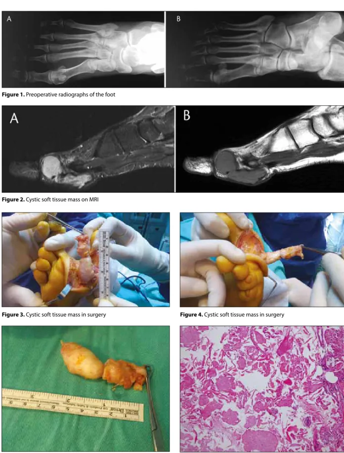
A 34-year-old female patient was admitted to our clinic with complaints of swelling at the base of her right foot and pain during walk-ing. The patient’s complaints lasted for about one year. According to the patient, there was no trauma or stinging with a sharp-pointed structure or body. The range of motion of the right foot and the toes were normal on physi-cal examination. No neurovascular deficiency was detected. There were no signs of infection. A soft tissue mass approximately 3.5×1 cm in size that was in the first web was detected by palpation on the plantar side of the right foot. No structural bone pathology and soft tis-sue pathology was identified on the foot ra-diographs (Figure 1). A cystic soft tissue mass without contrast enhancement was detected
by contrast-enhanced MRI (Figure 2). Surgi-cal excision was recommended to the patient. Approximately 7 cm longitudinal side right incision was done on the plantar of the foot in the first web. Skin and subcutaneous tissues were passed, the soft tissue mass was dissected and a wide excision was done (Figures 3, 4 and 5). Subcutaneous tissues and skin were closed. The excised mass was sent for pathological analysis. There were neither neurovascular complications nor wound complications after the surgery. The excised soft tissue mass was reported as an epidermoid cyst according to the pathology report (Figure 6). There were no signs of recurrence after the six-month follow-up time period.
DISCUSSION
Ozatwa et al. [4] reported a 65-year-old male patient with a giant epidermoid cyst that was passed through interosseous muscles of the foot, with a mass on the dorsal side of the foot. The giant epidermoid cyst has no interosseous extension in our case. Epidermoid cysts may be atypical and with emissions. For this reason, the delineation of boundaries of the cyst be-fore surgery is important for surgical excision so as to prevent complications that may occur postoperatively.
Harish et al. [5] reported a case of keratin granuloma due to a ruptured epidermal cyst that occurred in the foot of a 52-year-old woman. There was no epidermoid cyst rupture in our case. Ruptured epidermal cysts must be taken into consideration in the differential diagnoses.
608
doi: 10.2298/SARH1410607M
Malkoc M. et al. Giant Epidermal Cyst of the Foot
Ghani et al. [6] reported a case of severe heel pain that did not respond to noninvasive measures. MRI scan re-vealed a soft tissue mass, which was found to be an epi-dermal cyst after a complete surgical excision. The patient experienced full resolution of symptoms after excision of the epidermal cyst. There was no development of trauma,
and in the pathological diagnosis was epidermoid cysts in our case identically as the case reported by Ghani et al. [6].
Matsumoto et al. [7] reported a 44-year-old woman admitted to the authors’ institution for evaluation of two masses in the right forefoot. Standard radiographs showed foci of calcification within the mass. MRI scan and
macro-Figure 5. Excised soft tissue mass Figure 6. Cyst has a wall nearly similar to epidermis and is filled with laminated keratin (HE, ×200)
Figure 1. Preoperative radiographs of the foot
Figure 2. Cystic soft tissue mass on MRI
609
Srp Arh Celok Lek. 2014 Sep-Oct;142(9-10):607-609
www.srp-arh.rs
1. Freedberg IM, Eisen AZ, Wolff K, Austen KF, Goldsmith LA, Katz SI, editors. Fitzpatrick’s Dermatology in General Medicine. 6th ed. New York: McGraw-Hill; 2003.
2. William J, Berger T, Dirk E. Andrews’ Diseases of the Skin: Clinical Dermatology. 10th ed. Philadelphia: Saunders; 2005.
3. Wu KK. Epidermoid cysts of the foot: with or without bone involvement. J Foot Surg. 1992; 31(2):203-6.
4. Ozawa T, Harada T, Ishii M. Giant epidermal cyst extending from sole to dorsum of the foot by penetrating the interosseous muscles. J Dermatol. 2008; 35(1):25-8.
5. Harish S, Jan E, Ghert M, Alowami S, Finlay K. Pseudotumoral appearance of a ruptured epidermal cyst in the foot. Skeletal Radiol. 2006; 35(11):867-71.
6. Ghani S, Fazal MA. An unusual cause of intractable heel pain. J Foot Ankle Surg. 2011; 50(6):744-6.
7. Matsumoto K, Okabe H, Ishizawa M, Egawa M.
Intermetatarsophalangeal bursitis induced by a plantar epidermal cyst. Clin Orthop Relat Res. 2001; (385):151-6.
8. Ferguson K, Thomson AG, Moir JS. Case study: Epidermoid cyst following percutaneous Topaz coblation for plantar fasciitis. Foot (Edinb). 2012; 22(1):46-7.
9. Basterzi Y, Sari A, Ayhan S. Giant epidermoid cyst on the forefoot. Dermatol Surg. 2002; 28(7):639-40.
scopic findings detected during surgery revealed that the lesion was composed mainly of two different compositions: dorsal cystic masses and a solid mass in the plantar side of the fifth metatarsal head. Histologic and immunohis-tochemical examinations showed that the former was an intermetatarsophalangeal bursitis induced by keratinous material, whereas the latter was a ruptured epidermal cyst. There was no formation of bursitis because of epidermoid cysts rupture in our case. Bursitis as a result of epidermoid cysts rupture must be thought in the differential diagnosis.
Ferguson et al. [8] reported a case of epidermoid cyst formation following the Topaz coblation for plantar
fas-ciitis. There was no history of treatment modality for foot in our case. Early treatment modalities must be learned before surgery.
Başterzi et al. [9] reported a 23-year-old woman with a painless tumor-like 6.5×5.5×5.5 cm mass in the right forefoot that was totally excised; histological diagnosis was epidermoid cyst. The size of the epidermoid cyst that we excised was smaller than that reported by Başterzi et al. [9]. Also, a large volume epidermoid cyst can be seen.
The epidermoid cyst should be in the list of differential diagnosis for a large soft tissue mass of the foot and surgi-cal excision must be done in appropriate limits.
REFERENCES КРАТАК САДРЖАЈ Увод Епи дер мо ид на ин клу зи о на ци ста обич но је са ста вље-на од епи дер мал них еле ме вље-на та им план ти ра них у дер мал не сло је ве. Бо ле сни ци с из ра сли ном се обич но ви ђа ју у кли-нич ким ам бу лан та ма. Ве ћи на их се жа ли на ме ха кли-нич ке и естет ске про бле ме. При каз бо ле сни ка Же на ста ро сти 34 го ди не при мље на је на на шу кли ни ку због ото ка и бо ло ва у де сном сто па лу. Пал пи-ра њем је от кри ве на пот ко жна ма са у ре ги ји из ме ђу пр ва два но жна пр ста. Ренд ген ским сним ком сто па ла се ни је ви де ла ко шта на ле зи ја. На лаз маг нет не ре зо нан ци је је от крио ма су ме ког тки ва у ре ги ји из ме ђу пр ва два но жна пр ста. Из вр-ше на је екс ци зи ја ма се ме ког тки ва, а узо рак је упу ћен на па то ло шку ана ли зу. Пре ма па то ло шком на ла зу, у пи та њу је би ла епи дер мо ид на ци ста ве ли чи не 5×2×1,5 cm. То ком ше-сто ме сеч ног пе ри о да клиничког праћења по сле опе ра ци је код бо ле сни це ни је до шло до ком пли ка ци ја. Же на ви ше ни је има ла про блем из ра сли не у сто па лу и при ли ком кон трол ног пре гле да ни је се жа ли ла на би ло ка кве до дат не про бле ме. За кљу чак По треб но је да се у ди фе рен ци јал ној ди јаг но зи раз ми шља и о мо гућ но сти епи дер мо ид не ци сте код на ла за ве ли ке ма се ме ког тки ва, а хи рур шка екс ци зи ја тре ба да се ура ди уну тар аде кват них гра ни ца. Кључ не ре чи: епи дер мо ид на ци ста; сто па ло; та ба ни