RESEARCH ARTICLE
Prognostic significance of the recurrence pattern and risk
factors for recurrence in patients with proximal gastric cancer
who underwent curative gastrectomy
Ahmet Bilici&Fatih Selcukbiricik
Received: 8 January 2015 / Accepted: 2 March 2015 / Published online: 12 March 2015 # International Society of Oncology and BioMarkers (ISOBM) 2015
Abstract Proximal gastric cancer has a high propensity of early recurrence after curative resection due to high incidence of lymph node involvement. In the present study, we aimed to investigate the pattern and time of recurrence and to evaluate the risk factors for recurrence of patients with proximal gastric cancer. Between 2005 and 2013, 99 patients with recurrent proximal gastric cancer who underwent radical gastrectomy were retrospectively analyzed. The prognostic significance of the pattern and the time of recurrence and the relationship between the pattern of recurrence and the other clinicopatho-logical factors were evaluated. The median time to recurrence was 24 months; 45.5 % of patients relapsed within 2 years. Forty-three (43.4 %) patients indicated hematogenous recur-rence and 41 (41.4 %) patients revealed peritoneal recurrecur-rence with the most predominant patterns. The median progression-free survival (PFS) time for patients with locoregional recur-rence was significantly better than that of patients with peri-toneal recurrences, hematogenous recurrences, and distant lymph nodes (32.2 vs. 18.9 vs. 18.2 vs. 9.7 months,p= 0.005, respectively). Moreover, the median overall survival (OS) interval for patients with distant lymph nodes recurrence was significantly worse than that of patients with locoregional, peritoneal, and hematogenous recurrences (13.5 vs. 48.5 vs. 31.4 vs. 29.9 months,p=0.006, respectively). The presence of lymph node metastasis (p=0.004) and surgery type (p=0.04)
for PFS and the time of recurrence (p=0.033), lymph node metastasis (p=0.03), and surgery type (p=0.04) for OS were found to be independent prognostic factors by multivariate analysis. Logistic regression analysis indicated that the pres-ence of lymph node metastasis and surgery type were inde-pendent risk factors for predicting the occurrence of early recurrence (p=0.001, OR 0.48 and p=0.028, OR 0.41, respec-tively). The median OS time of early recurrence patients was significantly shorter than that of patients with late recurrence (16.6 vs. 55.2 months,p<0.001). Furthermore, proximal gas-trectomy, poorly differentiated histology, advanced pT stage, and lymph node metastasis were significantly associated with early recurrence. Our results showed that lymph node metas-tasis and surgery type were independent risk factors for pre-diction of early recurrence in proximal gastric cancer. Thus, total gastrectomy with regional lymph node dissection may be a suitable treatment option for proximal gastric cancer patients with tumors that have high risk features for recurrence.
Keywords Proximal gastric cancer . Recurrence pattern . Prognosis . Survival
Introduction
Despite a decrease in incidence and major advancements in the treatment of early gastric cancer, it remains the second most frequent cause of cancer death worldwide, and the survival time for advanced gastric cancer is still poor [1,2]. Surgery is the only curative treatment, but it has a high rate of locoregional recurrence and distant metastases [3,4]. There is no effective treatment for recurrence [5], and advanced gastric cancer has fatal outcomes and regarded as an incurable disease [6]. Therefore, identification of early recurrence may improve
A. Bilici
Department of Medical Oncology, Medical Faculty, Istanbul Medipol University, Istanbul, Turkey
F. Selcukbiricik
Department of Medical Oncology, Sisli Etfal Education and Research Hospital, Istanbul, Turkey
A. Bilici (*)
TEM Avrupa Otoyolu Goztepe Cikisi, No: 1, 34214 Bagcilar, Istanbul, Turkey
patients’ survival because effective treatment strategies could be initiated while patients’ performance status is still sufficient to receive effective treatments for metastatic gastric cancer [7]. The majority of recurrences after surgery of gastric cancer are diagnosed within 2 years. Therefore, some studies have indicated that 2 years should be defined as a cutoff value for early and late recurrences [8,9]. In most patients, the patterns of recurrence are locoregional recurrence, peritoneal seeding, and hematogenous metastasis [10,11]. In recent years, where-as proximal cancers have increwhere-ased in developed countries, non-proximal tumors have continued to be predominant in Japan and other parts of the world [12–14].
Proximal gastric cancers have worse prognosis compared with those with gastric cancer located in the other parts of the stomach because the diagnosis is carried out in at advanced stage due to high incidence of lymph node metastasis and dif-ferent anatomical characteristics [13,14]. There have been few studies exploring the patterns of proximal gastric cancer in the literature [15,16]. Recently, Li et al. found that there was a significant correlation between deeper invasion and a higher incidence of hematogenous metastases and peritoneal recur-rence. They showed that the histological type, pT stage, and lymph node metastasis were independent risk factors for over-all recurrence. In addition, negative lymph node counts were found to be another independent risk factor for early recurrence in their study [17]. In the current study, we aimed to investigate the pattern and time of recurrence of patients with proximal gastric cancer. Furthermore, the risk factors for recurrence and prognostic factors of recurrent patients were also analyzed.
Patients and methods
Between 2005 and 2013, a total of 99 patients with recurrent proximal gastric cancer who had undergone curative gastrec-tomy were included in the study. The primary tumor was staged according to the American Joint Committee on Cancer (AJCC) TNM staging classification for gastric cancer [18]. The clinicopathological findings were determined ac-cording to the Japanese Classification of Gastric Carcinoma (JCGC) [19]. D1lymph node dissection was defined as only
the perigastric nodes directly attached along the lesser curva-ture, and greater curvatures of the stomach are removed (sta-tions 1–6, N1 level). An incomplete N1 dissection was de-scribed as D0lymphadenectomy. D2dissection (N2 level)
was defined as the removal of nodes along the left gastric artery (station 7), common hepatic artery (station 8), celiac trunk (station 9), splenic hilus, and splenic artery (stations 10 and 11). D3dissection included the dissection of lymph
nodes at stations 12 through 14, along the hepatoduodenal ligament and the root of the mesentery (N3 level).
Proximal gastric cancer was described as adenocarcinoma of the proximal third of the stomach or gastroesophageal (GE)
junction. If the GE junction tumor extented >2 cm onto the stomach, it was accepted to be of gastric origin [20]. Tumors of the other part of stomach and diffusely involving the whole stomach were also excluded from study. Patients who had R1
or R2 resections and insufficient disease information were
excluded from data analysis. Patient data were retrospectively obtained from patients’ charts with respect to age, gender, resection type, tumor location, histopathology, tumor stage, tumor size, histological grade, lymph node involvement, depth of tumor invasion (pT stage), lymphatic and blood ves-sel invasion, resection margins, type of adjuvant chemothera-py and/or radiation therachemothera-py, responses to treatment, and sur-vival after written informed consent had been obtained from patients or their relatives.
The patterns of recurrence were classified as locoregional, peritoneal, hematogenous, or distant lymph node metastases. Locoregional recurrences were defined as tumors within the gastric bed, regional gastric lymph nodes, and remnant stomach at the anastomosis or gastric stump. Peritoneal recurrences were defined as those cases with a positive cytology in the ascitic fluid, carcinomatosis, or ovarian metastasis. Hematogenous re-currences were described as visceral metastases. Distant lymph node metastases were also described as distant lymph nodes outside the regional basin. All recurrences were categorized as early and late. Early recurrence was defined as that recurring within 2 years, whereas late recurrence was defined as that recurring more than 2 years after resection.
Medical histories and physical examinations were per-formed every 3 months in the first postoperative year, every 6 months in the second postoperative year, and annually there-after for at least 5 years during follow-up. Complete blood counts and biochemistry panels as well as tumor markers were examined every 3 months in the first and second years, and annually thereafter. Chest X-rays and abdominal CT scans were performed every 3 months in the first year, every 6 months in the second postoperative year and annually there-after for 5 years. Gastroscopy was performed annually to con-trol suspicious cancer in the gastric remnant.
A total of 87 patients (87.8 %) with lymph node metastasis or pT3-4 received adjuvant chemoradiotherapy (CRT) within
4 weeks after surgery. Adjuvant CRT consisted of 5-fluorouracil 425 mg/m2per day, plus leucovorin 20 mg/m2per day for 5 days, followed by 4500 cGy of radiation at 180 cGy per day given 5 days per week for 5 weeks, with modified doses of fluorouracil and leucovorin on the first four and the last 3 days of radiotherapy. Adjuvant CRT was not given to the 12 of 99 patients without lymph node metastasis (12.2 %).
Statistical analysis All data were analyzed with SPSS 17.0 (SPSS Inc., Chicago, IL, USA) software. The clinicopatholog-ical factors of the patients among recurrence groups were compared by means of the chi-squared test and Fisher’s exact test. The survival analyses and curves were established with
the Kaplan-Meier method and compared with the log-rank test. Progression-free survival (PFS) was defined as the time from curative surgery to recurrence or to the date of death or loss of follow-up. OS was described as the time from diagno-sis to the date of the patient’s death or loss of follow-up. Univariate and multivariate analyses were performed with the Cox proportional hazards model to evaluate the
importance of the recurrence type and other clinicopatholog-ical features as prognostic factors. To identify predictive fac-tors related to recurrence, logistic regression analysis was used. Multivariatep values were used to characterize the in-dependence of these factors. The 95 % confidence interval (CI) was used to quantify the relationship between survival time and each independent factor. Allp values were two-sided
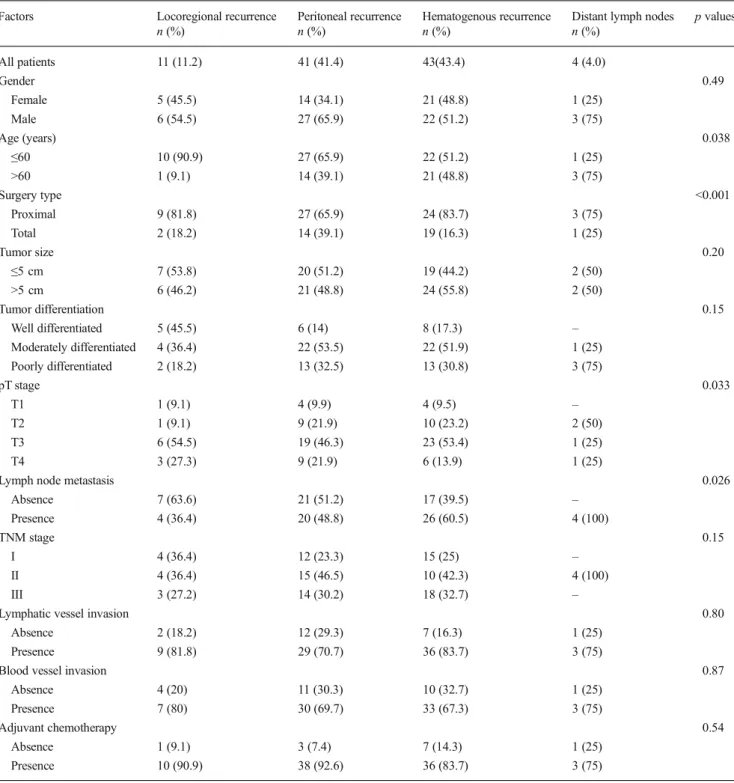
Table 1 Clinicopathological factors with respect to the recurrence patterns in patients with proximal gastric cancer who had curative gastrectomy
Factors Locoregional recurrence
n (%) Peritoneal recurrencen (%) Hematogenous recurrencen (%) Distant lymph nodesn (%) p values
All patients 11 (11.2) 41 (41.4) 43(43.4) 4 (4.0) Gender 0.49 Female 5 (45.5) 14 (34.1) 21 (48.8) 1 (25) Male 6 (54.5) 27 (65.9) 22 (51.2) 3 (75) Age (years) 0.038 ≤60 10 (90.9) 27 (65.9) 22 (51.2) 1 (25) >60 1 (9.1) 14 (39.1) 21 (48.8) 3 (75) Surgery type <0.001 Proximal 9 (81.8) 27 (65.9) 24 (83.7) 3 (75) Total 2 (18.2) 14 (39.1) 19 (16.3) 1 (25) Tumor size 0.20 ≤5 cm 7 (53.8) 20 (51.2) 19 (44.2) 2 (50) >5 cm 6 (46.2) 21 (48.8) 24 (55.8) 2 (50) Tumor differentiation 0.15 Well differentiated 5 (45.5) 6 (14) 8 (17.3) – Moderately differentiated 4 (36.4) 22 (53.5) 22 (51.9) 1 (25) Poorly differentiated 2 (18.2) 13 (32.5) 13 (30.8) 3 (75) pT stage 0.033 T1 1 (9.1) 4 (9.9) 4 (9.5) – T2 1 (9.1) 9 (21.9) 10 (23.2) 2 (50) T3 6 (54.5) 19 (46.3) 23 (53.4) 1 (25) T4 3 (27.3) 9 (21.9) 6 (13.9) 1 (25)
Lymph node metastasis 0.026
Absence 7 (63.6) 21 (51.2) 17 (39.5) – Presence 4 (36.4) 20 (48.8) 26 (60.5) 4 (100) TNM stage 0.15 I 4 (36.4) 12 (23.3) 15 (25) – II 4 (36.4) 15 (46.5) 10 (42.3) 4 (100) III 3 (27.2) 14 (30.2) 18 (32.7) –
Lymphatic vessel invasion 0.80
Absence 2 (18.2) 12 (29.3) 7 (16.3) 1 (25)
Presence 9 (81.8) 29 (70.7) 36 (83.7) 3 (75)
Blood vessel invasion 0.87
Absence 4 (20) 11 (30.3) 10 (32.7) 1 (25)
Presence 7 (80) 30 (69.7) 33 (67.3) 3 (75)
Adjuvant chemotherapy 0.54
Absence 1 (9.1) 3 (7.4) 7 (14.3) 1 (25)
in tests andp values less than 0.05 were considered statistical-ly significant.
Results
Forty-one patients (41.4 %) were female and 58 (58.6 %) were male, with a median age of 58 years (range 23 to 83 years). Thirty-one patients (31.4 %) were classified as stage I, 33
(33.3 %) as stage II, and 35 (35.3 %) as stage III. In 49 patients (49.4 %), the histology of the tumor was moderately differen-tiated; in 31 patients, tumors were poorly differendifferen-tiated; and in the remaining 19 patients, tumors were well differentiated. The majority of patients had pT3 tumor (n=49, 49.4 %). The mean tumor size was 6.5 + 2.9 cm (range 1–12 cm). Fifty-one (51 %) patients underwent D1lymph node
dissec-tion, while D2lymphadenectomy was performed in 41
pa-tients (41 %). D3lymp node dissection was also carried out 80 60 40 20 0 Time (months) 1.0 0.8 0.6 0.4 0.2 0.0 Probability survivi ng for pro gressio n-free (%) p=0.005 Distant lymph-node metastasis Peritoneal Loco-regional Hematogeneous Recurrence pattern Fig. 1 Progression-free survival
curves according to the patterns of recurrences 100 80 60 40 20 0 Time (months) 1.0 0.8 0.6 0.4 0.2 0.0 Prob
ability for Overall Surviving (%)
p=0.006
Distant lymph nodes Peritoneal Loco-regional Hematogenous
Recurrence pattern Fig. 2 Overall survival curves
with respect to recurrence patterns in patients with proxiamal gastric cancer
in only 8 patients (8 %). Moreover, the median number of dissected and metastatic lymph nodes were 19 (range 15–44) and 5 (range 0–23), respectively. Based on the presence of lymph node metastasis, 54 (54.5 %) patients were classified as node positive, and the remaining 45 patients (45.5 %) were node negative.
The median time to recurrence was 24 months (range 5– 101 months) and 45.5 % of patients had relapsed within 2 years. According to the recurrence pattern, 43 (43.4 %) pa-tients had hematogenous recurrence, and 41 (41.4 %) papa-tients revealed peritoneal recurrence, which were the most predom-inant patterns. The liver was the most commonly involved organ within the hematogenous pattern. In peritoneal rence patients, the majority of patients (92.6 %) had recur-rence at multiple peritoneal sites and three in the pelvic cavity and ovary. The remaining recurrence patterns were locoregional recurrence (11 patients, 11.2 %) and distant lymph nodes (4 patients, 4 %). Peritoneal and locoregional recurrences were significantly associated with younger age (p=0.038), and proximal resection was related to higher inci-dence of all recurrence patterns (p<0.001). Moreover, ad-vanced pT stage (T3–T4) was significantly correlated with hematogenous, peritoneal, and locoregional recurrence pat-terns (p=0.033). Hematogenous recurrence and distant lymph node metastasis had more lymph node positivity (p=0.026). The relationship between clinicopathological factors and re-currence patterns is summarized in Table1.
At the median follow-up of 27.5 months (range 6.5– 88 months), the median PFS time for patients with
locoregional recurrence was significantly better than that of patients with peritoneal recurrences, hematogenous recur-rences, and distant lymph nodes (32.2 vs. 18.9 vs. 18.2 vs. 9.7 months,p=0.005, respectively, Fig.1). Moreover, the me-dian OS interval for patients with distant lymph node recur-rence was significantly worse than that of patients with locoregional, peritoneal, and hematogenous recurrences (13.5 vs. 48.5 vs. 31.4 vs. 29.9 months,p=0.006, respectively, Fig.2).
The median OS time of early recurrence patients was sig-nificantly shorter than that of patients with late recurrence (16.6 vs. 55.2 months,p<0.001, Fig.3). There were signifi-cant differences between the early and late recurrence groups with respect to age, surgery type, tumor differentiation, pT stage, presence of lymph node metastasis, TNM stage and blood vessel invasion. Patients with early recurrences had older age (p=0.04), poorly differentiated tumors (p=0.04), increased lymph node involvement (p<0.011), advanced pT stage (p=0.025), advanced-staged disease (p<0.001), and blood vessel invasion (p=0.035) compared with patients with late recurrences. Whereas proximal gastrectomy was com-monly performed in the majority of patients with early recur-rence, total gastrectomy was commonly done in patients with late recurrence (p<0.001). The relationship between the sub-groups based on recurrences and clinicopathological factors is listed in Table 2. In both groups, the rate of patients who received adjuvant treatment was similar (93.2 % for early vs. 85.2 % for late,p=0.33). In addition, there was a significant difference between the early and late recurrence groups with
100 80 60 40 20 0 Time (months) 1.0 0.8 0.6 0.4 0.2 0.0 Probabili ty for Over all Su rviving (%) p<0.001 Late Recurrence Early Recurrence Fig. 3 The median overall
survival time of early recurrence patients was significantly shorter than that of patients with late recurrence (16.6 vs. 55.2 months, p<0.001) for patients with proximal gastric cancer
respect to the pattern of recurrence (p=0.017, Table 2). Patients with distant lymph node metastasis had early relapse, whereas locoregional recurrence tended to be late.
In the univariate analysis of factors associated with PFS, there were significant differences with respect to tumor
differentiation (p=0.04), surgery type (p=0.010), lymph node metastasis (p<0.001), TNM stage (p=0.0013), and pattern of recurrence (p=0.005). The univariate analysis for all recurrent gastric cancer patients showed that surgery type, presence of lymph node metastasis, TNM stage, pattern of recurrence (p= 0.006), and time of recurrence (<24 vs. >24 months,p<0.001) were significant prognostic factors for OS (Table3).
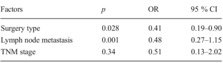
A multivariate analysis showed that only the presence of lymph node metastasis (p=0.004) and surgery type (p=0.04) were independent prognostic indicators for PFS. For OS, time of recurrence (p=0.033), presence of lymph node metastasis (p=0.03), and surgery type (p=0.04) were independent prog-nostic factors (Table3). In the logistic regression analysis, the presence of lymph node metastasis and surgery type were independent risk factors for predicting the occurrence of both overall and early recurrence (p=0.001, OR 0.48 and p=0.028, OR 0.41, respectively, Table4).
Discussion
Proximal gastric cancer is known to have unique clinicopath-ological features and is generally associated with higher inci-dence of recurrence compared with cancers of the other parts of stomach [21,22]. Therefore, the determination of risk fac-tors for recurrence and the pattern of recurrence may improve patient outcomes because effective treatment strategies may be initiated postoperatively, and eligible follow-up strategies may be produced to diagnose early recurrence. However, there are few papers which address prognostic factors for recurrence and the pattern of recurrence for proximal gastric cancer in the literature [15,16].
Recurrences are usually detected within 24 months of sur-gery in gastric cancer [8–10]. Likewise, Li et al. reported that 85.9 % of patients with proximal gastric cancer were recurred in 2 years [17]. In our study, early recurrence was detected in 45.5 % patients with proximal gastric cancer. This may be due to high number of patients with lymph node negativity (n= 45). Patients with early recurrences had older age, poorly dif-ferentiated tumor, increased lymph node involvement, ad-vanced pT stage, adad-vanced stage disease, and blood vessel invasion compared with patients with late recurrences. In ad-dition, proximal gastrectomy was significantly related to early recurrence. The median OS time of early recurrent patients was significantly shorter than that of patients with late recur-rence, and the prognostic importance of time of recurrence was proved by both univariate and multivariate analysis for OS. Our results were thus different from those of Li et al. [17]. Kim et al. reported that hematogenous and locoregional recurrence were the most recurrent types for proximal gastrec-tomy, whereas hematogenous spread was the dominant pattern after total gastrectomy. In addition, they showed that patients with proximal gastrectomy had a much higher recurrence rate
Table 2 Clinicopathological factors according to the recurrence times
in patients with proximal gastric cancer who had curative gastrectomy
Factors Early recurrence
n (%) Late recurrencen (%) p values
All patients 45 (45.5) 54 (54.5) Gender 0.84 Female 18 (40) 23 (42.6) Male 27 (60) 31 (57.4) Age (years) 0.04 ≤60 22 (48.9) 38 (70.4) >60 23 (51.1) 16 (29.6) Surgery type <0.001 Proximal 28 (62.2) 17 (31.5) Total 17 (37.8) 37 (68.5) Tumor size 0.99 ≤5 cm 21 (53.8) 28 (51.2) >5 cm 24 (46.2) 26 (48.8) Tumor differentiation 0.04 Well differentiated 4 (8.8) 15 (27.8) Moderately differentiated 25 (55.6) 24 (44.4) Poorly differentiated 16 (35.6) 15 (27.8) pT stage 0.025 T1 1 (2.2) 8 (14.8) T2 7 (15.6) 15 (27.8) T3 24 (53.3) 25 (46.3) T4 13 (28.9) 6 (11.1)
Lymph node metastasis <0.001
N0 6 (13.3) 39 (72.2) N1 14 (31.1) 10 (18.5) N2 15 (33.3) 5 (9.3) N3 10 (22.2) – TNM stage <0.001 I 4 (9.0) 27 (50.0) II 13 (28.8) 20 (37.0) III 28 (62.2) 7 (13.0)
Lymphatic vessel invasion 0.05
Absence 7 (15.6) 15 (32.6)
Presence 38 (84.4) 29 (67.4)
Blood vessel invasion 0.035
Absence 8 (17.8) 18 (33.3)
Presence 37 (82.2) 36 (66.7)
Adjuvant chemotherapy 0.33
Absence 4 (6.8) 8 (14.8)
than patients with total gastrectomy. The authors concluded that this might be related to inadequate extensive lymph node
dissection for proximal gastrectomy [23]. Therefore, Li et al. in their study including 135 recurrent proximal gastric cancer
Table 3 Univariate and multivariate analysis of patients with proximal gastric cancer for overall survival (OS) according to clinicopathological factors
HR hazards ratio, CI
confidence interval,NR
not reached
Factors Median OS time
(months)
Univariatep values Multivariatep values HR
(95 % CI) Gender 0.70 Male 31.4 Female 40.3 Age (year) 0.086 ≤60 41.9 >60 27.4 Surgery type 0.001 0.04 0.68 Proximal 19.9 (0.29–1.10) Total 34.5 Tumor size 0.97 ≤5 cm 30.9 >5 cm 27.6 Tumor differentiation 0.46 Well differentiated 40.5 Moderately differentiated 32.0 Poorly differentiated 24.0 pT stage 0.85 T1 NR T2 32.0 T3 31.1 T4 27.4
Lymph node metastasis <0.001 0.03 2.53
Absence 48.5 (0.98–5.49) Presence 19.9 TNM stage 0.004 0.32 0.77 I 40.5 (0.45–1.29) II 31.1 III 18.8
Lymphatic vessel invasion 0.28
Absence 41.9
Presence 31.4
Blood vessel invasion 0.15
Absence 55.2 Presence 31.1 Adjuvant treatment 0.91 Absence 31.4 Presence 38.4 Recurrence pattern 0.006 0.22 1.28 Locoregional 48.5 (0.85–1.92) Peritoneal 31.4 Hematogenous 29.9
Distant lymph node metastasis 13.5
The time of recurrence <0.001 0.033 0.49
Early 16.6 (0.31–0.96)
patients recommended that proximal gastrectomy should not be performed to be rational surgical approach in patients with advanced pT stage [17]. In our study, hematogenous recur-rence and peritoneal recurrecur-rence were the predominant patterns of recurrence. We detected a significant relationship with re-spect to surgery type. Proximal gastrectomy was significantly associated with locoregional and peritoneal recurrence pat-terns, which was compatible with previous reports [17,21,
24,25] but was not consistent with the study of Kim et al. [23]. In a study performed by Li et al., the authors demonstrated that tumor differentiation, pT stage, lymph node metastasis, and count of negative lymph nodes were risk factors for over-all and early recurrence for patients with proximal gastric can-cer. In the multivariate analysis, tumor differentiation, pT stage, and negative lymph node count were remained to be independent predictive factors for recurrence in their study [17]. In the present study, the univariate analysis determined that tumor differentiation, surgery type, lymph node metasta-sis, TNM stage, and the pattern and time of recurrence for OS were significant prognostic factors. Subsequently, a multivar-iate analysis showed that only the presence of lymph node metastasis and surgery type were independent prognostic in-dicators for PFS. For OS, the time of recurrence, the presence of lymph node metastasis, and surgery type were independent prognostic factors. In the logistic regression analysis, the pres-ence of lymph node metastasis and surgery type were inde-pendent risk factors for predicting the occurrence of both over-all and early recurrences. Thus, our results were comparable with previous reports [17,23] with regard to lymph node metastasis and tumor differentiation. However, our results were different from their study with regard to the pattern of recurrence, surgery type, and the time of recurrence and TNM stage.
In contrast to the above reports [17,21,23–25], we ana-lyzed survival according to the pattern of recurrence. The me-dian PFS time for patients with locoregional recurrence was significantly better than that of patients with the other recur-rence patterns. Moreover, the median OS interval for patients with distant lymph node recurrence was significantly worse than that of patients with locoregional, peritoneal, and hema-togenous recurrences in the current study. This may be related to the significant relationship between the pattern of recur-rence and age, surgery type, and pT stage. Li et al. also
reported that the pT stage, negative lymph node count, and surgery type were associated with the recurrence pattern. Furthermore, pT stage was also related with higher incidence of hematogenous metastases and peritoneal recurrence [17]. Our results were thus compatible with their study.
Proximal gastrectomy, poorly differentiated histology, ad-vanced pT stage (T3/T4), and the presence of lymph node me-tastasis were found to provide additional information in postop-erative surveillance for early recurrence after curative surgery for patients with proximal gastric cancer according to our re-sults. Consequently, these factors may be related to a more ag-gressive nature of tumor biology. Therefore, it may be recom-mended that patients with these factors should closely be follow-ed up for recurrence after postoperative adjuvant treatment.
The short follow-up intervals and a relatively small sample size were important limitations of our study, which might have influenced the results. The other limitation was the retrospec-tive nature of this study. Although our results should be con-firmed by prospective studies with larger sample sizes which will analyze different follow-up protocols for proximal gastric cancer, we believe that these results contribute to the knowl-edge of proximal gastric cancer because distinct prognostic factors were detected for both early and late recurrence groups and analyzed survivals according to the recurrence pattern differently from previous studies.
In conclusion, our study demonstrated that the presence of lymph node metastasis and surgery type were independent risk factors for predicting the occurrence of early recurrence in patients with proximal gastric cancer. Moreover, proximal gastrectomy, poorly differentiated histology, advanced pT stage and lymph node metastasis were found to be related to early recurrence. Therefore, total gastrectomy with extensive lymph node dissection was thought to be a more suitable treatment approach for proximal gastric cancer patients who had tumors characterized by advanced pT stage, poorly differ-entiated histology, and lymph node metastasis. These factors may provide additional information to explore the biological aggressiveness of proximal gastric cancer as a beneficial prog-nostic marker, which is important in predicting recurrence during follow-up and may guide to develop more appropriate and aggressive adjuvant therapies.
Conflict of interest None
References
1. Siegel R, Naishadham D, Jemal A. Cancer statistics, 2013. CA
Cancer J Clin. 2013;63:11–30.
2. Crew KD, Neugut AI. Epidemiology of gastric cancer. World J Gastroenterol. 2006;12:354–62.
3. Whiting J, Sano T, Saka M, Fukagawa T, Katai H, Sasako M.
Follow-up of gastric cancer: a review. Gastric Cancer. 2006;9:74–81.
Table 4 Logistic regression analysis of the predictive factors for
recurrence in patients with proximal gastric carcinoma
Factors p OR 95 % CI
Surgery type 0.028 0.41 0.19–0.90
Lymph node metastasis 0.001 0.48 0.27–1.15
TNM stage 0.34 0.51 0.13–2.02
4. Yoo CH, Noh SH, Shin DW, Choi SH, Min JS. Recurrence following
curative resection for gastric carcinoma. Br J Surg. 2000;87:236–42.
5. Huang KH, Chen JH, Wu CW, Lo SS, Hsieh MC, Li AF, et al. Factors affecting recurrence in node-negative advanced gastric
can-cer. J Gastroenterol Hepatol. 2009;24:1522–6.
6. Marrelli D, Roviello F, de Manzoni G, Morgagni P, Di Leo A, Saragoni L, et al. Different patterns of recurrence in gastric cancer
depending on Lauren’s histological type: longitudinal study. World J
Surg. 2002;26:1160–5.
7. Böhner H, Zimmer T, Hopfenmüller W, Berger G, Buhr HJ.
Detection and prognosis of recurrent gastric cancer—is routine
follow-up after gastrectomy worthwhile? Hepatogastroenterology.
2000;47:1489–94.
8. Kodera Y, Ito S, Yamamura Y, Mochizuki Y, Fujiwara M, Hibi K, et al. Follow-up surveillance for recurrence after curative gastric can-cer surgery lacks survival benefit. Ann Surg Oncol. 2003;10:898– 902.
9. Lai JF, Kim S, Kim K, Li C, Oh SJ, Hyung WJ, et al. Prediction of recurrence of early gastric cancer after curative resection. Ann Surg Oncol. 2009;16:1896–902.
10. Shiraishi N, Inomata M, Osawa N, Yasuda K, Adachi Y, Kitano S. Early and late recurrence after gastrectomy for gastric carcinoma. Univariate and multivariate analyses. Cancer. 2000;89:255–61. 11. Yokota T, Saito T, Teshima S, Yamada Y, Iwamoto K, Takahashi M,
et al. Early and late recurrences after gastrectomy for gastric cancer: a multiple logistic regression analysis. Ups J Med Sci. 2002;107:17– 22.
12. Schwarz RE, Zagala-Nevarez K. Recurrence patterns after radical gastrectomy for gastric cancer: prognostic factors and implications for postoperative adjuvant therapy. Ann Surg Oncol. 2002;9(4):394– 400.
13. Yang L. Incidence and mortality of gastric cancer in China. World J
Gastroenterol. 2006;12:17–20.
14. Kubo A, Corley DA. Marked regional variation in adenocarcinomas of the esophagus and the gastric cardia in the United States. Cancer.
2002;95:2096–102.
15. Corley DA, Buffler PA. Oesophageal and gastric cardia adenocarci-nomas: analysis of regional variation using the Cancer Incidence in
Five Continents database. Int J Epidemiol. 2001;30:1415–25.
16. Ishikawa S, Shimada S, Miyanari N, Hirota M, Takamori H, Baba H. Pattern of lymph node involvement in proximal gastric cancer. World
J Surg. 2009;33:1687–92.
17. Li F, Zhang R, Liang H, Liu H, Quan J. The pattern and risk factors of recurrence of proximal gastric cancer after curative resection. J Surg Oncol. 2013;107:130–5.
18. AJCC (American Joint Committee on Cancer) Cancer Staging Manual, 7th ed, Edge, SB, Byrd, DR, Compton, CC, et al (Eds), Springer, New York 2010. p. 117.
19. Japanese Gastric Cancer Association. Japanese classification of gas-tric carcinoma. 2nd English ed. Gasgas-tric Cancer 1998; 1: 10-24. 20. Harrison LE, Karpeh MS, Brennan MF. Proximal gastric cancers
resected via a transabdominal-only approach. Results and compari-sons to distal adenocarcinoma of the stomach. Ann Surg. 1997;225:
678–83.
21. Saito H, Fukumoto Y, Osaki T, Fukuda K, Tatebe S, Tsujitani S, et al. Distinct recurrence pattern and outcome of adenocarcinoma of the gastric cardia in comparison with carcinoma of other regions of the stomach. World J Surg. 2006;30:1864–9.
22. Gulmann C, Hegarty H, Grace A, Leader M, Patchett S, Kay E. Differences in proximal (cardia) versus distal (antral) gastric carcino-genesis via the retinoblastoma pathway. World J Gastroenterol.
2004;10:17–21.
23. Kim JH, Park SS, Kim J, Boo YJ, Kim SJ, Mok YJ, et al. Surgical outcomes for gastric cancer in the upper third of the stomach. World J
Surg. 2006;30:1870–6.
24. Kunisaki C, Shimada H, Nomura M, Matsuda G, Otsuka Y, Ono H, et al. Surgical outcome in patients with gastric adenocarcinoma in the
upper third of the stomach. Surgery. 2005;137:165–71.
25. Zhang XF, Huang CM, Lu HS, Wu XY, Wang C, Guang GX, et al. Surgical treatment and prognosis of gastric cancer in 2,613 patients.