EVALUATION OF GASTRIC INFLAMMATORY PARAMETERS CAUSED BY PYLORIC RING FAILURE
MuhaMMed Zübeyr üçüncü1, bünyaMin Gürbulak2, Şükrü Çolak2
1TC Gelişim University, Healt Sciences Institute, Istanbul Turkey - 2Istanbul Training and Research Hospital, Department of
General Surgery, Fatih 34098, Istanbul, Turkey ABSTRACT
Introduction: We aimed to compare the inflammatory parameters of patients with have failure of pyloric ring with those of that have intact pyloric ring.
Material and methods: Patients who underwent upper gastrointestinal endoscopy between May and September 2017 in-cluded in this study. The data of the study were collected prospectively and analysed retrospectively.
Results: A total of 205 patients were included in this study. 69 (33.7 %) male and 136 (66.3%) female patients, the mean age was 40.1 ± 11.38 years. Endoscopically, failure of pyloric ring was detected in 86 patients and intact pyloric ring in 119 patients. The histopathological examination of antrum; chronic gastritis in 91 patients (44.4 %), active chronic gastritis in 89 patients (44.3%) and normal findings were found in 23 patients (11.2%). The histopathological examination of corpus; chronic gastritis in 105 patients (51.2%), active chronic gastritis in 25 patients (12.2%) and normal findings were found in 73 patients (35.6%). Comparing the patients that have intact and pyloric ring failure, the inflammation and activation scores and Helicobac-ter Pylori (H. Pylori ) density were higher in pyloric ring failure group (p:0.001, p: 0,018, p: 0,006). It was seen that in patients with pyloric ring failure had a significant decrease in inflammatory activation and H. Pylori density toward from antrum to the corpus, and this was statistically significant (p: 0.001, p: 0.001, p: 0.001).
Conclusion: Because of the high inflammatory histopathologic parameters caused by pyloric ring failure, these patients should be followed closely.
Keywords: Intestinal metaplasia, Bile reflux, Failures of pyloric ring, Gastric imflammation, Helicobacter Pylori, Gastric cancer. DOI: 10.19193/0393-6384_2019_3_229
Received November 30, 2018; Accepted March 20, 2019
Introduction
Duodeno gastric reflux (DGR) is defined as du-odenal content’s reflux from the duodenum to stom-ach(1). The effects of chronic biliary reflux have been
studied either by experimental models or by evalu-ating mucosal injury after surgical procedures such as gastric resection, pyloroplasty or gastroenteric anastomosis. In mucosal damage caused by DGR, the number of inflammatory cells increases in the gastric mucosa, parietal cells decrease, and glandular morphology changes due to hyperplasia in mucous cells(2). These events have been associated with
gas-tric intestinal metaplasia, reflux esophagitis, barrett esophagus and even adenocancer development(3-7).
Even the bile acids are shown to have an antibacterial effect against H. Pylori, there are conflicting results in the literature(8).
Therefore, the demonstration of the pathological effects of chronic bile reflux due to pyloric ring failure in patients without gastric resection combined by or absence of H. Pylori is clinically important and there are few studies in the literature with this regard. In this study, we aimed to compare the histopathological inflammation parameters which means that lympho-cytes and neutrophils infiltrate the mucosa in a char-acteristically manner in patients that intact and failure of pyloric ring in upper gastrointestinal endoscopy.
Material and methods
Patients who underwent upper gastrointestinal endoscopy between May and September 2017 in-cluded in this study. We retrospectively reviewed a prospective database of a tertiary referral center. The demographic, endoscopic, and histopathological
find-ings of the patients were compared. Endoscopically, status of pyloric ring (failure or intact) ,erythema, erosion, ulcer, metaplastic areas, presence of bile at antrum, corpus and cardiac regions of stomach were evaluated. Antrum and corpus biopsy specimens were buffered in 10% formalin solution and paraffin blocks were prepared and stained with Giemsa and Hema-toxylin-Eosin(9). H. Pylori density and gastric
histo-pathological findings were evaluated by the patholo-gist who did not know the clinical background and endoscopic findings of the patient.
Patients whom previously underwent upper gas-trointestinal or gastric surgery, patients with gastric cancer, patients who use non-steroid anti-inflamma-tory drug (NSAID), neuropathic diseases such as di-abetes, inflammatory bowel disease (IBD), patients with collagen vascular disease and those who had H. Pylori eradication in the last year were excluded from this study.
In this study, we did not use quantitative param-eters such as gastric pH monitoring, 24 hour gastric bile monitoring with devices such as bilitec 2000, hepatobiliary scintigraphy, amylase and bilirubin lev-els in gastric fluid. We planned to monitor and eval-uate the patients according to the histopathological findings of intact and pyloric sphicter failure in the upper gastrointestinal endoscopy performed by the experienced endoscopist.
We describe the pyloric ring failure that the di-ameter of pyloric ring allows at least two endoscopes could pass comfortably, which does not contraction during the procedure and or causing DGR. The intact pyloric ring was described as which can be opened by direct contact with the tip of the endoscope and contracted during the procedure. Again, there is no exact description pyloric insufficiency or failure in literature.
All procedures were performed under propofol and midazolom sedation anesthesia, to exclude to the provocative bile reflux caused by vomiting. Written informed consent was obtained from all patients in-cluded in this study. The information was collected in accordance with the Declaration of Helsinki. The eth-ical committee approval was obtained for this study.
Statistical Analysis
Descriptive statistical methods such as mean, standard deviation, frequency, and percentage were used in the evaluation of the study data. The distri-bution of variables was checked by the Kolmogor-ov-Simirnov test. Independent sampling t - test, Mann - Whitney U test and Wilcoxon test were used in the
analysis of quantitative data, chi - square test was used in the analysis of qualitative data. P <0.05 was considered statistically significant.
Results
Upper gastrointestinal endoscopy was per-formed in 205 patients with dyspeptic complaints. 69 (33.7%) of the patients were male and 136 (66.3%) were female. The mean age of the patients was 40.1 ± 11.38. In the upper gastrointestinal endoscopy, py-loric ring failure in 86 patients and intact pypy-loric ring were detected in 119 patients. In the histopathologi-cal examination of antral biopsy, the chronic gastritis was found in 91 (44.4%), active chronic gastritis in 89 (44.3%) and normal findings in 23 (11.2%) patients. The chronic gastritis in 105 (51.2%), active chron-ic gastritis in 25 (12.2%) and normal findings in 73 (35.6%) were detected in the corpus biopsy.
There was no statistically significant differ-ence between the groups in terms of age and gender (p:0.084, p:0.249). There was no statistically signifi-cant difference between the groups in terms of indi-gestion, belching, nausea-vomiting, gas or bloating feeling (p> 0,05).
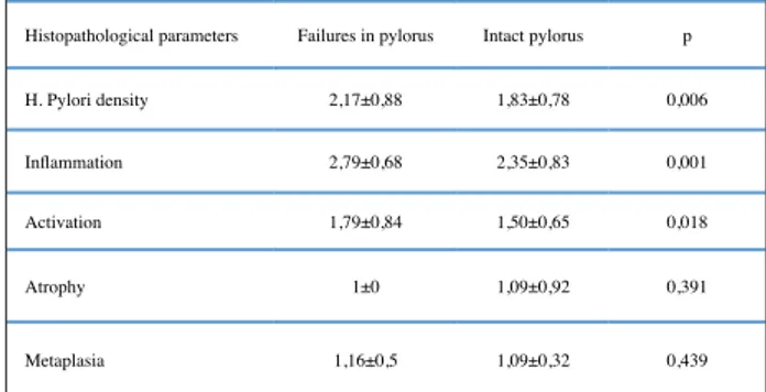
When the histopathological parameters of the antrum were compared in patients that have intact and pyloric ring failure, there was a statistically significant difference between the H. Pylori density, inflamma-tion and activainflamma-tion scores of the patients. But no sig-nificant difference was found in the atrophy and meta-plasia scores (p: 0.006, p: 0.001, p:0.018) (Table 1).
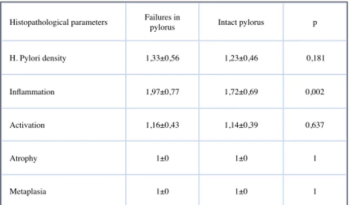
When the histopathological parameters of cor-pus were examined, only the inflammation scores were significantly different (p:0.002) (Table 2). When the data of 86 patients were examined who have py-loric ring failure, it was seen that the decrease in in-flammation, activation and H. Pylori density scores were statistically significant toward from antrum to the corpus. (p: 0.001, p: 0.001, p:0.001) (Table 3).
Histopathological parameters Failures in pylorus Intact pylorus p H. Pylori density 2,17±0,88 1,83±0,78 0,006 Inflammation 2,79±0,68 2,35±0,83 0,001 Activation 1,79±0,84 1,50±0,65 0,018
Atrophy 1±0 1,09±0,92 0,391
Metaplasia 1,16±0,5 1,09±0,32 0,439
Table 1: Histopathological findings of antrum in intact
Discussion
DGR is a risk factor for atrophy and intestinal metaplasia (IM) development(10). Duodenal fluid
in-creases inflammatory cells in the gastric mucosa and changes the glandular morphology by reducing pari-etal cells and mucous cell hyperplasia. These changes leading to esophagitis, gastritis, gastric and duodenal ulcers(2). When gastric microenvironment resembles
to duodenum, intestinal metaplasia develops in the gastric mucosa to reduce the mucosal damage caused by alkaline fluid.
In our study, the IM scores of patients with py-loric ring failure were high, but not statistically sig-nificant. We think that this is due to DGR caused by pyloric ring failure. DGR is physiological in the early morning and postprandial period(11). However,
exces-sive DGR causes gastritis, esophagitis, ulcers, gastric polyps, metaplasia, esophageal and gastric cancers(12).
In 30 to 40% of patients, DGR may be associated with reflux esophagitis or gastroesophageal reflux (GER). DGR is widespread in asymptomatic cases, as well as in patients with gastric and duodenal ulcer, pulmonary disease and whom underwent cholecys-tectomy(13). Endoscopically, pyloric ring failure and
duodenal gastric reflux, erythema and erosion, gastric atrophy, petechiae, gastric plea thickening, metapla-sia, and history of gastric surgery may be associated with DGR diagnosis(14).
Gastric mucosal damage is caused by the release of vasoactive materials such as histamine due to mast cell degranulation, resulting in vascular congestion in lamina propria(15). These findings can also be seen in
other conditions such as H. pylori infection.
Patients which used proton pump inhibitors(P-PI) have been shown to have higher gastric pH, which in turn increases mucosal damage and suggesting that DGR-induced inflammatory scores may be higher in patients that pyloric ring failure. In other words, gastric acid has a protective effect against DGR and GER. In particular, patients which used PPI, and who have pyloric ring failure should be followed closely and carefully for gastric cancer(16).
It has been shown that enzymes such as trypsin and PLA2 in biliary and pancreatic juice in DGR cause damage to gastric mucosa(17). Bile reflux causes antral
G-cell hyperplasia and hypergastrinemia and reduces somatostatin release(18,19). Decreased somatostatin
lev-els increases the hypergastrinemia and hypergatrinemia further increases the bile reflux(20). Cholecystokinin-2
(CCK-2) is a gastrin-specific receptor and is expressed in gastric pariatel cells and ECL cells(21). CCK-2
re-ceptors are exposed in normal esophagus, reflux es-ophagitis, barrette esophagus and adenocarcinoma(22).
It is known that H. Pylori is caused by precancerous lesions. Therefore, it was seen that the intensity of H. Pylori was increased in patients with have pyloric ring failure and it was accompanied synergistically with precancerous lesions such as atrophic gastritis and in-testinal metaplasia. Therefore, hypergastrinemia due to chronic biliary reflux, gastric epithelial hyperplasia and gastric gland expansions are caused by the same mechanism. Previously, only endoscopic examination of DGR was reported to be intuitive and subjective, and endoscopic findings would not be sufficient(23). In
our study, we found that histopathological scores were higher in patients which have pyloric ring failure by endoscopically.
We consider that, the status of the pyloric ring (failures or intact ) can be evaluated during the proce-dure due to the simplicity of the method and that the endoscopic findings alone are adequate for predicting risky patients.
Biliary reflux can not be diagnosed in the up-per gastrointestinal endoscopy with the presence of bile in the stomach. Fuchs et al. described DGR in gastric fluid with high pH, demonstrating bilirubin and pancreatic enzymes(24). 99mEHIDA (99m
Tc-ethyl hepatic iminodiacetic acid) scintigraphy showed 78.7% of cases diagnosed with DGR23 Bile acids have
a surfactant effect and this effect is necessary for lipid
Histopathological parameters Failures in pylorus Intact pylorus p H. Pylori density 1,33±0,56 1,23±0,46 0,181 Inflammation 1,97±0,77 1,72±0,69 0,002 Activation 1,16±0,43 1,14±0,39 0,637
Atrophy 1±0 1±0 1
Metaplasia 1±0 1±0 1
Table 2: Histopathological findings of corpus in intact and
failure pyloric ring.
Histopathological parameters Antrum Corpus p H. Pylori density 2,17±0,88 1,33±0,56 <0,001 Inflammation 2,79±0,68 1,97±0,77 <0,001 Activation 1,79±0,84 1,16±0,43 <0,001
Table 3: Comparison of histopathological findings of
absorption. This effect is due to hydrophilic-hydro-phobic balance. If the surfactant effect is too strong, the effect is cytotoxic. The hydrophilic and hydropho-bic balance shifts hydrophohydropho-bic to increase the toxic effect on the epithelium(25,26). The diagnosis of bile
reflux should be made carefully, excluding H. Pylori infection, NSAID with alcohol use, and other factors that cause mucosal inflammation.
After distal gastrectomy, it is thought that bilroth 2 increases the risk of remnant gastric cancer, accord-ing to bilroth 1, and it is thought to be the effect of bile acids in duodenal fluid(27). It was showed in an
exper-imental study, that rats infected H. Pylori and under-went pyloroplasty, have positive correlation between metaplasia, dysplasia and cancer development. Ne-oplasia was observed in 40% of rats that underwent pylorplasty(28). Metastasis is a multi-step process and
epithelial cells need to be transformed into mesenchy-mal cells (Epitelial-mesenchymesenchy-mal transition-EMT). Loss of epithelial proteins such as E-cadherin and increased mesenchymal proteins such as N-cadher-in and vimentN-cadher-in have been associated with advanced stage and poor prognosis in gastric cancer(29).
Normal gastric mucosa and gastric cancers ex-press a high rate of bile acid receptor, G-protein-cou-pled receptor (GPBAR1), which is strongly associat-ed with N-cadherin that EMT(30).
In the absence of biliary fluid in the stomach, DGR can not be diagnosed or 24 hour pH monitor-izations or scintigraphic gastric aspiration and radi-ological studies may be required. We also think that it is important to specify the status of the pyloric ring and mucosal changes such as atrophy, metaplasia, as well as biliary fluid in stomach for the diagnosis of DGR. Because histopathologic findings of DGR may be seen in other situations such as H. Pylori infection, NSAID use. It is important to use simple, reliable and easily reproducible methods for the diagnosis of DGR.
As we have shown in our study, the presence of pyloric ring failures and the histopathological changes such as inflammation and activation can be combined to diagnose of DGR even if the absence of biliary flu-id in the stomach.
The study suffered from several limitations. Due to the gastric fluid amylase and bilirubin levels were not measured, the level of damage due to the pancre-atic or bile secretions were not shown.
Since it is a risk factor for carcinogenesis, mul-tiple biopsies should be taken from patients with pyloric failure in upper gastrointestinal endoscopy and should be follow-up close for the carcinogenesis
cascade. We recommend the addition of pyloric ring failure to DGR-associated risk factors which has de-scribed in the literature.
Conclusion
DGR is a risk factor for atrophy and IM develop-ment. Because of higher histopathological inflamma-tion scores caused by DGR which patients that have pyloric ring failure, it is important that patients have close follow-up with multiple biopsies by yearly.
References
1) Fein M, Fuchs KH, Bohrer T, Freys SM, Thiede A. Fiberoptic technique for 14-hour bile reflux monitoring. Dig Dis Sci 1996; 41: 216- 225.
2) Mittal BR, Ibrarullah M, Agarwal DK, Maini A, Ali W, Sikora SS, et al. Comparative evaluation of scintigraphy and upper gastrointestinal tract endoscopy for detection of duodenogastric reflux. Ann Nucl Med 1994; 8: 183-186.
3) Dixon MF, Mapstone NP, Neville PM, Moayyedi P, Axon AT. Bile reflux gastritis and intestinal metaplasia at the cardia. Gut 2002; 51: 351-355.
4) Dixon MF, Neville PM, Mapstone NP, Moayyedi P, Axon AT. Bile reflux gastritis and Barrett's oesophagus: further evidence of a role for duodenogastro-oesophage-al reflux? Gut 2001; 49: 359-363.
5) Dresner SM, Griffin SM, Wayman J, Bennett MK, Hayes N, Raimes SA. Human model of duodenogas-tro-oesophageal reflux in the development of Barrett's metaplasia. Br J Surg 2003; 90: 1120-1128.
6) Oberg S, Peters JH, DeMeester TR, Lord RV, Johansson J, DeMeester SR, et al. Determinants of intestinal meta-plasia within the columnar-lined esophagus. Arch Surg 2000; 135: 651-5.
7) Goldman A, Shahidullah M, Goldman D, Khailova L, Watts G, Delamere N et al. A novel mechanism of acid and bile acid-induced DNA damage involving Na+/H+ exchanger: Implication for Barrett’s oesophagus. Gut 2010; 59: 1606-1616.
8) Itoh M, Wada K, Tan S, Kitano Y, Kai J, Makino I. An-tibacterial action of the bile acids against Helicobacter pylori and changes in its ultrastructural morphology: effect of unconjugated dihydroxy bile acid. J Gastroen-terol 1999; 34: 571-576.
9) Yang HB, Sheu BS, Su LJ, Chien CH, Lin XZ. Clinical application of gastric histology to monitor treatment of dual therapy in H. pylori eradication. Dig Dis Sci 1997; 42: 1835-1840.
10) Nakamura M, Haruma K, Kamada T, Mihara M, Yoshi-hara M, Imagawa M, et al. Duodenogastric reflux is as-sociated with antral metaplastic gastritis. Gastrointest Endosc 2001; 53: 53-59.
11) Tzaneva M. Effects of duodenogastric reflux on gastrin cells, somatostatin cells and serotonin cells in human an-tral gastric mucosa. Pathol Res Pract 2004; 200: 431-438.
12) Lirón R, Parrilla P, Martinez de Haro LF, et al. Quanti-fication of duodenogastric reflux in Barrett’s esophagus. Am J Gastroenterol 1997; 92: 32-36
13) Romagnoli R, Collard JM, Serra AM. Is the DGR Bilitec profile different in GERD patients with and without Bar-rett’ s esophagus? In: Giuli R, Scarpignato C, Collard JM, editors. The Duodenogastroesophageal reflux. Par-is: John Libbey pre; 2006. P. 445-449.
14) Matsuhisa T, Arakawa T, Watanabe T, Tokutomi T, Sakurai K, Okamura S, et al. Relation between bile acid reflux into the stomach and the risk of atrophic gastritis and intestinal metaplasia: a multicenter study of 2283 cases. Dig Endosc. 2013: 25(5): 519-525.
15) Bechi P, Amorosi A, Mazzanti R, et al. Reflux-related gastric mucosal injury is associated with increased mu-cosal histamine content in humans. Gastroenterology 1993; 104: 1057-1063.
16) Wetscher GJ, Hinder RA, Smyrk T, et al. Gastric acid blockade with omeprazole promotes gastric carcino-genesis induced by duodenogastric reflux. Dig Dis Sci 1999; 44: 1132- 1135.
17) Mason RC. Duodenogastric reflux in rat gastric carcino-ma. Br J Surg 1986; 73: 801-803.
18) Wetscher GJ, Hinder RA, Kretchmar D, Perdikis G, Smyrk T, Klingler PJ, et al. Duodenogastric reflux caus-es growth stimulation of foregut mucosa potentiated by gastric acid blockade. Dig Dis Sci 1996; 41: 2166-2173. 19) Kaminishi M, Sadatsuki H, Johjima Y, et al. A new mod-el for production of chronic gastric ulcer by duodeno-gastric reflux in rats. Gastroenterology 1987; 92: 1913-1918.
20) Thompson JN, Barr JA, Collier N, et al. Basal, sham feed and pentagastrin stimulated gastric acid, pepsin and electrolytes after omeprazole 20 mg and 40 mg daily. Gut 1985; 26: 1018-1024.
21) Tommeras K, Hammer P, Sundler F, Borch K, Mårdh S, Cabero JL. Immunolocalization of cholecystokinin-2 receptors in rat gastric mucosa. Scand J Gastroenterol 2002; 37: 1017-1024.
22) Haigh CR, Attwood SE, Thompson DG, Jankowski JA, Kirton CM, Pritchard DM, et al. Gastrin induces prolif-eration in Barrett's metaplasia through activation of the CCK2 receptor. Gastroenterology 2003; 124: 615-625. 23) Chen TF, Yadav PK, Wu RJ, et al. Comparative
evalua-tion of intragastric bile acids and hepatobiliary scintig-raphy in the diagnosis of duodenogastric reflux. World J Gastroenterol 2013; 19(14): 2187-2196.
24) Fuchs K-H, Maroske J, Fein M, Ritter MP, Heimbuch-er J, et al. Variability in thecomposition of physiologic duodenogastric reflux. J GastrointestSurg 1999; 3: 386-396.
25) Heuman DM. Quantitative estimation of the hydrophili-chydrophobic balance of mixed bile salt solutions. J Li-pid Res 1989; 30: 719-730.
26) Sagawa H, Tazuma S, Kajiyama G. Protection against hydrophobic bile salt-induced cell membrane damage by liposomes and hydrophilic bile salts. Am J Physiol 1993; 264: G835-839
27) Orlando R III, Welch JP. Carcinoma of the stomach after gastric operation. Am. J. Surg. 1981; 141: 487-491. 28) Araujo JC, Carvalho JJ, Serra HO. Influence of
duo-denogastric reflux in the gastric mucosa histological changes of rats infected with Helicobacter pylori. Rev Col Bras Cir 2016; 43(4): 235-42.
29) Ye X, Weinberg RA. Epithelial-Mesenchymal Plasticity: A Central Regulator of Cancer Progression. Trends Cell Biol 2015; 25:675-686.
30) Carino A, Graziosi L, D'Amore C, Cipriani S, Marchi-anò S, Marino E, et al. The bile acid receptor GPBAR1 (TGR5) is expressed in human gastric cancers and pro-motes epithelial-mesenchymal transition in gastric can-cer cell lines. Oncotarget 2016; 20; 7(38): 61021-61035.
–––––––––
Corresponding Author: bünyaMin Gürbulak, MD
Istanbul Training and Research Hospital, Department of General Surgery, Fatih 34098, Istanbul
Turkey Zip Code:34098 Fatih; Istanbul-Turkey E-mail: [email protected]