INSTITUTE OF NATURAL AND APPLIED SCIENCES
PURIFICATION AND CHARACTERIZATION OF β-GALACTOSIDASE FROM Enterobacter sp. 3TP2A
Bestoon AHMED SHAIKHAN
MASTER THESIS
DEPARTMENT OF BIOLOGY
DIYARBAKIR January - 2016
FEN BİLİMLERİ ENSTİTÜSÜ
Enterobacter sp.3TP2A BAKTERİSİNDEN İZOLE EDİLEN β-GALAKTOSİDAZ ENZİMİNİN SAFLAŞTIRILMASI VE KARAKTERİZASYONU
Bestoon AHMED SHAIKHAN
YÜKSEK LİSANS TEZİ BİYOLOJİ ANABİLİM DALI
DİYARBAKIR Ocak-2016
UNIVERSITY OF DICLE
INSTITUTE OF NATURAL AND APPLIED SCIENCES DIYARBAKIR
‘‘Purification and characterization of β-galactosidase from Enterobacter sp. 3TP2A, Submitted by Bestoon AHMED SHAIKHAN in partial fulfillment of the requirements for the degree of Master of Science in Molecular Biology.
Examination Committee:
Title Name & Surname Signature
Chairman (Supervisor): Prof. Dr. Dr. Kemal GÜVEN Member :Assist. Prof. Dr. Selahattin TEKEŞ Member :Assist. Prof. Dr. Nesrin HAŞİMİ
Date of Thesis Defense: 19 /01 /2016
I approve accuracy of the above information.
Assoc. Prof. Dr. Mehmet YILDIRIM MANAGER OF THE INSTITUTE
I
ACKNOWLEDGEMENTS
I would like to express my gratitude to all those who gave me the possibility to complete this thesis. I am deeply indebted to my supervisor Prof. Dr. Kemal GÜVEN whose help and supports, stimulating suggestions and encouragement helped me in all the time of research. His trust, continuous support and scientific excitement inspired me in the most important moments of making right decisions.
I extent my sincere appreciation to Assoc. Prof. Dr. Reyhan GÜL GÜVEN for her motivation and advices.
I wish to express my thanks and gratitude to the presidency of the Dicle University. My thanks to the head of the Biology Department Prof. Dr. Selçuk ERTEKİN for his support and providing facilities required for this study.
I wish to express my special thanks to Res. Assist. Dr. Fatma MATPAN BEKLER for her assistance in the all steps of this research and greatly indebted to Dr. Ömer ACER for his assistance and advices.
I wish to express my thanks to Prof. Dr. Kadri GÜL the head of the Microbiology Department in medical school.
I would also like to thank Mr. Dlsoz M. Rashid and Mr. Bzhar Hussen for thier extensive help. Also my thanks to Nanakaly Hospital and Dicle University Hospital Central Laboratory technicians for their help for their cooperation.
Finally, I would like to express my special thanks to my wife (AWAZ Mohammed) and my family members, my dear father, my brothers and sisters, my children (Kamand and Kinaz) for their constant encouragement and support and all whom I love.
II TABLE OF CONTENTS Page no. ACKNOWLEDGEMENTS……… I ABSTRACT... VI ÖZET………... VIII LIST OF TABLES... X LIST OF FIGURES………... XI LIST OF APPENDIX………... XII ABBREVIATION AND SYMBOLS... XIII
1. INTRODUCTION ……….……..… 1
2. PREVIOUS STUDY……… 5
2.1. The genus Enterobacter……….……… 5
2.1.1. Enterobacter cloacae………. 5
2.2. Industrial enzymes in biotechnology………... 8
2.2.1. The use of β-galactosidase in biotechnology………. 11
2.3. Properties of β-galactosidase………. 14
2.3.1. Psychrophilic β-galactosidase……… 18
2.3.2. Mesophilic β-galactosidase……… 18
2.3.3. Thermophilic β-galactosidase……… 20
2.4. Bacterial sources of β-galactosidase……….. 21
2.5. Purification of β-galactosidase……….. 24
III
2.5.2. Ammonium sulphate precipitation………. 25
2.5.3. Dialysis……… 25
2.5.4. Ultrafiltration……… 25
2.5.5. Gel permeation chromatography………...……...……….. 26
2.5.6. Affinity chromatography……….……….. 26
2.6. Lactose hydrolysis……….……. 28
2.7. Summary about subject……….. 30
3. MATERIAL AND METHODS………... 45
3.1. Materials………. 45
3.1.1. Chemicals……… 45
3.1.2. Equipments………. 45
3.1.3. Bacterial strain……… 45
3.1.4. Media………. 45
3.1.5. Buffers and reagents……….. 45
3.2. Methods………...….. 46
3.2.1. Classification of 3TP2A strain as Enterobacter cloacae by MALDI…. 46
3.2.2. Cultivation of strain and preparation of crude enzyme……….…. 46
3.2.3. β-galactosidase assay………...………... 46
3.2.4. Protein assay (Lowry method)………..……….………. 47
3.2.5. Time course of bacterial growth and production of β-galactosidase in the presence and absence of lactose………..…. 48
IV
3.2.6. Effect of temperature on β-galactosidase activity………...….. 48
3.2.7. Effect of pH on β-galactosidase activity……… 49
3.2.8. Effect of lactose concentration on production of β-galactosidase…….. 49
3.2.9. Purification of β-galactosidase……….. 49
3.2.9.1. Production of crude extract………..………….. 49
3.2.9.2. Ammonium sulphate precipitation………..……… 50
3.2.9.3. Gel permeation chromatography……… 50
3.2.9.4. Affinity chromatography ……….. 51
3.2.10. Characterization of β-galactosidase……… 51
3.2.10.1. SDS and native-PAGE ……….…. 51
3.2.10.2. Thermal stability……….... 52
3.2.10.3. pH stability……… 52
3.2.10.4. The effect of inhibitors on β-galactosidase activity ……….. 52
3.2.10.5. Effect of metals on β-galactosidase activity……….. 53
3.2.10.6. Effect of substrate concentration (o-NPG) on β-galactosidase activity.. 53
3.2.11. Lactose hydrolysis……….. 53
4. RESEACH FINDINGS……… 55
4.1. Classification of 3TP2A strain as Enterobacter cloacae by MALDI… 55
4.2. Identification of Enterobacter sp.3TP2A by 16S rRNA……….. 55
V
4.3. Time course of bacterial growth and production of β-galactosidase…. 57
in the presence and absence of lactose 4.3.1. Growth of Enterobacter cloacae……….. 57
4.3.2. Enzyme activity……….. 59
4.4. Effect of lactose concentration on production of β-galactosidase…….. 59
4.5. Effect of temperature on β-galactosidase activity……….….. 60
4.6. Effect of PH on β-galactosidase crude enzyme………. 61
4.7. Purification of β-galactosidase enzyme………...….. 62
4.8. Enzyme characterization………..…... 63
4.8.1. Native and SDS-PAGE……… 63
4.8.2. Thermal stability……….… 64
4.8.3. pH stability……….. 65
4.8.4. Effect of metals on purified β-galactosidase activity………... 66
4.8.5. The effect of inhibitors on purified β-galactosidase activity……….... 67
4.8.6. Effect of substrate concentration (o-NPG) on β-galactosidase activity.. 68
4.9. Lactose hydrolysis……….. 69
5. DISCUSSION AND CONCLUSION………...……… 71
6. REFERENCES……….… 83
APPENDIX ………...……….…... 97
VI ABSTRACT
PURIFICATION AND CHARACTERIZATION OF β-GALACTOSIDASE FROM Enterobacter sp. 3TP2A
MASTER THESIS
Bestoon AHMED SHAIKHAN DEPARTMENT OF BIOLOGY
INSTITUTE OF NATURAL AND APPLIED SCIENCES UNIVERSITY OF DICLE
2016
In this study, a mesophilic Enterobacter sp. 3TP2A isolated from petroleum station in Batman in the southeast of Turkey was identified and found to produce a high amount of mesophilic β-galactosidase. The lactose was found to increase the β-galactosidase production to a great extent, meaning that this enzyme is inducible. An intracellular β-galactosidase from Enterobacter sp. 3TP2A was purified and characterized. The enzyme was purified by ammonium sulphate precipitation 70%, dialysis, ultrafiltration and finally using sephadex G-75 chromatography method. The enzyme was purified to 17.3-fold after gel permeation chromatography with a yield of approximately 11%. The purified enzyme was found to be stable in pH 8.0 and temperature of 35 oC. The molecular weight of the purified enzyme was found to be about 60 kDa by both SDS-PAGE and native-PAGE.
The effects of various metal ions at different concentrations of CaCl2, MgCl2, ZnCl2, CuCl2, and EDTA (1, 2, 5, 10 and 20 mM) were tested. EDTA and Cu2+ had an inhibitory effect on the β-galactosidase purified from Enterobacter sp. 3TP2A. EDTA inhibited the enzyme activity (upto 76%) and Cu2+ had strong inhibitory effect on β-galactosidase even at low concentrations (96.9%). However, Mg2+ caused activation of the purified enzyme. Ca2+ did not effect enzyme activity to a great extent, causing deactivation of the enzyme at 20 mM (only 16%), while Zn2+ at 1, 2 and 5 mM inhibited enzyme
VII
activity (32, 27, 8%, respectively). Increase in the concentration of Mg2+ causing activation upto 47% and also inhibition by EDTA show that the enzyme is metal-dependent or a metalloenzyme. Also determining the effect of different concentration of inhibitors on purified enzyme; PCMB (0.2, 0.4,1, 2 mM) , Iodo, DTT, β-mer, N-Ethyl, (1, 2, 4, 8 mM). The enzyme was completely inhibited by N-Ethyl (100%), but not affected by DTT. The enzyme was slightly affected by β-mer enhancing β-galactosidase activity at 8mM with 14% . The Iodo had a slight effect on β-galactosidase activity (upto 13%). PCMB inhibited the enzymatic activity to a great extent upto approx. 87%.
The Lineweaver-Burk plot was linear, suggesting a simple Michealis-Menten kinetics. The Vmax was found as 0.701 (μmol/ min mg) and Km was found as 0.104 mM. It was also found that the time for lactose hydrolysis continues up to 10 h with the reaction catalyzed by purified β-galactosidase.
The aim of this study was to purify and characterize the mesophilic β-galactosidase from Enterobacter sp. 3TP2A and then to test for use in biotechnology such as lactose hydrolysis. The results obtained indicated that this species may well be a good candidate.
Key words: β-galactosidase, Enterobacter cloacae 3TP2A, purification, characterization, inhibition and lactose hydrolysis.
VIII ÖZET
Enterobacter sp. 3TP2A BAKTERİSİNDEN İZOLE EDİLEN β-GALAKTOSİDAZ ENZİMİNİN SAFLAŞTIRILMASI VE KARAKTERİZASYONU
YÜKSEK LİSANS TEZİ Bestoon AHMED SHAIKHAN
DİCLE ÜNİVERSİTESİ FEN BİLİMLERİ ENSTİTÜSÜ
BİYOLOJİ ANABİLİM DALI
2016
Bu çalışmada, Türkiye’nin Güneydoğu Bölgesi’nde yer alan Batman ilindeki bir akaryakıt istasyonundan izole edilen mezofilik Enterobacter sp. 3TP2A bakterisinin yüksek miktarda mezofilik galaktosidaz enzimi ürettiği görülmüştür. Ayrıca, laktozun β-galaktosidaz üretimini önemli ölçüde arttırdığı görülmüş olup bu bulgu, bu enzimin endüke edilebilir bir enzim olduğunu göstermektedir. Enterobacter sp. 3TP2A bakterisinden izole edilen intraselüler β-galaktosidaz saflaştırılıp karakterize edildi. Saflaştırma işlemi %70 amonyum sülfat çöktürmesi, diyaliz, ultrafiltrasyon ve son olarak da Sephadex G-75 kromatografisi yöntemi kullanılarak gerçekleştirilmiştir. Enzim, jel geçirgenlik kromatografisi kullanılarak yaklaşık %11 verimle 17.3 kat saflaştırıldı. Saflaştırılan enzimin pH 8.0 ve 35 oC’de stabil olduğu ve moleküler ağırlığının ise SDS-PAGE ve native-PAGE metodlarına göre yaklaşık 60 kDa olduğu tespit edilmiştir.
Çeşitli metal iyonların farklı CaCl2, MgCl2, ZnCl2, CuCl2 ve EDTA (1, 2, 5, 10 ve 20 mM) konsantrasyonlardaki etkileri incelenmiştir. EDTA ve Cu2+’nın Enterobacter sp. 3TP2A’dan saflaştırılan β-galaktosidaz üzerinde inhibe edici bir etkiye sahip oldukları belirlenmiştir. EDTA’nın enzim aktivitesini %76’ya kadar inhibe ettiği ve Cu2+’nın β-galaktosidaz üzerindeki inhibe edici etkisinin düşük konsantrasyonlarda bile güçlü olduğu görülmüştür (96.9%). Bununla birlikte, Mg2+’nın saflaştırılan enzimin aktivasyonuna sebep
IX olduğu görülmüştür. Ca2+
ise enzim aktivitesini önemli ölçüde etkilememiş olup 20 mM (yalnızca 16%)’de enzimin deaktivasyonuna sebep olmuştur. Ancak, Zn2+ 1, 2 ve 5 mM’de enzimin aktivasyonuna sebep olmuştur (sırasıyla, %32, %27, %8). Mg2+ konsantrasyonundaki artışın 47%’ye kadar enzim aktivasyonuna sebep olması ve EDTA tarafından inhibe edilmesi, bu enzimin metal-bağımlı veya metaloenzim olduğunu göstermektedir. Buna ilaveten, farklı konsantrasyonlardaki inhibitörlerin saflaştırılan enzim üzerindeki etkileri şu konsantrasyonlarda incelenmiştir: PCMB (0,2, 0,4, 1, 2 mM) , Iodo, DTT, β-mer, N-etil, (1, 2, 4, 8 mM). Enzim, N-etil tarafından tamamen inhibe edilmesine karşın (%100), DTT tarafından etkilenmemiştir. Enzim, β-galaktosidaz aktivitesini 8 mM’de %14 oranında arttıran β-mer tarafından hafifçe etkilenmiştir. Iodo da β-galaktosidaz aktivitesini hafifçe etkilemiştir (%13’e kadar). PCMB ise enzimatik aktiviteyi %87’lere varan inhibisyonla büyük oranlarda etkilemiştir.
Lineweaver-Burk grafiği lineer olarak belirlenmiş olup bu sonuç basit bir Michealis-Menten kinetiğine işaret etmektedir. Vmax 0.701 (μmol/ dak mg) ve Km 0.104 mM olarak tespit edilmiştir. Ayrıca, laktoz hidrolizi süresinin, saflaştırılan β-galaktosidaz tarafından katalize edilen reaksiyondan dolayı, 10 saate kadar çıktığı gözlemlenmiştir.
Bu çalışmanın amacı Enterobacter sp. 3TP2A’dan mezofilik β-galaktozidaz’ı saflaştırmak, karakterize etmek ve laktoz hidrolizi gibi biyoteknoloji alanında kullanımını test etmekti. Elde edilen bulgular bu enzimin iyi bir aday olabileceğini göstermiştir.
Anahtar kelimeler: β-galaktosidaz, Enterobacter cloacae 3TP2A, saflaştırma, karakterizasyon, inhibisyon ve laktoz hidrolizi
X LIST OF TABLES
Table no. Page
Table 2.1. Classification of Enterobacter cloacae ... 7
Table 2.2. Biotechnological applications of β-galactosidase ... 12
Table 2.3. Alist of sources of supplier for commercial β-galactosidase... 17
preparations Table 2.4. Results of some species and genera with lactose fermentation…………. 20
Table 2.5. Some properties of β-galactosidase from bacterial sources... 22
Table 2.6. Bacterial sources of β-galactosidase... 23
Table 2.7. Various β-galactosidase applications... 29
Table 4.1. Identified the strain by Bruker Daltonic MALDI Biotyper... 55
Table 4.2. 16S rRNA gen sequencing... 56
Table 4.3. Steps of β-galactosidae purification... 63
Table 4.4. Effect of metal ions on the activity of purified β-galactosidase... 67
Table 4.5. Effect of inhibitors on the activity of purified β-galactosidase... 68
XI LIST OF FIGURES
Figure no. Page
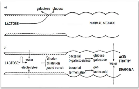
Figure 2.1. a) Mechanism of lactose hydrolysis... 13
Figure 2.1. b) Pathogenesis of diarrhaea in lactose intolerance... 13
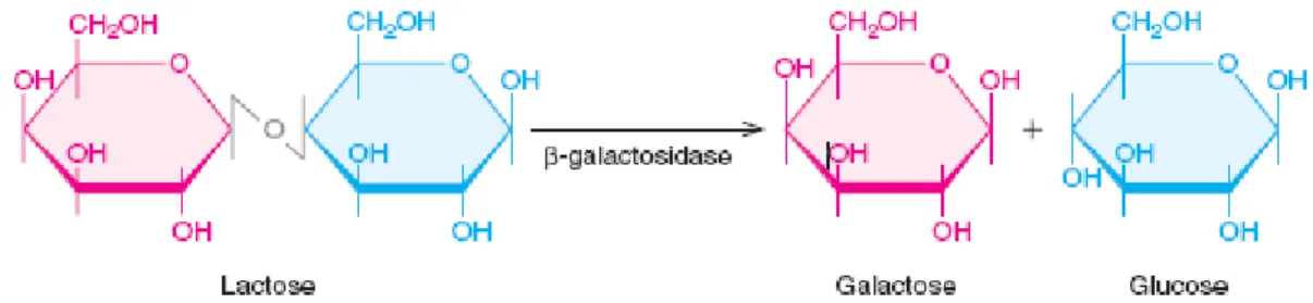
Figure 2.2. β-galactosidase hydrolysis of lactose ... 14
Figure 2.3. Graphic summarizing the β-galactosidase roles in the cell... 16
Figure 3.1. Gel permeation chromatography... 50
Figure 4.1. Identification of Enterobacter sp. 3TP2A by 16S rRNA... 57
Figure 4.2. a) Effect of incubation time on growth of Enterobacter sp. 3TP2A .. 58
Figure 4.2. b) Effect of incubation time on the production... 59
of β-galactosidase enzyme Figure 4.3. Effect of lactose concentration on production... 60
of β-galactosidase Figure 4.4. Specific activity of β-galactosidase at variuos temperature... 61
Figure 4.5. Specific activity of β-galactosidase at variuos pH... 62
Figure 4.6. Native and SDS-PAGE... 64
Figure 4.7. Effect of thermal stability... 65
Figure 4.8. Effect of pH on stability... 66
Figure 4.9. Lineweaver-Burk plot of the purified β-galactosidase... 69
XII LIST OF APPENDIX
Appedix Page
Appedix A List of chemicals... 97
Appedix B List of equipments... 98
Appedix C Media... 99
XIII
ABBREVIATION AND SYMBOLS
α : Alpha
APS : Amonium per-sulphate
o A : Angstrom β : Beta β-mer : Beta-mercaptoethanol % : Percent BNG : 6-Bromo-2-naphthyle-β-D-galactopyronoside BSA : Bovine serum albumin
CaCl2 : Calcium Chloride
CBB : Coomassie brilliant blue
cm : Centimeter
CuCl2 : Copper chloride
Da : Dalton
DTT : Dithiothreitol
EDTA : Ethyldiamine tetra aceticacid FCR : Folin-ciocalteu reagent g/L : Gram/litre h : Hour Iodo : Iodoacetamide IPTG : Isopropyβ-D-1-thiogalactopyronoside kDa : Kilodalton Km : Mechaelis constant L : Litre M : Molarity mg : Milligram MgCl2 : Magniesium chloride min : Minute
XIV
mL : Mililitre
µL : Microlitre
mM : Milimolar
NaCl : Sodium chloride
NaoH : Sodium hydroxide Na2CO3 : Sodium bicarbonate NB : Nutrient broth N-Ethyl : N-Ethylmaleimide nm : Nanometer o C : Degrees centegrade OD : Optical density
ONPG :o-Nitrophenol- β-D-galactopyronoside
PABTG : p-aminobenzoic-1-thio-β-D-galactopyranoside PCMB : p-Chloromercuribenzoic acid
pH : Power of hydrogen
rpm : Rotation per minute
SDS : Sodium dedocyl sulphate
SDS-PAGE : Sodium dedocyle sulphatepolyacrilamide gel electrophoresis TEMED : Tetramethylethylenediamine
U : Unit
UV : Ultraviolet
U/µL : Unit/microlitre
V : Volt
Vmax : Maximum reaction velosity
w/v : Weight/volume
1
1.INTRODUCTION
Enterobacter species are important human opportunistic pathogens, which are in charge of nosocomial infections such as urinary tract infections (UTI), neonatal meningitis, cholecystitis and osteomyelitis (Sanders et al. 1997, Ren et al. 2010).
Species of the Enterobacter are extensively encountered in nature, but they are also pathogens: E. cloacae and E. hormaechei are most often secluded from human clinical specimens. Therefore, the most common Enterobacter sp. is E. cloacae , affecting only nosocomial infections and on the antibiotic resistance features of these microorganisms, there has been a lot of publishing. In spite of the significance of E. cloacae as a nosocomial pathogen, the factors and the pathogenic mechanisms which are contributing in such disease associated with the E. cloacae complex have not been understood yet; this could be caused by the insufficiency and the dispersion of information obtainable. Its capacity to form biofilms and to secrete different cytotoxins (enterotoxins, pore-forming toxins, hemolysins ) is important for its pathogenicity (Mezzatesta et al. 2012, Davin-Regli 2015).
β-galactosidase or lactase (EC.3.2.1.23) hydrolyzes the sugar lactose of milk into monosaccharides, glucose and galactose and also catalyzes mixture of different galactosides. Among enzymes which are of few industrially importance, β-galactosidase discovers wide application in several main areas, pharmaceutical, health, food, technology and environment. Concerning health issues large number of population undergoes from lactose intolerance syndrome all over the world. Primary lactose intolerance has a high degree of race dependence that in the USA exists about 95%-100% of American Indians, 80%- 90% of Blacks, Mediterraneans, Asians and Jews and 50% of individuals of northern and central European origin. The reduction or loss of lactase activity in the intestinal brush border causes Lactase deficiency. When lactose is not indigested and when it passes to the large intestine, it will be converted into acids and carbone dioxide (CO2) through fermentation by intestinal microflora, so this will cause giddiness, headache and nausea, it could also cause tissue dehydration, poor calcium absorption, generation of hydrogen and carbon dioxide gases, abdominal pain, diarrhea, bloating, flatulence, blanching, and cramps
2
as well (Kaur et al. 2006, Chen et al. 2009, Amir and Whorwell 2009, Patil et al. 2011, Ghatak et al. 2013, Princely et al. 2013).
More applications of β-galactosidases, such as the preventing lactose from being crystallized in frozen and condensed milk products, whey can cause reduction of water pollution, and also it will increase the sweetening properties of lactose (Soliman 2008).
The lactose hydrolyzing enzyme, β-galactosidase will make the reaction between the disaccharide molecules (lactose) and water easier, after that the oxygen bridge will be cleaved and will result in the production of two simple sugars (glucose and galactose) (Kara 2004) . The extent of these indications is variable and indeed most individuals can endure a moderate amount of lactose in their diet (Lifran et al. 2000).
β-galactosidase can be obtained from various sources such as plants, animals and microorganisms. But microorganisms are regarded as an appropriate source for industrial applications. Among bacteria, yeast and fungi, bacteria are the most suitable because they are generally regarded as safe (GRAS). To produce -galactosidase, it is important to select a microorganism with great potentiality (Todorova-Balvay et al. 2006, Osiriphum and Jaturapiree 2009, Natarajan et al. 2012).
β-galactosidase is of highly technological importance. Microorganisms have various advantages over other obtainable sources such as easy handling, higher multiplication degree, and high production revenue. Because of commercial interest in β-galactosidase, a huge number of microorganisms have been measured as potential sources of this enzyme (Panesar et al. 2010).
Despite producing many commercial β-galactosidases, mostly from yeast and fungi, the practical application of these enzymes still has various technical problems. For example, the most of the discovered β-galactosidases (optimum temperatures above 30°C) do not act well for hydrolysing lactose at low temperatures of 0-10°C at which milk is usually stored and kept to prevent it from being spoiled (Asraf and Gunasekaran 2010).
3
On the other hand, a transgalactosylation reaction can also be catalyzed by lactase enzyme, which cause the formation of di, tri, or higher galacto-oligosaccharides (GOS). GOS were found to stimulate the growth and establishment of Bifidobacteria in the human intestine and overpower potentially harmful bacteria such as Clostridia and Bacteriodes species in the gut and are now considered as a prebiotic food ingredient (Hsu et al. 2007).
In addition to the microorganisms used to produce the β-galactosidases, they have various nutritional requirements and thus they produce enzymes rather than β-galactosidases like proteolytic and lipolytic enzymes which can produce inferior organoleptic properties or other quality defects in milk/milk products (El-Kader et al. 2012). In the present study, a strain (3TP2A) of E. cloacae identified by Bruker Daltonik MALDI Biotyper, as well as by 16S rRNA gene sequence analysis was used to purify mesophilic β-galactosidase, after which the purified enzyme was characterized and tested for use in lactose hydrolysis.
5 2. PREVIOUS STUDIES
2.1.The genus Enterobacter
Enterobacter is gram-negative genus which is a common bacteria, is facultative an aerobic, rod-shaped, non-spore-forming belongs to the family Enterobacteriaceae. There are two famous sorts of Enterobacter: Enterobacter cloacae and E. aerogenes which have taken on clinical importance as unprincipled bacteria and have appeared from intensive care patients pathogenic as nosocomial pathogens, particularly on mechanical ventilation (Mezzatesta et al. 2012).
E. cloacae and E. aerogenes have already been reported as significant opportunistic and multi-resistant bacterial pathogens for humans throughout the last three decades in hospital zones. The spreading of Enterobacter sp. is related to the occurrence of terminated regulatory cascades that powerfully control the membrane permeability confirming the bacterial protecting and the expressing of detoxifying enzymes involved in antibiotic inactivation or degradation. In addition, these bacterial species can acquire many genetic mobile basics that strongly give antibiotic resistance. Furthermore, this specific fitness help them to colonize some environments and hosts quickly and efficiently get used to their metabolism and physiology to exterior conditions and environmental stresses (Davin-Regli 2015).
2.1.1. Enterobacter cloacae
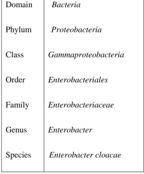
E. cloacae was first called as Bacterium cloacae and as Cloaca cloacae but when the genus was established it was renamed as E. cloacae(Table 2.1). E. cloacae is ever-present in nature and is known to be causes of many diseases in various plants like onion, ginger, papaya, and macadamia (Gillespie and Hawkey 2006,Nishijima et al. 2007).
The properties of Enterobacter are similar to Klebsiella and could be recognized by motility and certain biochemical reactions. The motility properties are used to differentiate
6
motile E. cloacae from non-motile Kleibsella sp. whereas they could not be distinguished biochemically and morphologically from each other (Muhialdin 2014).
E. cloacae is among the difficult-treated pathogens which are basically resistant to penicillins, cephalosporins (first and second-generation) and amoxicillin/clavulanic acid (AMC) owing to the chromosomal formation of Amp C β-lactamases (cAmpC) (Hilty et al. 2013). E. cloacae, as the typical species of Enterobacter, is a dominant nosocomial pathogen because of showing antibiotics highly resistance (Ren et al. 2010).
E. cloacae is a universal bacteria that lives in aquatic and terrestrial environments (water, sewage, soil, and food).The species appears as commensal microflora in the intestinal areas of humans and animals. It is also pathogen in plants and insects. This variety of habitats is reflected by the genetic diversity of E .cloacae. Recently, (multi locus sequencing typing) MLST and (Pulsed-field Gel Electrophoresis) PFGE epidemiological methods data showed world circulation of several widespread clonal complexes (Mezzatesta et al. 2012, Izdebski et al. 2015).
7
Table 2.1. Classification of Enterobacter cloacae (from Humann et al. 2011).
E. cloacae cause various medical contaminations, intravenous and other hospital equipment’s and devices (Dugleux et al. 1991). This bacterium can colonize on operative cleaning solutions and various surgical instruments causing nosocomial outbreaks (Wang et al. 2000). It has been reported for the past decades that the bacteria are considered as nosocomial pathogens in newborn units and several infection outbreaks (Fernandez-Baca et al. 2001, Pestourie et al. 2014).
Nowadays variability among strains is less common and outbreaks resulted from clonal E. cloacae hyper-producing Am PC lactamase and ESBL(extended spectrum β-lactamase) carrier isolates are defined from neonate specimens, adults urines/feces samples or from environmental samples (Pestourie et al. 2014).
Domain Phylum Class Order Family Genus Species Bacteria Proteobacteria Gammaproteobacteria Enterobacteriales Enterobacteriaceae Enterobacter Enterobacter cloacae
8
The genetic heterogeneity of the nomenspecies E. cloacae is famous. E. nimipressuralis, E. kobei, E. hormaechei, E. asburiae, E. dissolvens, E. cancerogenus and E. asburiae are basically associated with it and are involved in the so-called E. cloacae complex (Hoffman and Roggenkamp 2003).
2.2. Industrial enzymes in biotechnology
Enzymes have been industrialized since 1874 for the first time by a chemist Christian Hansen. The chemist employed the first enzyme used for industrial purposes called rennet which was derived from dried claves, stomachs with saline solution (Binod et al. 2013).
The term enzyme which means “in leaven” originated in a Greek word 𝜀𝜐𝜁𝜐𝜇o, that term was first used by a professor of physiology, Wilhelm Friedrich K¨uhne, at the University of Heidelberg in 1877. Currently it is one of the most widely used molecules after civilization of the ancient human (Gurung et al. 2013).
The sixth edition of the International Union of Biochemistry (I.U.B.) and Molecular Biology was published in 1992, has 3196 different enzymes. The international union of biochemistry introduced the standards for classifying enzymes which recommend that enzyme names originated in both the type of reaction catalyzed and the substrate acted upon. Enzymes could be divided into six parts based on the enzyme commission, oxidoreductase (EC 1), transferase (EC 2), hydrolase (EC 3), lyase (EC 4), isomerase (EC 5) and ligase (EC 6) (Gurung et al. 2013).
Biotechnology is currently using a wide range of enzymes manufactured on a commercial scale utilizing purposely screened microbial. The bio-catalysts of enzymes have an important role in all stages of biochemical reactions and metabolism. The contribution of certain enzymes as organic catalysts is of special interest in different processes on industrial scales. Superior enzymes are such microbial enzymes that are derived from microorganisms and they are employed for application purposes in industrial scales. However, the enzymes have been obtained from microorganisms since the 20th
9
century, characterization of properties, studies on their isolation, their application in bio-industry and manufacturing on bench-scale to pilot-scale have been developed. Moreover, a number of enzymes originated microbial sources have been employed commercially in various processes. Certain microorganisms such as bacteria, fungi and yeasts have been found in several global studies for the economically viable prepared bio-synthesis of many enzymes contributing in various commercial applications (Nigam 2013).
Enzymes could be used as biocatalysts in conventional catalytic reactions either in immobilized forms or in free and this is based on the specificity of enzyme. Biotechnology has been developed in a way that different enzymes which are being designed or purposely engineered based on the requirements of a procedure. Particular catalytic reactions are performed by several established class of enzymes which have also established their roles in designated bio-processes. Furthermore, several new enzymes have been designed with the involvement of biochemical-reaction engineering and protein-engineering. Certain molecular techniques have recently been engaged to improve and develop the performance and quality of microbial enzymes for their industrial applications more widely (Chirumamilla et al. 2001).
Microbial enzymes play an essential role in the industrial developing bioprocesses. Applications are currently involved in several markets, for instance textiles and detergents, chemical, pharmaceuticals, beverages and food, biofuels, leather, paper and pulp. At present, there is a necessity for improving more versatile enzymes in an attempt to develop more sustainable and industrial economically competitive production. Novel molecular techniques and diversity in microbial, such as genomics and metagenomics, are being exploited to determine new microbial enzymes with catalytic properties which later could be modified/ improved by various approaches based on rational, semi-rational and random directed development. The industrial enzymes are mostly found in recombinant forms synthesized in both bacteria and fungi (Adrio and Demain 2014).
Microorganisms can produce two types of enzymes; the one is retained inside the cell wall called intracellular while the other one is released into the growth medium called
10
extracellular. Glucose isomerase acts as the intracellular enzyme when it converts glucose into fructose and plays a vital role in the food industry. This type of enzyme needs to be activated by a particular molecule. In contrast, an average signal molecule cannot activate the enzyme since it cannot pass through the cell wall (Prasad 2014).
The extracellular enzymes are mostly used in industry including certain vital enzymes for instance, β-galactosidases, proteases, xylanases, keratinases and amylases.
Amylase: this is an essential enzyme due to its specific application in the starch conversion process in the industry (Nigam 2013).
Xylanase: Hemicellulose constitutes mainly of agricultural residues and plants along with pectin, lignin and cellulose (Nigam and Pandey 2009).
Keratinases: Keratin is considered as fibrous structural and insoluble protein that is a constituent of wool and feathers. The protein could chiefly be found as by-product keratinous wastes which represent the main source of proteins and amino acids that may possible be used as a source of nitrogen for plants or for animal feeds (Gushterova et al. 2005).
Proteases: They are hydrolytic enzymes, belong to the largest group of enzymes and are the most commercially-applicable enzymes, among the enzymes within this group the microbial proteases have been widely studied. Proteases prepared from microbial systems are of three types: acidic, neutral and alkaline. Alkaline proteases are well-organized under alkaline pH conditions and involve a serine residue at their active site (Gupta et al. 2002, Mukherjee et al. 2008, Vijayalakshmi et al. 2011).
11 2.2.1. The use of β-galactosidases in biotechnology
β-galactosidases is applied in the industrial in two ways; the use of immobilized enzyme bioreactors or utilizing β-galactosidase in a solution called free enzyme method (Gänzle et al 2008). The free enzyme technique is easily applied while reusing soluble enzyme is difficult due to its high costs of enzyme provisions and also high disadvantages (Gosling et al. 2010, Oliveira et al. 2011).
The provided entire cells can be applied as an alternative way to make less the technical and economical efforts relating to the extraction and purification/ isolation of enzyme. The main disadvantage is that lactose permeates through cell membranes very poorly; however chemical agents, such as chemical agents or detergents could be used to improve the microbial cells permeability (Panesar et al. 2006).
The hydrolyzed products such as hydrolysis of lactose derived from whey are sweeter; therefore they could be applied for developing additives for animal and human food. In addition to this, bioethanol could be obtained from the converted monosaccharaides after fermentation by utilizing proper microorganism and gasoline could also be blended with the produced bioethanol following proper purification. Accordingly, the promising process of hydrolyzed lactose has been found in several discoveries for many years for several reasons. However, the less fermentable lactose comparing to other sugars is an obstacle for the utilization of whey as it results in crystalizing at low temperature. The whey lactose could be hydrolyzed to galactose and glucose for improving that issue (Jurado et al. 2002, Matioli et al. 2003, Das et al. 2015).
The hydrolysis of lactose in whey and other dairy products can be accomplished in two approaches; acidic hydrolysis and enzymatic hydrolysis. The enzymatic hydrolysis is more desired than the other way as it allows milder conditions of temperature and pH, without causing any bad flavor, color and odor. Moreover, the acidic method results in protein denaturation finding in lactose solution and produces uninterested by products
12
(Sener et al. 2006, Demirhan et al. 2010). Table 2.2. Shows the biotechnological applications of β-galactosidase in bacteria.
Table 2.2 Biotechnological applications of β-galactosidases: lactose hydrolysis in milk and cheese whey and synthesis of GOS or other lactose/galactose derivatives by transgalactosylation reactions. (c) Cold-active enzymes; (m) mesophilic; (t) thermophilic enzyme (Oliveira et al. 2012).
β-galactosidases
Hydrolysis by β-galactosidases Transgalactosylation by β-galactosidases Milk
Acid whey
GOS Other lactose or
galactose derivatives Sweet whey Lactobacillus planetarium. Bifidobacterium infantis. Geobacillus staerothermophilus. Bacillus staerothermophilus(t) Arthrobacter sp(c). Bacillus staerothermophilus(t) Alicyclobacillus acidocaldarius(t). Lactobacillus bulgaricus Lactosucrose: Bacillus circulans Galactosyl trehalose Escherichia coli Galactosylmannitol, Galactosyle sorbose, Galactosyle salisin: Enterobacter cloacae B5 (m)
Applications of β-galactosidase are involved in dairy industry for managing the lactose intolerance (Mozumder et al. 2011). Lack of ability to hydrolyze a sugar type,
13
lactose, found in dairy products called lactose intolerance; this is a worldwide population problem due to the deficiency of β-galactosidase enzyme (Vasiljevic and Jelen 2002).
Mechanism of lactose hydrolysis and absorption and pathogenesis of diarrhea in intolerance is displayed in (Figure 2.1) the chief symptoms of lactose intolerance contain gassiness, swelling, diarrhea and abdominal pain. The undigested lactose passing from the small intestine into the colon will cause these symptoms. In the colon, the fermentation of unabsorbed lactose is caused by the bacteria normally present there will produce short chain fatty acids and gases (carbon dioxide and hydrogen). Flatulence, bloating and swelling pain might be resulted from Gas production. Unabsorbed lactose also has an osmotic effect in the gastrointestinal tract, drawing liquid into the lumen and resulting in diarrhea (de Vresa et al. 2001).
Figure 2.1. a) Mechanism of lactose hydrolysis and absorption.
14 2.3. Properties of β-galactosidase
β-galactosidase, an enzyme which has both historical and scientific importance. It is found in many organisms. The β-galactosidase encoded by the lac Z gene of the lac operon of E. coli has been very widely studied. It catalyzes β-D-galactopyranoside collapse and galactosyl transfer. Jacob and Monod used it to study introduction of gene expression of the lac operon. They won a Nobel Prize for this work (Huber2013).
It was investigated that the β-galactosidase is one of the most important enzyme used in food processing, which catalyses the lactose hydrolysis to its component monosaccharides, glucose and galactose (Figure 2.2). It is most frequently identified as lactase. The enzyme has been insulated and cleansed from a wide range of microorganisms but it is most commonly used. β-galactosidases are resulting from yeasts and fungal bases. The optimum pH for lactose hydrolysis is the main variance between yeast and fungal enzyme. The β-galactosidase application for lactose hydrolysis in milk and whey has technological, nutritional and environmental applications to human life (Panesar et al. 2006).
Figure 2.2: β -galactosidase hydrolysis of lactose (Weaver 2004).
β-galactosidase is a tetramer which is composed of four chains of indistinguishable polypeptide and each one formed in 1023 amino acids. The earliest structure of crystal was settled in a crystalized monoclinic formula with four asymmetric tetramers. The structure later will be refined to a single orthorhombic tetramer crystal with 1.7 oA resolution in the asymmetric unit. The technical superior of last form has been used for following practical
15
and structural studies. In single monomer, the 1023 amino acids constitute five domains with well-defined structure. The third domain centrally located (residues 334–627) is known as TIM (triose phosphate isomerase) or a8b8 barrel with the active place forming a deep pit at the end of this barrel on C-terminal (Juers et al. 2002).
β-galactosidase has three enzymatic activities (Figure 2.3), ( Juers et al. 2012). The first one, it can catalyze the lactose to glucose and galactose, which can then enter glycolysis. Secondly, the enzyme can catalyze the transgalactosylation of lactose to allolactose and the last one, the allolactose can be cleaved to the monosaccharides. It is allolactose which fixes to lac Z repressor and makes the positive feedback loop by which the amount of β-galactosidase is regulated in the cell. In many regards, β-galactosidase is best documented for its reaction with X-gal (5-bromo-4-chloro-3-indoyl-β-D-galactopyranoside).X-gal is a soluble colorless compound that involves galactose binded to a replaced indole. The specificity of β-galactose is great for the galactose part of its substrates but β-galactose has little specificity for the remainder. Thus, it hydrolyzes X-gal, releasing the substituted indole that unexpectedly dimerizes to give an insoluble, forcefully blue result. On growth medium containing X-gal, colonies of Escherichia coli that have an active β-galactosidase become blue because of this reaction.
16 From Huber et al. (1976).
Figure 2.3: Graphic summarizing the β-galactosidase roles in the cell. The enzyme can hydrolyze lactose to
glucose and galactose, it can transgalactosylate to form allolactose, and can hydrolyze allolactose. The presence of lactose can cause the synthesis of allolactose which binds to the lac repressor and reduces its affinity for the lac operon. This allows the synthesis of β-galactosidase, the product of the lac Z gene in turn.
17
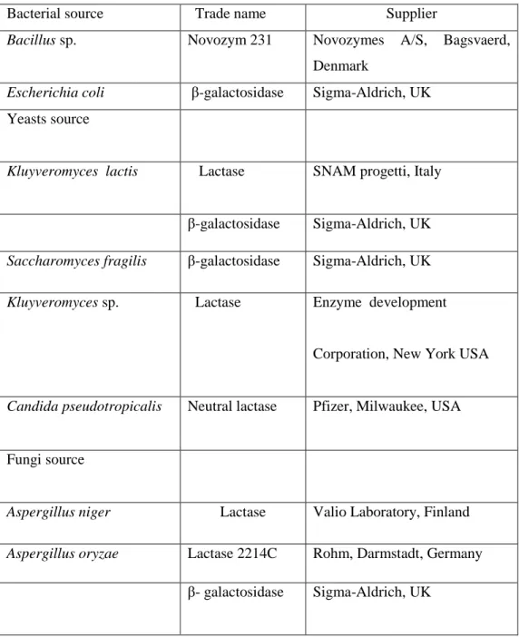
As shown in Table 2.3. There are many sources of suppliers of β-galactosidases from bacteria to fungi as commercial preparations.
Table 2.3:A list of sources and suppliers for commercial β-galactosidase preparations.
Bacterial source Trade name Supplier
Bacillus sp. Novozym 231 Novozymes A/S, Bagsvaerd, Denmark
Escherichia coli β-galactosidase Sigma-Aldrich, UK Yeasts source
Kluyveromyces lactis Lactase
SNAM progetti, Italy
β-galactosidase Sigma-Aldrich, UK
Saccharomyces fragilis β-galactosidase Sigma-Aldrich, UK
Kluyveromyces sp. Lactase Enzyme development
Corporation, New York USA
Candida pseudotropicalis Neutral lactase Pfizer, Milwaukee, USA
Fungi source
Aspergillus niger Lactase Valio Laboratory, Finland
Aspergillus oryzae Lactase 2214C Rohm, Darmstadt, Germany
β- galactosidase Sigma-Aldrich, UK
18 2.3.1. Psychrophilic β-galactosidase
Microorganisms of Psychrotrophic and Psychrophilic play a crucial role in our environment as the temperature of our planet is mostly cold (under 5°C). These microorganisms can generally be applied in biotechnological processes under low temperature, however they are considered as the chilled food spoilage. The metabolic processes can be retained at the high rates in the cold condition when the enzymes from psychrophiles show high activity at low temperature. The enzyme activity is usually reflected in low optimum temperature compared with such corresponding enzymes from thermophiles/mesophiles. These enzymes are known as thermolabile because they are inactivated rapidly and show a high specific activity at higher temperatures, in conjunction with their manufacturers they can have an critical character as biocatalysts in food processing and in biotechnology (Karasová et al. 2002).
The isolated enzymes of cold-adapted β-galactosidase from psychrophilic microorganisms are active at the range of temperature between 0 and 30 oC. These potential enzymes are not only employed in biotechnological purposes but also in the applications of industry. The properties of enzyme structure with high flexibility make the cold-adapted enzymes to be unfolded at low to moderate temperatures, and this can cause the inactivation of enzyme. This enzyme inactivation at moderate temperature .The inactivation of enzymes at moderate temperatures is suitable in experimental laboratories; however it also confines reactions to low temperatures (Fan et al. 2015).
2.3.2. Mesophilic β-galactosidase
It has been reported that the β-galactosidases are derived mainly from mesophilic microorganisms; accordingly, low thermo-stability of these enzymes are the main drawback. Some studies approved that reactions catalyzed at high temperature could have two advantages. The initial reaction rate becomes higher by improving reaction kinetics, and also increased substrate solubility can cause a higher volumetric throughput (Kong et al. 2014).
19
The enzymes of E.coli beside the β-galactosidases as a molecular genetics tool have been deeply studied after discovering the lactose operon. However, the industrial enzyme usage is restricted in the applications of food due to not considering safe, but it is still commercially used for analytical applications. Thus, microorganisms selection based on not harm for human use and also capability for synthesizing high β-galactosidase throughput are essential duty. In this regard, the rich sources of β-galactosidases for the efficient usage in food applications are Lactic acid bacteria including a diverse group of Lactobacilli, Streptococci and Lactococci,and Bifidobacterium, which are called GRAS organisms (Generally Recognized as Safe) (Princely et al. 2013).
Jacob et al. (1961) Investigated that β-galactosidase derived E. coli showing a particular place in the practice of molecular biology and the history. It has a key player in the regulation of gene expression in Jacob and Monod’s development of the operon model. A distinguishable blue reaction product indicates its presence and that could be a good worker in certain molecular biology procedures such as cloning.
E. coli β-galactosidase is a single tetramer composed of four symmetrical 1023-amino acid chains, each of which contains five domains. The active site located on the third one that comprises an eight-stranded α/β barrel. This site involves elements from other subunits and other domains. The polypeptide chains with the N-terminal region formed from interfaces of the single subunit. These all features together offers a basic structure for the known property of α-complementation. Covalent galactosyl formation makes the progression of catalytic activity when it intermediates with Glu537, and comprises ‘deep’ and ‘shallow’ modes of binding substrate (Matthews 2005).
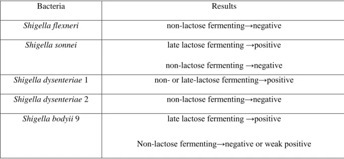
Late lactose-fermenting strains of the following usually prompt lactose-fermenting species and genera , namely, Escherichia coli ,Citrobacter,cloacae, and Klebsiella, are o-NPG positive. Also o-o-NPG positive are certain lactose-variable or non-lactose- fermenting genera and species, namely, Dispar, Hafnia, Serratia, Vibrio and Aeromonas. Non-lactose fermenting species and genera, Salmonella, Proteus , Providencia, Alkalescens
20
, Pseudomonas and Moraxella are o-NPG Negative. Those species and genera which gave variable results to o-NPG tests are set out below Table 2.4. (Lapageand Jayaraman 1964).
Table 2.4: Results of some species and genera with lactose fermentation.
Bacteria Results
Shigella flexneri non-lactose fermenting→negative
Shigella sonnei late lactose fermenting →positive
non-lactose fermenting →negative Shigella dysenteriae 1 non- or late-lactose fermenting→positive Shigella dysenteriae 2 non-lactose fermenting→negative
Shigella bodyii 9 late lactose fermenting →positive
Non-lactose fermenting→negative or weak positive
Modified from Lapageand Jayaraman (1964).
The most isolated bacteria as producer was diagnosed as Bacillus subtilis. Furthermore, certain environmental and nutritional conditions for the enzyme production via the selected isolate were studied of changed media comprise as pH of the medium 7.0, yeast extract 10 g/L, lactose 5 g/L and the incubation for 48 hours at the temperature 30 °C was considered as the most appropriate conditions for the β-galactosidase production (El-Kader et al. 2012).
2.3.3. Thermophilic β-galactosidases
A number of isolated β-galactosidases described as thermostable have been formerly achieved from both mesophilic Archaeabacteria and Eubacteria.They were distinguished and utilized for lactose hydrolysis and in GOS production (Petzelbauer et al 2001, Ansari et al 2012). Though, these thermostable enzymes are mostly produced at very low amount by archaeabacteria and thermophilic bacteria; for which reason they could not
21
be purified easily. Therefore, they could be synthesized on a large scale at industrial scale by employing recombinant techniques in mesophilic hosts (Demirjian et al. 2001, Ansari et al. 2012). The efficacy of recombinant β-galactosidases with thermostable achieved from Geobacillus stearothermophilus, Sulfolobus solfataricus, Thermotoga maritima, Pyrococcus furiosus and Thermus sp. had already been reported in the production of GOS pursuing at high temperatures. They demonstrated few advantages over innate enzymes comprising large-scale production, developments in their activity and their simplicity of purification (Akiyama et al. 2001, Bruins et al. 2001, Placier et al. 2009).
2.4. Bacterial sources of β-galactosidase
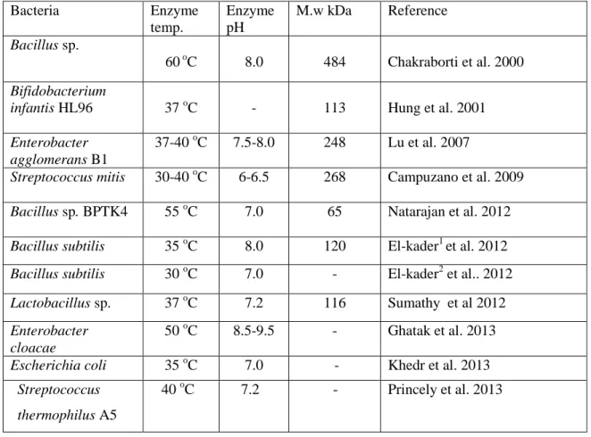
The β-galactosidase enzyme can be synthesized by a large group of bacteria such as Bacillus stearothermophilus and Streptococcus thermophilus, which are potentially employed as bacterial sources. The enzyme derived from Escherichia coli could be used as a model for comprehending how the catalytic mechanism of β-galactosidase acting, whereas the host coliform causes toxicity problems and make it does not suitable in food applications. For this reason, the enzyme from E. coli is not normally preferred in the industry of food (Panesar et al 2010).Bacterial sources are desired due to their fermentation easily, high stability and activities of the enzyme. Several bacterial species have been employed by a number of dairy industries for producing β-galactosidase enzyme; these species belonging to genera of Bifidobacterium and Lactobacillus (Sumathyet al. 2012). A few β-galactosidases properties from bacterial sources are presented in (Table 2.5).
22
Table 2.5 : Some properties of β-galactosidases from bacterial sources.
Bacteria Enzyme temp. Enzyme pH M.w kDa Reference Bacillus sp. 60 oC 8.0 484 Chakraborti et al. 2000 Bifidobacterium
infantis HL96 37 oC - 113 Hung et al. 2001
Enterobacter agglomerans B1
37-40 oC 7.5-8.0 248 Lu et al. 2007
Streptococcus mitis 30-40 oC 6-6.5 268 Campuzano et al. 2009 Bacillus sp. BPTK4 55 oC 7.0 65 Natarajan et al. 2012 Bacillus subtilis 35 oC 8.0 120 El-kader1 et al. 2012 Bacillus subtilis 30 oC 7.0 - El-kader2 et al.. 2012 Lactobacillus sp. 37 oC 7.2 116 Sumathy et al 2012 Enterobacter
cloacae
50 oC 8.5-9.5 - Ghatak et al. 2013
Escherichia coli 35 oC 7.0 - Khedr et al. 2013
Streptococcus thermophilus A5
40 oC 7.2 - Princely et al. 2013
Accounting for 0.1–1% of the bacterial species in environment could be cultivated by employing conventional methods. Hence, an auspicious source is considered as the metagenome used for new β-galactosidases discovery with industrially favorable properties (Erich et al. 2015).
The optimized ultrasonication approaches has been used for disrupting the extra cells of E. coli for the β-galactosidase release (Panesar et al. 2010). The cultures of Lactobacillus delbrueckii subsp. bulgaricus were treated for the β-galactosidase release using a high-pressure homogenizer, a high-speed bead mill and sonication (Bury et al. 2001). Bacterial sources of β-galactosidases are shown in (Table 2.6).
23 Table 2.6: Bacterial sources of β-galactosidase.
Bacteria
Alicyclobacillus acidocaldarius subsp. rittmannii Arthrobacter sp.
Bacillus acidocaldarius, B. circulans, B. coagulans, B.subtilis, B. megaterum, B. stearothermophilus
Bacteriodes polypragmatus
Bifidobacterium bifidum, B. infantis
Clostridium acetobutylicum, C. thermosulfurogens Corynebacterium murisepticum
Enterobacter agglomerans, E. cloaceae Escherichia coli
Klebsiella pneumoniae
Lactobacillus acidophilus, L. bulgaricus,L. helviticus, L. kefiranofaciens, L. lactis, L. sporogenes, L. themophilus, L.delbrueckii
Leuconostoc citrovorum
Pediococcus acidilacti, P. pento Propioionibacterium shermanii Pseudomonas fluorescens Pseudoalteromonas haloplanktis
Streptococcus cremoris, S. lactis, S. thermophius Sulfolobus solfatarius
Thermoanaerobacter sp. Thermus rubus, T. aquaticus Trichoderma reesei
Vibrio cholera
Xanthomonas campestris
24 2.5. Purification of β-galactosidase
Different techniques are currently applied for the improvement and purification of
several bioproducts. The selection of unit operations appropriate for each process stage along with its intrinsic characteristics is required for a purified bioproduct achievement. Various feasible strategies could also be combined with a few techniques. If the unit operations are employed very well, the resulting by product and the development of the process will be impacted. The whole downstream processing costs around two-thirds of entire process costs (Lemes et al. 2014). The production of bioproducts in the industry is not a simple task as it needs a big effort in process improvement. Therefore, the purification strategies are necessary to be investigated because of employing low-cost technologies. The traditional methods are involved in isolating and purifying proteins and include several steps, such as the precipitation of ammonium sulfate, dialysis, ion and affinity chromatography and finally the product concentration (Oliveira et al. 2012). Hence, there is a requirement to examine the optimization and application of alternative methods that could increase the purification processefficiency, and also improve the separation process with a low-cost (Ahmad et al. 2010).
A number of techniques and procedures have been published for the β-galactosidase purification. Though, the evaluation of a single purification sequence is specified in the most existing studies in the literature and it is also mot considered the technique with peculiarities hindering the scale-up method and compromise the efficiency of the process (Lemes et al. 2014).
2.5.1. Sonication
The bacterial cellwalls can be disrupted by one of the most widely used techniques called sonication. One of an effective way of cell breakage is the production of sound waves with high frequency for applying to microbial cells. The differences in local transient pressure can break cell walls and the mechanism of micro-cavitation is believed to involve in. The cell breakage efficiency can be influenced by several factors such as, the volume of material processed, the exposure duration and the power output of the instrument. The
25
treated volume in a given time is not preferred specifically, for instance, using high pressure extrusion. The produced heat is necessary to be avoided by cooling. Ultrasound is referred to frequency of sound waves higher than 15-20 kHz, and this result in cell inactivation, whereas the application of higher acustic power inputs along with the ultrasound can result in microbial cells destruction in suspension (Dinnison 1999, Kara 2004, Prasad 2014).
2.5.2. Ammonium sulfate precipitation
This is aspecific step carried out in a more general method termed salting out. The widely used technique is the addition of neutral salts for precipitation of fractionating proteins. The protein is not denatured after the precipitation and also its activity is improved by the pellet re-dissolving. Moreover, the proteins can be stabilized by adding the salts for avoiding bacterial contamination, proteolysis or denaturation. Therefore, the salting out is considered as a critical step carrying out either before or after centrifugation to store an extract overnight (Amersham 2001).
2.5.3. Dialysis
Dialysis can be described as the solutions of different concentrations diffusethrough semi-permeable membranes acting as the boundary between them. Certain average pore size can be found in the membrane acting as an inert sieve. The distributed fibers randomly can form the pores synthesizing the semi-permeable dialysis tubing or the dialysis membrane, which is normally produced from cellulose acetate and contains pores ranging from 1 to 20 nm in diameter (Harris 1989).
2.5.4. Ultrafiltration
Ultrafiltration is a technique related to dialysis, and can also be used to desalt protein solutions, effect buffer exchange, or concentrate protein solutions. It is more expensive than dialysis, however, as special equipment and membranes are required. In this technique, pressure is applied to the solution to cause a bulk flow of water and dissolved
26
low molecular weight solutes, through the membrane, while high molecular weight solutes are retained (Dennison 1999).
2.5.5. Gel permeation chromatography (GPC)
A form of partition chromatography which is used for separating molecules of various sizes calledGel permeation chromatography (GPC). This type of chromatography can be simply termed as gel chromatography or in other forms such as, molecular sieve chromatography, gel exclusion chromatography and gel filtration. This chromatography type basically works on the partitioned molecules among stationary phase of defined porosity and solvent. A porous gel matrix in bead form surrounded by solvent in a packed column is used for the separation process ( Swadesh 2000,Amersham 2001).
2.5.6. Affinity chromatography
The above aforementioned chromatographic methods are relied on the protein with the gross physicochemical properties. Though, activity of protein is biologically more subtle and also relies on the very complementary specific and steric relationship between a substrate (or inhibitor) and the active site, or a ligand and a binding site. This biospecific correlation between a protein and a ligand can be exploited by affinity chromatography to choose an interested protein in a single step from a crude mixture (Harris 1989).
The β-galactosidase purification was achieved from gram-negative of Pseudoalteromonas haloplanktis TAE 79 strain to homogeneity. The sequence of the NH2 -terminal amino acid and nucleotide of the purified enzyme specify 1,038 amino acids with a calculated Mr of 118,068 constitute the β-galactosidase enzyme subunit. Furthermore, this enzyme has certain structural properties with β-galactosidase from Escherichia coli which includes 51% amino sequence identity, related subunit mass, comparable optimal pH value, protection of amino acid residues comprised in catalysis and needed for divalent metal ions, and it also varies by changing optimum activity towards low temperatures and stability at lower thermal, and by a higher catalytic efficiency on natural and synthetic substrates.
27
Furthermore, the enzyme is also characterized by activation of particular thermodynamic parameters and by a higher pI (7.8). The β-galactosidase from P. haloplanktis was released in E. coli, and also properties presented by the recombinant enzyme are similar to those of the wild-type enzyme. Intrinsic fluorescence spectroscopy is employed to monitor unfolding heat-induced and show values of lower melting point for recombinant β-galactosidase associated with the mesophilic enzyme and P. haloplanktis wild-type. The assessed lactose hydrolysis in milk reveals β-galactosidase derived P. haloplanktis which is capable to outperform the current β-galactosidase employed commercially from Kluyveromyces marxianus var. lactis, and it has been suggested to hydrolyze lactose using the cold-adapted β-galactosidase in processing dairy products in refrigerated plants. (Hoyoux et al. 2001).
The purification of β-galactosidase has also been accomplished from psychotropic
such as Pseudoalteromonas sp. and the enzyme isolated form Antarctica. A rapid purification scheme could be used to achieve a high throughput of purification by employing extraction in an aqueous two-phase system ultrafiltration techniques and interaction of hydrophobic chromatography (Fernandeset al. 2002).
The E. coli K-12 strain LC110 β-galactosidase has been purified and characterized. Strain LC110 is a Lac+ revertant of a mutant with a removal of the lac Z β-galactosidase gene. Its new evolved β-galactosidase (ebg) activity was shown to be due to a discrete protein, immunologically unrelated to lac Z β-galactosidase (Arraj and Campbell 1975).
Enterobacter agglomerans B1 was served to obtain a newly purified and a homodimer of 248 kDa transglycosylating β-galactosidase. The optimum temperature and pH for the hydrolysis of o-NPGal were 37-40 oC and 7.5-8.0, while 114 and 0.06 mM were the Km values for lactose and o-NPGal. The enzyme formed galacto oligosaccharides in around 38% yield at the concentration of lactose 12.5% (w/v). Once o-NPGal is used as a donor, glycosyl can be catalyzed by the enzyme and transfer to a series of acceptors, comprising aromatic glycosides, cyclitol, hexahydroxy alcohol, β or α-disaccharides, hexose and pentose. Therefore, this makes the enzyme to contribute as a robust synthetic
28
tool for the preparation of chemicals containing galactose. Thermal asymmetric interlaced PCR (TAIL-PCR) and degenerate PCR were capable to clone the gene encoding this enzyme. It demonstrated that the expressed 1029 amino-acid protein in Escherichia coli was encoded by an open reading frame (ORF) of 3090 nucleotides. The Natural and recombinant enzymes reported the same activities of transferase (Lu et al. 2007).
2.6. Lactose hydrolysis by β-galactosidase
Lactose is a disaccharide sugar composed of galactose and glucose that is originated in milk. Lactose-free milk and dairy products have a significant market, which can be gained by enzymatic hydrolysis using β-galactosidases. The sweetness and solubility of lactose is faint compared to other sugars, including glucose, galactose, fructose and sucrose (Gänzle et al. 2008, Oliveira et al. 2011). That is why, lactose hydrolysis reduces precipitation problems and improves the sweetening control, thus increasing the food applications of lactose solutions, for example sucrose or starch syrups are substituted by lactose solutions in the confectionary and ice-cream industries. (Siso 1996, Oliveira et al. 2011).
Cheese whey and milk are the main sources of lactose, which is a primary by-product of cheese manufacturing. During the by-production of 1 kg of cheese about 9 L of whey stream are created, amounting to over 160 million ton of whey produced worldwide each year (Guimarães et al. 2010). The capacity of Whey's organic is extraordinary (biochemical oxygen demand of 30–50 g/L and chemical oxygen demand of 60–80 g/L), largely because of the lactose content (Guimarães et al. 2010). In this regard, lactose hydrolysis by β-galactosidases again plays a vital part by extending whey's applications. Particularly, whey fermentations can be prolonged away from somewhat limited capacities of lactose-consuming microorganisms, to the use of lactose-hydrolyzed whey as feedstock for the production of added-value molecules or bulk supplies by lactose-negative microbes. For instance, Saccharomyces cerevisiae wild strains (lactose negative) can be used to
29
produce ethanol from hydrolyzed whey, although catabolite repression-resistant mutants should be applied in order to avoid glucose-galactose diauxy (Oliveira et al. 2011).
The lactose hydrolysis can be accomplished by employing either enzyme or acid hydrolysis. The acid hydrolysis includes lactose heating with acid reagents for instance H2SO4, tetraoxosulphate (VI) acid, a cationic exchange resin as acid form called a solid acid or as a free acid form. This is a slight complex process from a mechanistic perspective. This is partly because of further degradation of the monosaccharide products into uninterested chemicals, in which the expected side reactions based on, among additional factors, the composition of lactose such as whey permeate and its source (Cote et al. 2004). The hydrolysis of lactose with free enzyme is not applicable because its high costs and the final products can be contaminated with interference of foreign protein (Olafadehan et al. 2010).
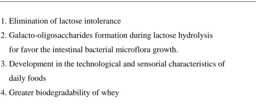
Table 2.7: Shows various β-galactosidase applications (Jurado et al. 2002, Kara 2004).
1. Elimination of lactose intolerance
2. Galacto-oligosaccharides formation during lactose hydrolysis for favor the intestinal bacterial microflora growth.
3. Development in the technological and sensorial characteristics of daily foods