OYUN CERRAHİSİ
Detection of Helicobacter pylori in adenoid tissue by
real-time polymerase chain reaction
Gerçek zamanlı polimeraz zincir reaksiyonu ile adenoid dokuda
Helicobacter pylori tespiti
Mustafa Kaymakçı, M.D.,1 Mehtap Aydın, M.D.,2 Selçuk Yazıcı, M.D.,3 Özlem Sağır, M.D.,4 Özer Erdem Gür, M.D.,5 Murat Sayan, M.D.6
Objectives: This study aims to investigate the presence of Helicobacter pylori (H. pylori) using the real-time polymerase chain reaction (PCR) method in the adenoid tissues in children undergoing surgical operation due to adenoid hypertrophy. Patients and Methods: Adenoid tissues of 23 children (8 girls, 15 boys; mean age 6.2 years; range 3 to 9 years) who were operated with the diagnosis of adenoid hypertrophy, tonsil + adenoid hypertrophy or tonsil + adenoid hypertrophy + serous otitis media in our clinic between January 2012 and April 2012 were examined.
Results: Of 23 patients, H. pylori was detected in the adenoid tissues of two (8.7%). Regurgitation was present in seven patients. However, no regurgitation was found in H. pylori-positive patients.
Conclusion: In this study the presence of H. pylori in patients with adenoid hypertrophy has been demonstrated using PCR method. To be able to support the hypothesis that H. pylori has a place in etiology of adenoid hypertrophy, multicenter studies are warranted.
Key Words: Adenoid; helicobacter pylori; hypertrophy.
Amaç: Bu çalışmada adenoid hipertrofi nedeniyle cerrahi yapılan çocukların adenoid dokularında gerçek zamanlı poli-meraz zincir reaksiyonu (PZR) yöntemi ile Helicobacter pylori (H. pylori) varlığı araştırıldı.
Hastalar ve Yöntemler: 2012 Ocak - 2012 Nisan tarihleri arasında kliniğimizde adenoid hipertrofisi, tonsil + adeoid hipertrofisi veya tonsil + adenoid hipertrofisi + seröz otitis media tanısı ile ameliyat edilen 23 çocuğun (8 kız, 15 erkek; ort. yaş 6.2 yıl; dağılım 3-9 yıl) adenoid dokuları incelendi.
Bulgular: Yirmi-üç hastanın ikisinin (%8.7) adenoid dokularında H. pylori saptandı. Yedi hastada regürjitasyon vardı. Ancak, H. pylori pozitif hastalarda regürjitasyona rastlanmadı.
Sonuç: Bu çalışmada, adenoid hipertirofisi olan hastalarda H. pylori varlığı PCR yöntemi ile gösterilmiştir. H.pylori’nin adenoid hipertrofisinin etiolojisinde yerini alması için çok merkezli çalışmalar ile bu hipotezin desteklenmesi gerekmek-tedir.
Anahtar Sözcükler: Adenoid; helicobacter pylori; hipertofi.
1Department of Otolaryngology, Medical Faculty of Balıkesir University, Balıkesir, Turkey
2Department of Infectious Disease and Clinical Microbiology, Medical Faculty of Balıkesir University, Balıkesir, Turkey 3Department of Pediatrics, Medical Faculty of Balıkesir University, Balıkesir, Turkey
4Department of Anesthesiology and Reanimation, Medical Faculty of Balıkesir University, Balıkesir, Turkey 5Department of Otolaryngology, Antalya Training and Research Hospital, Antalya, Turkey 6Department of Central Laboratory, PCR Unit, Medical Faculty of Kocaeli University, Kocaeli, Turkey
Received / Geliş tarihi: October 04, 2013 Accepted / Kabul tarihi: December 09, 2013 Correspondence / İletişim adresi: Özer Erdem Gür, M.D. Antalya Eğitim ve Araştırma
Hastanesi Kulak Burun Boğaz Hastalıkları Kliniği, 07100 Antalya, Turkey. Tel: +90 532 - 599 61 75 e-mail (e-posta): [email protected] Available online at
www.kbbihtisas.org
doi: 10.5606/kbbihtisas.2014.28445 QR (Quick Response) Code
Adenoid hypertrophy is an important problem, being one of the leading causes of upper respiratory tract obstruction in children.[1] Bacterial infections transmitted by the respiratory route are the most common cause of adenoid hypertrophy. In recent years, there have been debates on the association of gastroesophageal or laryngoesophageal reflux with colonization of Helicobacter pylori (H. pylori) in the upper respiratory tract and its role in the development of infections.[2,3] Helicobacter pylori is one of the most common causative agents of chronic bacterial infections worldwide. Helicobacter pylori, which is a gram-negative bacillus, influences more than half of the child and adult population.[4] The prevalence of H. pylori is not homogenous worldwide and has been reported in cross-sectional studies to range from 9% to 57%.[5] Its prevalence can reach up to 80% in developing countries such as India.[6] Different detection methods also contribute to differences in prevalence reported from different regions.
Helicobacter pylori infection is acquired in childhood before the age of 10 years, and it persists lifelong unless antibiotic treatment is used.[7] Despite information about the risk factors that influence colonization of H. pylori and about pathogenesis, there are points that remain to be elucidated. Helicobacter pylori has been associated primarily with gastritis, gastric and/or duodenal ulcer, and gastric cancer. Moreover, there is a collection of literature data about positive or negative associations between H. pylori and some diseases including functional dyspepsia, gastroesophageal reflux, iron deficiency, growth retardation, idiopathic thrombocytopenic purpura, and asthma, which have not been clearly defined yet.[5,7-10] The presence of H. pylori has been demonstrated in the oral cavity including dental plaque, oral lesions and saliva, and this condition has been reported as a potential source of infection and contamination.[11-14] Helicobacter pylori has also been shown to be present in high ratios in nasal polyps, laryngeal samples, and vocal cord lesions using real-time polymerase chain reaction (PCR) method.[15,16] There are conflicting results about the presence of H. pylori in tonsil and adenoid tissues. Minocha et al.[17] demonstrated a decrease in gastric H. pylori colonization in patients who underwent tonsillectomy and claimed that tonsil tissue might serve as a reservoir for this bacterium.
Contrarily, in a recent study, Toros et al.[18] failed to detect H. pylori colonization in tonsil and adenoid tissues and suggested that searching for H. pylori colonization in the aerodigestive tract had no clinical benefit.
The present study aimed to investigate the presence of H. pylori using real-time PCR method in the adenoid tissues of children who underwent surgical operation due to adenoid hypertrophy.
PATIENTS AND METHODS
Adenoid tissues of 23 children (8 girls and 15 boys; mean age 6.2 years; range 3 to 9 years), who underwent surgical operation due to adenoid hypertrophy, tonsil+adenoid hypertrophy, or tonsil+adenoid hypertrophy+serous otitis media, were examined. The medical history of all patients was obtained and detailed physical examinations were performed before the surgical procedures. Symptoms indicating the presence of gastroesophageal reflux (night cough, sleep apnea, dysphagia, frequent throat clearing, and regurgitation) were particularly questioned.
The presence of adenoid hypertrophy was confirmed by X-ray and endoscopic examination. The patients were operated on under general anesthesia. Informed consents were obtained from the families of the children, and surgically removed adenoid tissues were examined for the presence of H. pylori using real-time PCR method. Qiagen Minelute Virus Spin Kit protocol and QIAamp DNA Mini Kit protocol (QIAGEN GmbH, Hilden, Germany) were used for DNA isolation from the tissue samples. The 156 base-pair fragment of the ureC gene of H. pylori was amplified using the real-time PCR method with primer pair H. pylori-F (TCGGTAAAGACACCAGAAAA) and H. pylori-R (TCATAATACCCGCATCACAG). The main reaction mixture was prepared to include 1 mL (5 mM) primer F, 1 mL (5 mM) primer R, 10 mL template, and 0.5 mL dH2O for each reaction (25 mL).
Conditions for real-time PCR were 15 minutes at 95 °C and then 30 seconds at 95 °C, 45 seconds at 54 °C, and 30 seconds at 72 °C; it was performed for 50 cycles. The PCR product was detected during the reaction using SYBR Green, a fluorescent stain. Confirmation of the amplified DNA fragments being the targeted region was made by melting curve analysis.
RESULTS
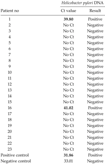
Helicobacter pylori was demonstrated to be present in the adenoid tissues of two (8.7%) of 23 patients. Regurgitation was present in seven patients. However, regurgitation was not found in the patients who were positive for H. pylori. The real time PCR results of 23 adenoid tissues, as well as positive and negative controls of the test, are presented in Table 1.
DISCUSSION
Tissue based methods, which are being used to detect H. pylori, include rapid urease test (RUT), histological examination, culture, and PCR. Although twisted and curved shape image in histological examination is useful to show the presence of H. pylori, Bitar et al.[19] reported negative results using two-step (nested) PCR in
the adenoid tissue samples that showed positive results in histological analysis. Rapid urease test appears to have higher specificity and sensitivity as compared to histological examination.[20] Nevertheless, studies performed on adenoid tissue using RUT method have reported high rates of false positivity.[19] Although culture has the highest specificity (100%) in establishing the diagnosis, its sensitivity is low.[20] It has been shown that PCR method has almost 100% sensitivity and specificity in the detection, identification and quantitation of H. pylori in biological samples.[21-23] Real-time PCR method has been reported to increase the likelihood of detecting H. pylori in the samples that produce false negative results histologically.[24] In additional that real time PCR offers increased sensitivity and specificity in a rapid format, in comparison to traditional gel-based PCR assays, Unlike traditional systems, which rely upon endpoint analysis, real time PCR assays visualize the reaction as it is taking place allowing quantification and reaction analysis.
Since real time PCR reactions are performed in a closed system (no gel analysis needed) the risk of contamination has been substantially reduced.[25,26] Although PCR has technical superiorities and its commercial kits have become easily available, it is expensive to be routinely used and is being used primarily for academic research.[20] In the present study, we investigated the presence of H. pylori in the adenoid tissue using real-time PCR method and found a positivity of 8.7%. Contradictory results have been reported in the studies investigating H. pylori in the adenoid tissue. Bulut et al.[27] investigated the presence of H. pylori in 118 tissue samples (71 tonsil and 47 adenoid tissues) from 71 children, of whom 28 had recurrent tonsillitis and 43 had adenotonsillar hypertrophy. They detected H. pylori in 24.6% of the samples using PCR analysis. Moreover, they showed virulence markers cytotoxin-associated (cagA) gene in 58.6% of the positive samples. The higher rate of cagA + H. pylori in cases with adenotonsillar hypertrophy than those with recurrent adenotonsillitis has raised the thought that H. pylori might play a role in the development of adenotonsillar hypertrophy. Farivar et al.[28] assessed the presence of H. pylori using real time PCR method in 103 archival paraffin-embedded tonsillar tissues, which were obtained from patients with chronic tonsillitis, and demonstrated H. pylori DNA in 21.35% of the samples.
Table 1. Results of Helicobacter pylori detection in adenoid tissue via real-time polymerase chain reaction method
Helicobacter pylori DNA
Patient no Ct value Result
1 39.80 Positive 2 No Ct Negative 3 No Ct Negative 4 No Ct Negative 5 No Ct Negative 6 No Ct Negative 7 No Ct Negative 8 No Ct Negative 9 No Ct Negative 10 No Ct Negative 11 No Ct Negative 12 No Ct Negative 13 No Ct Negative 14 No Ct Negative 15 No Ct Negative 16 41.02 Positive 17 No Ct Negative 18 No Ct Negative 19 No Ct Negative 20 No Ct Negative 21 No Ct Negative 22 No Ct Negative 23 No Ct Negative
Positive control 31.86 Positive Negative control 33.01 Negative
In their study, Ağırdır et al.[29] investigated the presence of H. pylori with the Campylobacter-like organism (CLO) test in 45 patients undergoing adenoidectomy and myringotomy with the diagnosis of chronic otitis media and adenoid hypertrophy. They detected H. pylori in the middle ear effusions in 66.6% and in the adenoid tissue in 33.3% of 30 patients with middle ear effusion. They found H. pylori positivity to be 26.6% in the adenoid tissues of the group without middle ear effusion (n=15). In addition, they reported that none of their patients had a positive CLO test in the wash out liquid of the middle ear. In a similar study conducted by Yılmaz et al.,[30] the presence of H. pylori was investigated using real-time PCR in the middle ear effusions of patients with adenoid hypertrophy and chronic otitis; they detected positivity in 12 (67%) of 18 patients; however, they failed to detect H. pylori in the adenoid tissues of this group of patients. In that particular study, they detected H. pylori in the adenoid tissue of only one patient out of 20 who had adenoid hypertrophy alone. In the study by Eyigör et al.,[31] the presence of H. pylori was assessed with RUT and PCR methods in 35 adenoid and 20 tonsil tissue samples obtained from 47 patients with chronic tonsillitis and adenoid hypertrophy. While they found positivity rate to be 5.5% using RUT, they detected H. pylori in none of the patients using PCR. This study investigated the presence of H. pylori using real-time PCR method in the adenoid tissues of 23 children who underwent surgical operation due to adenoid hypertrophy and H. pylori was demonstrated to be present in the adenoid tissues of two (8.7%) of 23 patients. In conclusion, H. pylori may be implicated in the etiology of adenoid hypertrophy, but the number of cases is insufficient to be able to give a definite ratio. Different patient groups and different methods used in the studies make comparison difficult. The results might have been influenced by different prevalence rates from different geographical regions. Further multicenter and large-scale studies on this issue are needed.
Declaration of conflicting interests
The authors declared no conflicts of interest with respect to the authorship and/or publication of this article.
Funding
The authors received no financial support for the research and/or authorship of this article.
REFERENCES
1. Blum RH, McGowan FX Jr. Chronic upper airway obstruction and cardiac dysfunction: anatomy, pathophysiology and anesthetic implications. Paediatr Anaesth 2004;14:75-83.
2. Tauber S, Gross M, Issing WJ. Association of laryngopharyngeal symptoms with gastroesophageal reflux disease. Laryngoscope 2002;112:879-86.
3. Kurtaran H, Uyar ME, Kasapoglu B, Turkay C, Yilmaz T, Akcay A, et al. Role of Helicobacter pylori in pathogenesis of upper respiratory system diseases. J Natl Med Assoc 2008;100:1224-30.
4. Wallis-Crespo MC, Crespo A. Helicobacter pylori infection in pediatric population: epidemiology, pathophysiology, and therapy. Fetal Pediatr Pathol 2004;23:11-28.
5. Spee LA, Madderom MB, Pijpers M, van Leeuwen Y, Berger MY. Association between helicobacter pylori and gastrointestinal symptoms in children. Pediatrics 2010;125:e651-69.
6. Poddar U, Yachha SK. Helicobacter pylori in children: an Indian perspective. Indian Pediatr 2007;44:761-70. 7. Pacifico L, Anania C, Osborn JF, Ferraro F, Chiesa
C. Consequences of Helicobacter pylori infection in children. World J Gastroenterol 2010;16:5181-94. 8. Windle HJ, Kelleher D, Crabtree JE. Childhood
Helicobacter pylori infection and growth impairment in developing countries: a vicious cycle? Pediatrics 2007;119:e754-9.
9. Moyaert H, Franceschi F, Roccarina D, Ducatelle R, Haesebrouck F, Gasbarrini A. Extragastric manifestations of Helicobacter pylori infection: other Helicobacters. Helicobacter 2008;13 Suppl 1:47-57. 10. Sýkora J, Rowland M. Helicobacter pylori in pediatrics.
Helicobacter 2011;16 Suppl 1:59-64.
11. Li C, Musich PR, Ha T, Ferguson DA Jr, Patel NR, Chi DS, et al. High prevalence of Helicobacter pylori in saliva demonstrated by a novel PCR assay. J Clin Pathol 1995;48:662-6.
12. Desai HG, Gill HH, Shankaran K, Mehta PR, Prabhu SR. Dental plaque: a permanent reservoir of Helicobacter pylori? Scand J Gastroenterol 1991;26:1205-8.
13. Mravak-Stipetić M, Gall-Troselj K, Lukac J, Kusić Z, Pavelić K, Pavelić J. Detection of Helicobacter pylori in various oral lesions by nested polymerase chain reaction (PCR). J Oral Pathol Med 1998;27:1-3.
14. Cammarota G, Tursi A, Montalto M, Papa A, Veneto G, Bernardi S, et al. Role of dental plaque in the transmission of Helicobacter pylori infection. J Clin Gastroenterol 1996;22:174-7.
15. Ozyurt M, Gungor A, Ergunay K, Cekin E, Erkul E, Haznedaroglu T. Real-time PCR detection of Helicobacter pylori and virulence-associated cagA in nasal polyps and laryngeal disorders. Otolaryngol Head Neck Surg 2009;141:131-5.
16. Tiba M, Fawaz S, Osman H. Helicobacter pylori and its role in vocal folds' minimal lesions. Clin Respir J 2010;4:237-40.
17. Minocha A, Raczkowski CA, Richards RJ. Is a history of tonsillectomy associated with a decreased risk of Helicobacter pylori infection? J Clin Gastroenterol 1997;25:580-2.
18. Toros SZ, Toros AB, Kaya KS, Deveci I, Özel L, Naiboğlu B, et al. A study to detect Helicobacter pylori in adenotonsillar tissue. Ear Nose Throat J 2011;90:E32. 19. Bitar MA, Soweid A, Mahfouz R, Zaatari G, Fuleihan N.
Is Helicobacter pylori really present in the adenoids of children? Eur Arch Otorhinolaryngol 2005;262:987-92. 20. Guarner J, Kalach N, Elitsur Y, Koletzko S.
Helicobacter pylori diagnostic tests in children: review of the literature from 1999 to 2009. Eur J Pediatr 2010;169:15-25.
21. He Q, Wang JP, Osato M, Lachman LB. Real-time quantitative PCR for detection of Helicobacter pylori. J Clin Microbiol 2002;40:3720-8.
22. Liu H, Rahman A, Semino-Mora C, Doi SQ, Dubois A. Specific and sensitive detection of H. pylori in biological specimens by real-time RT-PCR and in situ hybridization. PLoS One 2008;3:e2689.
23. Ruzsovics A, Molnar B, Tulassay Z. Review article: Deoxyribonucleic acid-based diagnostic techniques to detect Helicobacter pylori. Aliment Pharmacol Ther 2004;19:1137-46.
24. Ramírez-Lázaro MJ, Lario S, Casalots A, Sanfeliu E, Boix L, García-Iglesias P, et al. Real-time PCR improves
Helicobacter pylori detection in patients with peptic ulcer bleeding. PLoS One 2011;6:e20009.
25. Arya M, Shergill IS, Williamson M, Gommersall L, Arya N, Patel HR. Basic principles of real-time quantitative PCR. Expert Rev Mol Diagn 2005;5:209-19. 26. Bustin SA, Nolan T. Pitfalls of quantitative real-time
reverse-transcription polymerase chain reaction. J Biomol Tech 2004;15:155-66.
27. Bulut Y, Agacayak A, Karlidag T, Toraman ZA, Yilmaz M. Association of cagA+ Helicobacter pylori with adenotonsillar hypertrophy. Tohoku J Exp Med 2006;209:229-33.
28. Farivar TN, Pahlevan A, Johari P, Safdarian F, Mehr MA, Najafipour R, et al. Assessment of helicobacter pylori prevalence by scorpion real-time PCR in chronic tonsillitis patients. J Glob Infect Dis 2012;4:38-42. 29. Agirdir BV, Bozova S, Derin AT, Turhan M. Chronic
otitis media with effusion and Helicobacter pylori. Int J Pediatr Otorhinolaryngol 2006;70:829-34.
30. Yilmaz MD, Aktepe O, Cetinkol Y, Altuntaş A. Does Helicobacter pylori have role in development of otitis media with effusion? Int J Pediatr Otorhinolaryngol 2005;69:745-9.
31. Eyigor M, Eyigor H, Gultekin B, Aydin N. Detection of Helicobacter pylori in adenotonsiller tissue specimens by rapid urease test and polymerase chain reaction. Eur Arch Otorhinolaryngol 2009;266:1611-3.