PEPTIDE NANOFIBERS FOR
ENGINEERING TISSUES AND IMMUNE SYSTEM
A DISSERTATION SUBMITTED TO
MATERIALS SCIENCE AND NANOTECHNOLOGY PROGRAM OF THE GRADUATE SCHOOL OF ENGINEERING AND SCIENCE
OF BILKENT UNIVERSITY
IN PARTIAL FULFILLMENT OF THE REQUIREMENTS FOR THE DEGREE OF
DOCTOR OF PHILOSOPHY
By
RASHAD MAMMADOV February, 2014
I certify that I have read this thesis and that in my opinion it is fully adequate, in scope and in quality, as a thesis of the degree of Doctor of Philosophy.
……….
Assoc. Prof. Dr. Ayşe Begüm Tekinay (Principal Advisor)
I certify that I have read this thesis and that in my opinion it is fully adequate, in scope and in quality, as a thesis of the degree of Doctor of Philosophy.
……….
Assoc. Prof. Dr. Mustafa Özgür Güler (Co-advisor)
I certify that I have read this thesis and that in my opinion it is fully adequate, in scope and in quality, as a thesis of the degree of Doctor of Philosophy.
………. Prof. Dr. Mahinur Akkaya
I certify that I have read this thesis and that in my opinion it is fully adequate, in scope and in quality, as a thesis of the degree of Doctor of Philosophy.
………. Assoc. Prof. Dr. Bahri Aydın
I certify that I have read this thesis and that in my opinion it is fully adequate, in scope and in quality, as a thesis of the degree of Doctor of Philosophy.
………. Assist. Prof. Dr. Aykutlu Dâna
I certify that I have read this thesis and that in my opinion it is fully adequate, in scope and in quality, as a thesis of the degree of Doctor of Philosophy.
………. Assist. Prof. Dr. Emrah Özensoy
Approved for the Graduate School of Engineering and Science:
………. Prof. Dr. Levent Onural Director of the Graduate School
i
ABSTRACT
PEPTIDE NANOFIBERS FOR
ENGINEERING TISSUES AND IMMUNE SYSTEM
Rashad MammadovPhD in Materials Science and Nanotechnology Supervisor: Assoc. Prof. Dr. Ayşe Begüm Tekinay Co-supervisor: Assoc. Prof. Dr. Mustafa Özgür Güler February, 2014
Interdisciplinary work at the interface of biology and materials science is important for finding cures to complex diseases. Achievements in materials science allow us to control materials at nanoscale and design them according to specific therapeutic purposes. This includes incorporating biophysical and biochemical signals into materials to make them biologically functional. These signals are sensed by cells in normal or pathological cases and influence their decision-making process, which eventually alters cellular behavior. However, cellular environment is so complex in terms of these signals that recapitulating it with synthetic materials is unattainable considering our limited resources. Therefore, we need to distinguish those signals that are structurally simple, but at the same time biologically critical, that would drive cellular behavior to desired outcome.
In this thesis, I will describe peptide nanofiber systems for tissue engineering and vaccinology applications. First system is inspired from heparan sulfate (HS) – a natural polymer in extracellular matrix – that bind to growth factors and regulate their functioning, therefore central for induction of various physiological processes. Peptide nanofibers with right composition of bioactive chemical functional groups from HS showed specific interaction with growth factors and induced endothelial cells to form blood vessels similar to natural matrices carrying HS. Considering mentioned features, these peptide nanofibers could be useful for effective regeneration of tissues. Secondly, the peptide nanofiber system carrying pathogenic DNA motives, which is an infection signal, was developed. While non-immunogenic by itself, these nanofibers shifted immune response against pathogenic DNA towards a context that is useful for fighting intracellular pathogens and cancer.
Overall, this thesis demonstrates that structurally simple but appropriate biophysical and biochemical signals could be synergistic for inducing desired biological processes at the nanoscale.
Keywords: peptide amphiphiles, nanofibers, biomaterials, tissue engineering, drug delivery, immunomodulation.
ii
ÖZET
DOKU VE İMMÜN SİSTEMDE MÜHENDİSLİK İÇİN PEPTİT
NANOFİBERLER
Rashad Mammadov
Malzeme Bilimi ve Nanoteknoloji, Doktora Tez danışmanı: Assoc. Prof. Dr. Ayşe Begüm Tekinay
Eş Danışman: Assoc. Prof. Dr. Mustafa Özgür Güler Şubat, 2014
Biyoloji ve malzeme biliminin disiplinlerarası çalışması kompleks hastalıklara çare bulunması için önemlidir. Malzeme biliminin geldiği nokta bize malzemeleri nanoölçekte kontrol etmemizi ve onları spesifik tıbbi amaçlara yönelik tasarlamamızı mümkün kılıyor. Bu biofiziksel ve biyokimyasal sinyalleri malzemelere onları biyolojik olarak fonksiyonel yapmak için ekleyebilmemizi içeriyor. Bu sinyaller normal veya hastalık durumunda hücreler tarafından algılanarak onların karar verme süreçlerini etkiliyor ve sonunda hücre davranışında değişikliğe yol açıyorlar. Ancak hücre ortamı bu sinyaller açısından o kadar karmaşıktır ki sentetik malzemelerle bunları taklit etmemiz – kaynaklarımızın sınırlılığını göze alınca – ulaşılabilir değildir. Bu yüzden yapısal olarak sade fakat aynı zamanda biyolojik olarak kritik ve hücre davranışını arzu edilen yöne doğru çekecek sinyalleri ayırt etmemiz gerekiyor. Bu tezde doku mühendisliği ve bağışıklık uygulamaları için peptit nanofiber sistemler açıklanmıştır. İlk sistem hücrelerarası matrisde büyüme faktörlerine bağlanan ve onların fonksiyonlarını düzenleyen, bu yüzden fizyolojik süreçlerin çalıştırılması için merkezi olan polimerden – heparan sülfattan (HS) esinlenilmiştir. HS’tan doğru kimyasal fonksiyonel grupları taşıyan peptit nanofiberler büyüme faktörlerine karşı spesifik etkileşim göstermiş ve HS içeren doğal matrisler gibi endotel hücreleri damar oluştumaya yönlendirmiştir. Bu özellikleri göz önünde bulundurduğumuzda bu peptit nanofiberler dokuların efektif rejenerasyonu için faydalı olabilir. İkinci olarak bir enfeksiyon sinyali olan patojenik DNA’dan motifler taşıyan peptit nanofiberler sistemler geliştirilmiştir. Kendi başına immünojenik olmamasına rağmen, bu nanofiberler patojenik DNA’ya karşı immün tepkiyi hücreiçi patojenler ve kansere karşı savaşmasına yararlı olabilecek bir kontekste yönlendirmiştir.
Bütünlükte, bu tez yapısal olarak sade fakat uygun biyofiziksel ve biyokimyasal sinyallerin arzuedilen biyolojik süreçleri çalıştırabilmemiz için sinerji oluşturabileceğini gösteriyor.
Anahtar kelimeler: peptit amfifil, nanofiber, biyomalzeme, doku mühendisliği, ilaç taşıma, immünomodülasyon.
iii
Babam Nizam’a…
(1933-2013)
iv
ACKNOWLEDGEMENTS
This thesis became a possibility due to support of my supervisors. Since accepting my request to perform PhD dissertation work under their supervision, Prof. Tekinay and Prof. Guler opened lab opportunities to me, supported me to participate in symposiums and enrolled me in interesting projects from which I learned many. I would like to acknowledge these and their patience to let me grow.
I acknowledge Dr. Aykutlu Dana for assisting me in AFM studies, as well as his evaluating me as a member of thesis progress committee regularly. Special thanks also to Dr. Mahinur Akkaya, member of thesis progress committee, whose joy and cheerfulness smoothened process. I appreciate Dr. Bahri Aydın for his collaboration in corneal angiogenesis assay and Dr. Handan Kayhan for flow cytometry work. I performed most of my dissertation work by using UNAM facilities. I would like to thank to everyone contributed to the making and working of UNAM. Operators (engineers) of facilities are indispensable for the working of UNAM and contributed to this thesis much also. I would like to express my gratitudes, especially to Zeynep Erdoğan and Mustafa Güler for their contributions.
TÜBİTAK deserves great portion of acknowledgements due to its financial support. I received my stipend from TÜBİTAK BİDEB 2215 - PhD fellowship programme during PhD period. Also, our experimental work was supported by TÜBİTAK projects 110M355 and 112T042.
NBT (Prof. Tekinay’s group) and BML (Prof. Guler’s group) lab members were there when I needed them. Certain parts of my dissertation work are fruits of collaborations with lab members like Büşra, Göksu, Şehmus, Sıla, Ahmet Emin and Melis. I would like to thank for their kindness and contributions to this thesis.
I appreciate my old friends Seymur, Ruslan and Tural who were a great consolation for me during this period.
v
In UNAM, I also met with Dr. Mecit Yaman, to whom I feel extremely grateful especially for the friendship he provided me. The books he recommended me to read became a way of communication between us after all. Now, I would like to remind “Bununla beraber hayatımın bir safhasında ufak bir eser yazmaya muvaffak oldum.”. My grandfather would have been happy, had he seen my finished dissertation. My early imaginations and ideas about science came from him, who was a physics professor.
I understood my parents after my daughter Gülnare was born. I understood that what you owe to them cannot be paid. I appreciate their and my sister Arzu’s constant support, which boosted my psychology from time to time. I have also special thanks to my parents-in-law, who were equally supportive and encouraging.
At last, one was constantly with me in good days and bad days. I have very very special gratitudes to my wife Büşra who carried our rose and gave birth to her this year.
vi
TABLE OF CONTENTS
TABLEOFCONTENTS ... VI
LISTOFABBREVIATIONS ... IX
LISTOFFIGURES... XI
LISTOFTABLES ... XVI
1.INTRODUCTION ... 2
1.1. Self-assembling peptide amphiphile molecules ... 2
1.2. Materials for tissue engineering ... 7
1.2.1. Heparin and heparan sulfate glycosaminoglycans. ... 11
1.3.Materials for engineering immune response ... 12
1.3.1. Pathogen-specific immune context ... 13
1.3.2. How to drive immune response to desired context? ... 17
2.DESIGNOFHEPARIN-MIMETICPEPTIDENANOFIBERSFORINDUCTIONOFANGIOGENESIS. ... 20
2.1. Objective ... 21
2.2. Introduction ... 21
2.3. Results and Discussion ... 24
2.3.1. Structural characterization of peptide nanostructures... 32
2.3.2. Evaluation of in vitro angiogenic potential of peptide nanofibers ... 39
2.3.3. Interaction of VEGF with PA nanofibers ... 52
2.3.4. Gene expression analysis of angiogenic switch in endothelial cells... 59
2.3.5. In vivo neovascularization assay ... 63
2.4. Experimental Details ... 66
2.4.1. Materials. ... 66
2.4.2. Synthesis of Peptide Amphiphiles ... 67
2.4.3. Peptide Amphiphile Nanofiber Formation. ... 68
2.4.4. AFM Imaging of PA Nanofibers ... 68
2.4.5. SEM Imaging of PA Gels ... 68
2.4.6. TEM Imaging of PA Nanofibers and Nanofiber Size Measurements ... 69
2.4.7. SEM Imaging of Cells on the PA Gels ... 69
2.4.8. Isothermal Titration Calorimetry ... 69
2.4.9. Oscillatory Rheology ... 71
2.4.10. Circular Dichroism ... 71
2.4.11. Zeta Potential Measurement ... 71
2.4.12. Cell Lines and Cell Culture Reagents ... 72
2.4.13. In Vitro Angiogenesis Assay ... 72
2.4.14. Cell Proliferation Assay ... 73
vii
2.4.16. Growth Factor Release from PA Gels ... 73
2.4.17. Real-Time Gene Expression Study ... 74
2.4.18. Detection of VEGF Secretion by Endothelial Cells... 75
2.4.19. In Vivo Corneal Micropocket Angiogenesis Assay ... 75
2.5. Conclusion ... 76
3.SELECTIVEGROWTHFACTORBINDINGBYHEPARIN-MIMETICPEPTIDENANOFIBERS. ... 78
3.1. Objective ... 79
3.2. Introduction ... 79
3.3.Results and Discussion ... 81
3.3.1. Analysis of interaction of PA nanofibers with heparin-binding growth factors. ... 83
3.3.2. Role of Heparin-Binding Domains of Growth Factors in Their Interaction with HM-PA/K-PA Nanofibers. ... 90
3.3.3. HM-PA/K-PA nanofiber - growth factor interaction is translated to cellular activity... 96
3.4. Experimental details. ...101
3.4.1. Materials ... 101
3.4.2. Peptide Synthesis ... 102
3.4.3. Nanofiber Formation Mechanism ... 103
3.4.4. ELISA-Based Binding Assay ... 104
3.4.5. Atomic Force Microscopy (AFM) Imaging ... 104
3.4.6. Isothermal Titration Calorimetry (ITC) ... 105
3.4.7. Immunogold Staining and Transmission Electron Microscopy (TEM) Imaging. ... 105
3.4.8. HM-PA/K-PA Nanofiber versus Heparin Competition Assay ... 106
3.4.9. NGF Induced Neurite Extension Assay ... 107
3.5.Conclusion ...108
4.VIRUS-LIKENANOSTRUCTURESFORTUNINGIMMUNERESPONSE:SHAPEDOESMATTER ...109
4.1. Objective ...110
4.2. Introduction ...110
4.3. Results and Discussion ...114
4.3.1. Structural characterizations of PA/ODN complexes ... 118
4.3.2. Characterization of immune response to ODN nanostructure complexes ... 132
4.3.3. Uptake of nanostructure ODN complexes into immune cells ... 141
4.3.4. Characterization of protection provided by nanostructures to ODN against enzymatic degradation ... 144
4.4. Experimental details ...147
4.4.1. Materials ... 147
4.4.2. Peptide synthesis ... 148
4.4.3. Preparation of virus-like nanostructures ... 148
viii
4.4.5. Transmission Electron Microscopy (TEM) imaging ... 149
4.4.6. Atomic Force Microscopy (AFM) imaging ... 150
4.4.7. Circular Dichroism Spectroscopy ... 150
4.4.8. Polyacrylamide Gel Electrophoresis (PAGE) ... 151
4.4.9. Zeta Potential... 151
4.4.10. Animals ... 152
4.4.11. Splenocyte culture and stimulation experiment ... 152
4.4.12. ELISA ... 153
4.4.13. Internalization of ODNs into immune cells ... 153
4.4.14. Staining of surface markers and flow cytometry ... 154
4.4.15. DNAse assay ... 154
4.5. Conclusion ...154
5.EPILOGUE ...157
ix
LIST OF ABBREVIATIONS
AFM : Atomic Force Microscope
BMP : Bone Morphogenic Protein
BrdU : Bromodeoxyuridine
CD : Circular Dichroism
CS : Chondroitin sulfate
FACS : Fluorescence Activated Cell Sorter
FGF : Fibroblast Growth Factor
GAG : Glycosaminoglycan
GF : Growth Factor
HGF : Hepatocyte Growth Factor
HM-PA : Heparin Mimetic Peptide Amphiphile
HS : Heparan Sulfate
HSPG : Heparan Sulfate Proteoglycan
HUVEC : Human Umbilical Vein Endothelial Cell
ITC : Isothermal Titration Calorimetry
MHC : Major Histocompatibility Complex
NGF : Nerve Growth Factor
ODN : Oligodeoxynucleotide
PA : Peptide Amphiphile
PAGE : Polyacrylamide Gel Electrophoresis
SEM : Scanning Electron Microscope
TCP : Tissue Culture Plate
x
TLR : Toll-like receptor
xi
LIST OF FIGURES
Figure 1.1. A peptide amphiphile structure. ... 3
Figure 1.2. Time-dependent PA gel formation and reversion of process via changing pH. ... 4
Figure 1.3. Heparin-binding peptide amphiphile. ... 6
Figure 1.4. Coordination of complex physiological processes via signals sensed by cells. ... 8
Figure 1.5. Chemical structure of heparin and heparan sulfates showing major and minor disaccharide repeating units.. ... 12
Figure 1.6. Complexity of immune system, which is evolved to distinguish pathogen from self and to provide immune context relevant to nature of pathogens... 14
Figure 1.7. Illustration of key receptor-ligand interactions at the immunological synapse formed between an antigen presenting cell and a T-cell during T-cell activation. ... 17
Figure 2.1. Co-receptor function of HSPG in VEGF signalling. ... 23
Figure 2.3. Chemical structures of heparin and designed peptide amphiphiles. ... 25
Figure 2.4. LC-MS analysis of synthesized Heparin-mimetic PA (HM-PA) molecule ... 27
Figure 2.5. LC-MS analysis of synthesized SO3-PA molecule. ... 28
Figure 2.6. LC-MS analysis of synthesized D-PA molecule. ... 29
Figure 2.7. LC-MS analysis of synthesized K-PA molecule. ... 30
Figure 2.8. LC-MS analysis of synthesized H-PA molecule. ... 31
Figure 2.9. Scanning Electron Microscopy (SEM) imaging of dehydrated PA gels and collagen matrix. ... 33
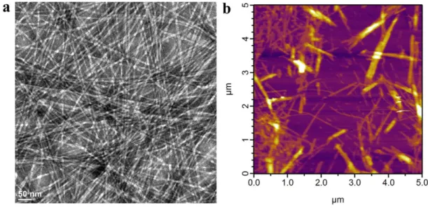
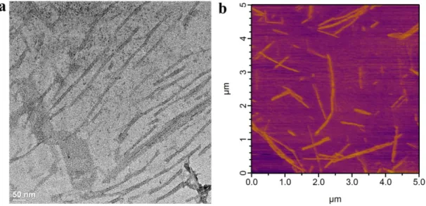
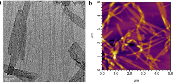
Figure 2.10. Transmission Electron Microscopy (TEM) and Atomic Force Microscopy (AFM) imaging of surfaces coated with HM-PA/K-PA formulation. ... 34
Figure 2.11. Transmission Electron Microscopy (TEM) and Atomic Force Microscopy (AFM) imaging of surfaces coated with SO3-PA/K-PA formulation. ... 34
Figure 2.12. Transmission Electron Microscopy (TEM) and Atomic Force Microscopy (AFM) imaging of surfaces coated with D-PA/K-PA formulation. ... 35
xii
Figure 2.13. Transmission Electron Microscopy (TEM) and Atomic Force
Microscopy (AFM) imaging of surfaces coated with K-PA/Heparin formulation. ... 35 Figure 2.14. Transmission Electron Microscopy (TEM) and Atomic Force
Microscopy (AFM) imaging of surfaces coated with H-PA pH=7 formulation.. ... 36 Figure 2.15. Circular dichroism analysis of heparin-mimetic and control PA
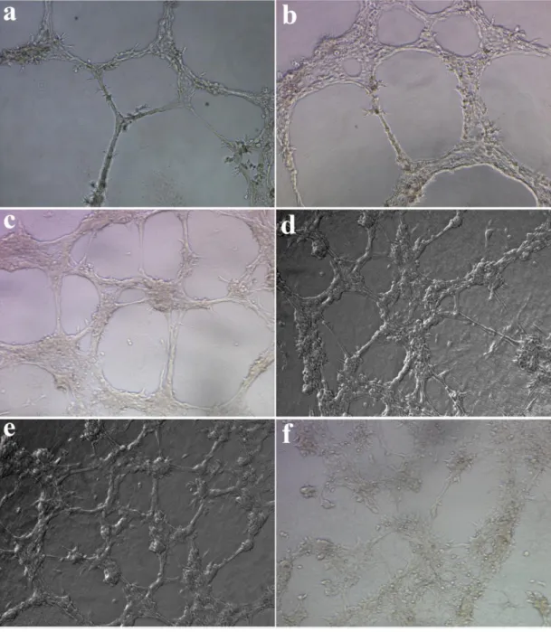
molecules. ... 37 Figure 2.16. Oscillatory rheology measurements of different PA gels. ... 38 Figure 2.17. In vitro angiogenesis assay - Matrigel, HM-PA and D-PA. ... 40 Figure 2.18. In vitro angiogenesis assay - SO3-PA, K-PA/heparin, H-PA and TCP.. 41
Figure 2.19. Measurement of binding constant and thermodynamic parameters in K-PA and heparin interaction by using ITC. ... 43 Figure 2.20. Quantification of lengths of tubes formed by endothelial cells
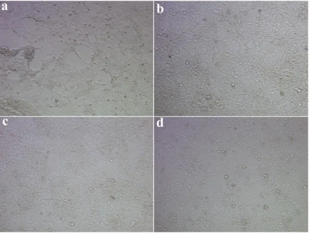
(HUVECs). ... 45 Figure 2.21. Viability of cells cultured on PA nanofiber matrices. ... 46 Figure 2.22. In vitro angiogenic performances of H5V (mouse endothelial cell line) cells on different PA nanofiber matrices. ... 47 Figure 2.23. Proliferation of endothelial cells on different PA nanofiber matrices and tissue culture plate (TCP). ... 48 Figure 2.24. Electron micrographs of endothelial cells on HM-PA nanofiber
scaffolds (a-d) and coverslip (e, f). ... 50 Figure 2.25. Electron micrographs of sprouts formed by endothelial cells on HM-PA nanofiber scaffolds. ... 51 Figure 2.26. Isothermal Titration Calorimetry (ITC) graphs for titration of Heparin-mimetic PA molecules in solution form with VEGF. ... 53 Figure 2.27. Isothermal Titration Calorimetry (ITC) graphs for titration of Heparin-mimetic PA (HM-PA) molecules in nanofiber form – mixed with K-PA – with VEGF.. ... 54 Figure 2.28. Release profile of vascular endothelial growth factor (VEGF) from various PA gels. ... 57 Figure 2.29. Zeta-potential measurements of PA combinations. ... 59 Figure 2.30. Investigation of expression profiles of angiogenic genes in endothelial cells (HUVEC) cultured on PA nanofiber matrices or tissue culture plate. ... 62
xiii
Figure 2.31. Determination of VEGF secretion from endothelial cells (HUVECs) cultured on PA nanofiber matrices or tissue culture plate. ... 63 Figure 2.32. Evaluation of in vivo bioactivity of HM-PA nanofibers by corneal angiogenesis assay. ... 64 Figure 2.33. Quantification of vascularized area in corneal angiogenesis assay. ... 65 Figure 2.34. Effect of HM-PA gel alone or with growth factors on corneal
angiogenesis. ... 66 Figure 2.35. Suggested mechanism for the induction of angiogenesis by bioactive HM-PA nanofibers. ... 77 Figure 3.1. Chemical structure of PA molecules and heparin; illustration of
nanofibers investigated for growth factor binding. ... 83 Figure 3.2. AFM images (5 µm x 5 µm) of PA nanofibers coated onto ELISA plates.. ... 84 Figure 3.3. Measurement of VEGF165 bindingto nanofiber coated surfaces by using
ELISA-based assay.. ... 86 Figure 3.4. Binding levels of different growth factors to HM-PA/K-PA or E-PA/K-PA nanofibers are measured by ELISA-based assay. ... 88 Figure 3.5. TEM images of immunogold stained HGF on HM-PA/K-PA nanofibers.. ... 89 Figure 3.6. EDX analysis of nanofiber aggregates shown in TEM images. ... 90 Figure 3.7. Measurement of affinity between VEGF165 and HM-PA/K-PA nanofibers
by using Isothermal Titration Calorimetry (ITC). ... 92 Figure 3.8. Measurement of affinity between VEGF121 and HM-PA/K-PA nanofibers
by using Isothermal Titration Calorimetry (ITC). ... 93 Figure 3.9. Competition assay between heparin and HM-PA/K-PA nanofibers for growth factor binding. ... 95 Figure 3.10. Competition assay between heparin or chondroitin sulfate (cs) and HM-PA/K-PA nanofibers for FGF-2 binding. ... 96 Figure 3.11. Neurite outgrowth performance of PC-12 cells on NGF treated and NGF-free substrates. ... 98 Figure 3.12. Neurite outgrowth response of PC-12 cells to washing away of unbound NGF from NGF treated substrates. ... 99
xiv
Figure 3.13. Immunostaining of PC-12 cells against β-III-Tubulin and Synaptophysin I on NGF treated surfaces. . ... 101 Figure 4.1. Potential therapeutic applications of CpG oligodeoxynucleotides (ODNs). ... 112 Figure 4.2. Schematic plot of designed virus-like nanostructures and tunability of immune response with these nanostructures. ... 113 Figure 4.3. Chemical representations of K-PA (C12-VVAGK) and P-PA (C12
-PPPGK) molecules used in this study. ... 115 Figure 4.4. LC-MS analysis of synthesized K-PA molecule. ... 116 Figure 4.5. LC-MS analysis of synthesized P-PA molecule. ... 117 Figure 4.6. Circular dichroic (CD) spectra of K-PA and P-PA molecules alone or mixed with immunostimulatory ODN. . ... 118 Figure 4.7. SAXS analysis and model fitting for self-assembled PA/ODN
nanostructures. ... 120 Figure 4.8. SAXS analysis and model fitting for self-assembled PA nanostructures.. ... 123 Figure 4.9. Imaging of PA/ODN nanostructures with TEM and AFM.. ... 124 Figure 4.10. STEM images of self-assembled K-PA/ODN peptide fibers. . ... 125 Figure 4.11. STEM images of self-assembled P-PA/ODN peptide spherical
nanostructures. . ... 126 Figure 4.12. AFM images of self-assembled K-PA/ODN and P-PA/ODN
nanostructures dried on mica and glass surfaces, respectively. . ... 127 Figure 4.13. TEM and STEM images of self-assembled K-PA and P-PA
nanostructures. ... 127 Figure 4.14. Determination of critical ODN/Peptide ratio by using polyacrylamide gel electrophoresis (PAGE). ... 130 Figure 4.15. Zeta Potential measurements of PA/ODN complexes for determination of critical ODN/Peptide ratio. . ... 131 Figure 4.16. Zeta potential values of PA molecules at various concentrations. K-PA shows higher zeta potential values than P-PA at similar concentrations. ... 132 Figure 4.17. Effect of nanostructures on (CpG ODN) dose-dependent IFNγ cytokine production by splenocytes. ... 133
xv
Figure 4.18. Effect of nanostructures on (CpG ODN) dose-dependent IL-6 cytokine production by splenocytes. ... 134 Figure 4.19. Effect of nanostructures on (CpG ODN) dose-dependent IL-12 cytokine production by splenocytes. ... 135 Figure 4.20. Nanostructures shift CpG-induced cytokine secretion profile of
splenocytes. ... 137 Figure 4.21. Effect of nanostructures on CpG-induced surface expression of co-stimulatory molecules. ... 139 Figure 4.22. Morphological changes in splenocytes upon stimulation with
K-PA/ODN, K-PA/cont.ODN, P-K-PA/ODN, P-PA/cont.ODN, ODN alone, media alone. ... 141 Figure 4.23. Uptake profile of FITC-ODN alone or bound with nanostructures into TLR9-expressing cell subsets in splenocytes... 143 Figure 4.24. Resistance of ODNs to DNAse were investigated with polyacrylamide gel electrophoresis (PAGE). ... 146 Figure 4.25. Time-dependent degradation of ODN in different formulations, plotted according to calculated band intensities... 147
xvi
LIST OF TABLES
Table 1.1. Effect of “K/B” and “D/A” ODN on the Immunogenicity of Engerix B. 19 Table 2.1. Nanofiber size measurements of PA gels. ... 36 Table 2.2. Loss tangents of PA gels (G''/ G'). ... 39 Table 2.3. Binding constants of VEGF – PA interaction at 30 °C measured by ITC. 55 Table 2.4. Gene expression profiles in endothelial cells cultured on PA nanofibers. 61 Table 2.5. Investigated genes taking role in angiogenesis process. ... 75 Table 3.1. Growth factors used in this study... 82 Table 4.1. Structural results obtained from fits to the SAXS data of K-PA/ODN complexes with elliptical cylinder model. ... 121 Table 4.2. Structural results obtained from fits to the SAXS data of P-PA/ODN complexes with oblate core shell sphere model. ... 121 Table 4.3. Structural results obtained from fits to the SAXS data of K-PA with elliptical cylinder model. ... 122 Table 4.4. Structural results obtained from fits to the SAXS data of P-PA with oblate core shell sphere model. ... 122
1
CHAPTER 1
2
1. INTRODUCTION
Biomaterials could provide solutions to various health problems. Today, as a result of economic and technological development, people live longer; communication and transportation are in unprecedented levels. However, increased life expectancy brought elevated incidence rate of chronic and degenerative diseases such as organ failures, cancer, neurodegenerative diseases, and chronic wounds, which cause extensive tissue damage. Also, enhanced communication and transportation cause pathogens to breach barriers against their intercontinental spread, increasing risk of pandemics. To find cures to these diseases, we need a paradigm shift in our approach. Interdisciplinary work of biology and materials science is promising in this regard. Recent achievements in materials science allow us to design materials with the purpose of curing pathophysiologies. Materials relevant to biology can be controlled, functionalized and characterized at nanoscale. Nanofibrous hydrogels for supporting cell adhesion and survival in damaged areas can be obtained with fiber sizes of a few nanometers. Drug delivery vehicles from polymeric nanoparticles and liposomes to gold nanoparticles can be obtained with different sizes and functionalized depending on specific purposes. In this thesis, we used self-assembling peptide amphiphile systems to design novel materials for tissue engineering and modulating immune response.
1.1. Self-assembling peptide amphiphile molecules
Peptide amphiphile molecules are composed of a hydrophilic peptide part and a hydrophobic alkyl tail covalently bound to each other (Figure 1.1a).1 Charged aminoacids are included in peptide part, which besides increasing solubility of molecule, allows controlling the self-assembly process. Upon neutralization of these charges with oppositely charged ions, pH change, macromolecules or another peptide amphiphile, they self-assemble into higher order nanostructures such as nanofibers and nanospheres through collapse of hydrophobic part inward and peptide part outward (Figure 1.1b).2 Nanofibers produced by this way are typically 5-15 nm in
3
diameter, which is fairly similar in size to fibers comprising natural extracellular matrices (ECM), therefore important to mimick ECM for regenerative medicine (Figure 1.1d.).
Figure 1.1. A peptide amphiphile structure. a. Chemical structure of a representative
PA with four rationally designed modules. b. Molecular graphics illustration of a PA molecule with a bioactive epitope and its self-assembly into nanofibers. Note that bioactive epitopes are exposed to surrounding media in aqueous solution. c. Scanning electron micrograph of the PA nanofiber network formed by adding cell media (DMEM) to the PA aqueous solution. d. Transmission electron micrograph of the PA nanofibers. (Reproduced with permission from ref. 3, copyright © 2010 John Wiley & Sons, Inc.).3
Peptide nanostructures are versatile materials that are amiable to engineering for presenting biofunctional ligands (Figure 1.1a). Nanoscale structure of fibers provide high surface area to volume ratio that allows them carry epitopes (biofunctional ligands) with high density due and make them more suitable for guiding cellular physiology. Entanglements of these nanofibers emerge as macroscopic self-supporting gels at adequate concentrations (Figure 1.1c, 1.2). Gel formation can be controlled via neutralization of charges via pH change or mixing with oppositely charged molecules. This makes PA gels suitable for encapsulation of cells, growth factors or small molecules in 3D environment for in vitro and in vivo applications.
4
Since gel formation can be controlled externally, they can be applied also as an injectable matrix to replace native extracellular matrix in damaged tissues via less invasive methods. Besides serving as scaffolds, these nanofibers provide an environment to cells invading into matrix where they can be manipulated with peptide signals exposed to aqueous solution from nanofibers, such as shown in Figure 1.1b.
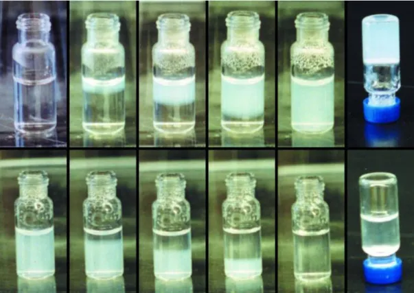
Figure 1.2. Time-dependent PA gel formation and reversion of process via changing
pH. In upper part of figure, PA molecule is dissolved in water at a concentration of 0.5% by weight at pH 8 and is exposed to HCl vapor. As the acid diffused into the solution a gel phase is formed, which self-supports upon inversion (far right). In lower part of figure, the same gel is treated with NH4OH vapor, which increases the
pH and disassembles the gel, returning it to a fully dissolved solution (Reproduced with permission from ref. 2, copyright © 2002 National Academy of Sciences, U.S.A.).2
5
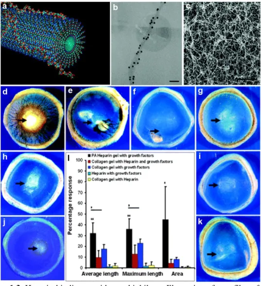
Peptide part can be engineered to carry epitopes from active domains of proteins such as growth factors and ECM proteins or to bind high molecular weight molecules such as heparin to generate functional nanofibers. All these properties make them very powerful tools for drug delivery and regenerative medicine. Peptide amphiphiles with heparin binding epitopes has been previously shown to exhibit strong binding to heparin.4-5 Heparin binding conferred these nanofibers a strong affinity to angiogenic growth factors. Probably due to the mentioned feature, these nanofiber gels induced in vitro and in vivo vascularization better than control PA gels and standard scaffolds such as collagen (Figure 1.3).4-5
PA nanofibers with laminin-derived epitope (IKVAV) induced neuronal differentiation of NSCs better than laminin, probably due to increased density of bioactive epitope on nanofibers.6 PAs carrying epitopes for TGF-β (transforming growth factor) binding derived from phage display library induced in vivo cartilage regeneration.7 In addition, several studies for tissue regeneration and drug delivery by using PA molecules were published in recent years making PA nanostructures a promising platform for regenerative medicine applications.
6
Figure 1.3. Heparin-binding peptide amphiphile. a. Illustration of nanofibers formed
by mixing heparin and PA. Heparin is presented by nanofibers into aqueous solution
b. TEM image showing bundle of nanofibers bound to heparin-gold nanoparticles
(black dots) (scale bar = 40 nm) c. SEM image of nanofiber network formed by heparin and PA (scale bar = 2 µm). d-k. In vivo angiogenesis assay. Rat cornea photographs 10 days after the placement of various materials at the site indicated by the black arrow. d. Heparin/PA nanofiber networks with growth factors induced extensive neovascularization. Collagen with heparin and growth factors (e) and collagen with growth factors (f) show some neovascularization. Heparin with growth factors (g), collagen with heparin (h), PA with growth factors (I), Heparin/PA without growth factors (j) and growth factors alone (k) showed little to no neovascularization. The bar graph (l) contains values for the average and maximum length of new blood vessels and the area of corneal neovascularization. A 100% value in the area measurement indicates that the cornea is completely covered, and a 100% value in the length parameters indicates that the new vessels are as long as the diameter of the cornea (bars are 95% confidence levels, * p < 0.05 when Heparin/PA gel was compared to collagen gel with growth factors, ** p < 0.005 when Heparin/PA gel with growth factors was compared to all of the other controls). (Reproduced with permission from ref. 5, copyright © 2006 ACS).5
7
1.2. Materials for tissue engineering
Organ failures and tissue damages require organ and tissue replacement, however organ donors are in scarcity. Another way is to unleash regenerative potential of our body, which naturally occurs during development of fetus. This requires the understanding of type of signals that are sensed by cells in their extracellular matrix and coax them into regenerative pathway. While biology provides us with this type of knowledge, regenerative medicine aims to find ways for presenting these cues to cells in an appropriate way. Tissue engineers exploit biomaterials decorated with these signals to direct cells to proliferate, differentiate or organize into desired tissue structures such as inducing endothelial cells to form blood vessels. Extracellular matrix (ECM) of cells has long been deemed as support material for cells, so main features sought in biomaterial scaffolds were mechanical properties.8 Physical properties such as porosity and stiffness have been emphasized extensively in material design for tissue engineering in the infancy of field. Moreover, these materials were expected to be `inert` - having minimal toxicity and immunogenicity.9 However, lacking necessary biochemical cues for instructing cells, effect of these materials in tissue engineering were limited. Aim of material design for tissue engineering in the current paradigm is to recapitulate biophysical and biochemical features of extracellular matrix, where cells live in their natural environment, to achieve instructing cells for specific destiny.10 However, financial considerations limit to project all complexity in ECM to designed biomaterial.11 Natural macromolecules in ECM are used extensively as biomaterials for tissue engineering since biological cues are inherent in them.10 However, these materials have also inherent problems regarding pathogen transmission, immunogenicity and purification. Thus, there is a need to design materials with similar functional sophistication as ECM but with simpler structural complexity. Growing understanding of principles of how cells recognize biophysical and biochemical signals in their environment, integrate them at the level of gene expression and make appropriate decisions (Figure 1.4) will pave the way to design synthetic – thus more defined - materials for tissue engineering.
8
Figure 1.4. Coordination of complex physiological processes via signals sensed by
cells. Cells recognize various physical and chemical signals in their environment through receptors on their membrane. This recognition is converted into signaling pathways, which eventually ends up with expression of different genes. Concerted actions of these genes influence cellular fate and induce various processes such as replication, migration or apoptosis. These cellular performances determine physiological processes at tissue level (Reproduced wıth permission from ref. 10, copyright © 2005 Macmillan Publishers Ltd.).10
9
What are those principles? Which distinguishable aspects of extracellular matrix are sensed by cells and what type of behavioral alterations in cells are observed as we change them? Immense amount of studies were published explaining how cells respond to biophysical signals such as stiffness, topography and size of individual fibers in ECM-like network. Stiffness of environment have shown to be a determining factor for mesenchymal stem cell (MSC) differentiation: with the increasing order of stiffness, MSCs were committed to neurogenic, myogenic and osteogenic pathways.12 Size of fibers forming scaffolds is another cue affecting cellular behavior. Natural ECM is formed by network collagen fibrils of size at nanoscale (50-500 nm).13-14 Smaller fiber size has larger surface area which might act synergistically with ligands carried on fibers. Endothelial cells showed more elongated phenotype, migration and capillary-like structures on micro/nano-fiber scaffold than one without nanofiber network.15 Nanofibers allowed stretching of endothelial cells between microfibers, which is known to be critical for their responsiveness to growth factors. Neural stem cells differentiated into different lineages according to size of fibers of scaffold they were cultured on.16 Considering available knowledge, scaffold stiffness, fiber size, porosity and topography can be adjusted according to purpose.
Recapitulating biochemical signals of natural ECM is incomparably difficult and expensive, when one considers complexity of network of these signals in ECM. Activation and performance of physiological mechanisms such as angiogenesis or neural regeneration depend on concerted act of numerous biological signals. Among these signals there are growth factors, cytokines, signaling epitopes on various structural proteins such as laminin and fibronectin, and glycosaminoglycans which bind and regulate activity of growth factors. Besides mere existence, their spatial and temporal presentation is also critical for effective regeneration. Since introducing all these ingredients into biomaterial scaffold and regulating their release from scaffolds spatiotemporally is an unattainable task, tissue engineers aim to identify critical elements in this network of biochemical signals, perturbations of which will induce regenerative pathway or desired bioactivity. In this context, conjugating integrin-binding epitopes of structural proteins, such RGD or IKVAV peptides, promote cell
10
adhesion to matrix.17-20 Another strategy is delivering critical growth factors in biomaterial scaffolds. Dose of growth factors sensed by cells is a critical issue, since higher doses of growth factors might cause unwanted effects.21 Physical encapsulation of growth factors may not be enough for slowing their release adequately, so they are conjugated to scaffolds via either non-covalent or covalent bonds. For this purpose, heparin (a highly sulfated glycoasminoglycan) has been conjugated to scaffolds, such as alginate and collagen, for binding to growth factors non-covalently and enhance their bioactivity.22-23 This binding slowed growth factor release from scaffold and improved potency of scaffold to induce angiogenesis. Other polymers carrying sulfate groups as heparin or affinity binding peptides also exhibited increased growth factor binding and performance regarding bioactivity.24-27 Chemical functional groups as biochemical signals are probably an irreducible form of complexity in extracellular matrix. Amazingly, it seems that they were enough to induce mesenchymal stem cell differentiation and by changing functional groups researchers were able to control cell fate.28 These functional groups are inspired from chemical structure of extracellular matrix of various tissues. Although exact mechanism was not clear, each functional group induced stem cell differentiation into the same tissue it was inspired from. For example, phosphates (from hydroxyapatites in bone matrix) induced differentiation into osteocytes.
Both biochemical signals and biophysical signals work in the context of each other in nature. Number of ligands bound by integrins was observed to be a function of matrix stiffness in 3d scaffolds.29 Optimal integrin binding by RGD ligands was responsible actually for induction of osteogenesis in mesenchymal stem cells at optimal stiffness. This shows that rather than focusing on biochemical signals or biophysical ones, using the right combination of simple signals from both might work synergistically and have tremendous effects on cellular behavior.
In Chapter 3 and 4 of this thesis, scaffold materials formed from networks of entangled nanofibers are described. We rendered building blocks of these nanofibers to carry chemical functional groups, which also exist on sulfated glycosaminoglycans (GAG) (please look to 1.2.1.). These building blocks are peptide amphiphile
11
molecules which form nanofibers when mixed with oppositely charged molecules via self-assembly. Nanofibers formed this way present chemical functional groups into environment just as GAG polymers. In chapter 3, I demonstrate that right composition of functional groups - sulfonate, hydroxyl and carboxylate together - render nanofibers avid to vascular endothelial growth factor (VEGF) and effective at inducing angiogenesis – new blood vessel formation. There, I also show that when this combination of functional groups lack one or two of these groups, functionality is severely impaired. In chapter 4, study on interaction of these nanofibers with growth factors is described. Functional groups on nanofibers made them affine to many growth factors such as VEGF, HGF and FGF-2, when compared to control nanofibers, which don’t have the same composition of chemical groups. Also, interestingly, they showed affinity to the same domain of growth factors where heparin binds, which is important for bioactivity of growth factors.
1.2.1. Heparin and heparan sulfate glycosaminoglycans.
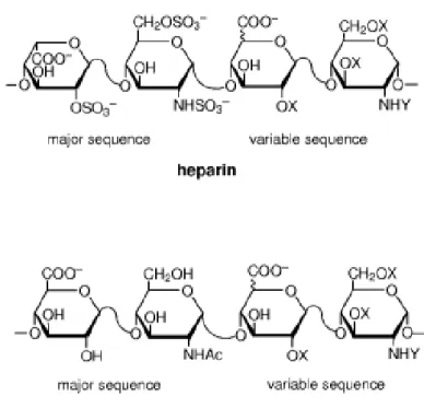
Glycosaminoglycans (GAGs) are polymers of disaccharides, carrying chemical functional groups on them (Figure 1.5). Biological roles of GAGs are not completely identified, however, it is obvious that most of them is related with their remarkable capability to bind to many proteins.30 Heparin and heparan sulfates are members of GAGs, which has been studied extensively for their property of binding various proteins and modulate their activity. Heparin is found in mast cell and basophilic granules and serve as anti-coagulant.30 It has the highest negative charge density of any known biological macromolecule, thanks to the sulfate and carboxylic acid groups found in its structure.31 Average number of sulfates per disaccharide is 2.7 (Figure 1.5).30 Heparan sulfates have similar structure but less negative charge density (average sulfate groups per disaccharide is less than 1).30 However, disaccharide units are more variable in heparan sulfates, making them more heteregenous than heparin in terms of domains.31 Different domains in heparan sulfates show different levels of sulfation probably allowing them to perform more complex functions (Figure 1.5). Highly sulfated domains are assumed to take role in
12
protein binding. Heparan sulfates are found in extracellular matrix and membrane of cells. They bind to growth factors there and protect them from enzymatic degradation, provide a reservoir of growth factors to cells and assist growth factors in their interaction with cognate receptors. Heparin is extensively used in tissue engineering because of high degree of sulfation of its monomers, which increase ability of scaffolds with heparin to bind to growth factors.
Figure 1.5. Chemical structure of heparin and heparan sulfates showing major and
minor disaccharide repeating units. (X=H or SO3
-, Y=Ac-, SO3
-, or H) (Reproduced with permission from ref. 31, copyright © 2002 John Wiley & Sons, Inc.)31
.
1.3. Materials for engineering immune response
Last part of this thesis is about materials for directing immune activity and developing vaccines against infectious diseases. Conventional vaccinology relies on introducing inactivated or live-attenuated form of pathogens into patients in an attempt to educate immune system about how to fight with the active pathogen. Basics of this strategy are fairly unchanged since invention of vaccination (“vacca”
13
in Latin means cow) by remarkable observation of Edward Jenner that infection with cowpox provides immunity against smallpox, in 1796. Although this strategy generates successful immune response against pathogens, it has several disadvantages. First of all, in cases of pandemic, mass production could be hampered by low growth of pathogens or scarcity of resources. Second but not least, using pathogen itself is not a defined formulation, so brings unwanted side effects and risk of becoming virulent of pathogen. For this reason, we need rational design of vaccines, which will drive immune response towards desired context (considering type of cytokines and costimulatory molecules expressed, type of cells activated) and induce long-lasting immune response, without compromising safety.32 However, this is challenging because our knowledge of these two subjects is not sufficient.33
1.3.1. Pathogen-specific immune context
One question is which type of immune response is required to protect an individual from each pathogen. Immune system has evolved to protect organism against diverse pathogens, while being tolerant to self. Having a similar level of sophistication allows immune system to solve this problem (Figure 1.6). Pathogens that are able to pass first-line barriers such as skin (e.g. in cases of tissue damage) encounter with innate immune system. Innate immune system cells inspect pathogen entry sites of body such as skin or mucosal surfaces and destroy pathogens through phagocytosis or secreting antimicrobial substances upon recognition.
14
Figure 1.6. Complexity of immune system, which is evolved to distinguish pathogen
from self and to provide immune context relevant to nature of pathogens. Dendritic cells (DC) distinguishes between foreign and self antigens according to microenvironmental signals. Along with other innate immune cells, their response to these signals determines the outcome of antigen recognition by T and B cells. a. Dendritic cells recognize immunogenic signals from infected or immunized, dying cells through danger receptors on them (TLRs, CLRs, NLRs, RLRs, SRs) and tolerogenic signals from dying self-cells or cellular debris generated by homeostatic turnover; these produce a continuous spectrum of output responses ranging from strong induction of effector-phase immunity to strong induction of tolerance, with the exact outcome determined by the integration of inputs by the dendritic cell. In response to these ‘danger’ or tolerizing signals, dendritic cells (and other innate cells) create the immunological context for antigen recognition by secreting cytokines, expressing diverse adhesive, co-stimulatory or regulatory receptors that provide cues to responding lymphocytes. b. According to mentioned cues, B cells can undergo somatic hypermutation, become short-lived plasmablasts, or differentiate into long-lived memory B cells or plasma cells while T cells can differentiate into effector cells or memory cells with distinct homing and functional capacities; effector cells can have diverse functions (Th1, Th2, Th17 and so on) depending on the context set by dendritic cells. Regulatory feedback loops are engaged even in highly inflammatory contexts, as part of the natural control system regulating immunity, and primed effector cells can be driven to anergic/exhausted states similar to tolerance at later stages of an immune response. c. Peripheral tolerance is maintained by a distinct set of signals: In tolerogenic contexts, T cells are driven into several different states of non-responsiveness that prevent effector responses against self or harmless environmental antigens. (Reproduced with permission from ref. 33, copyright © 2013 Macmillan Publishers Ltd.).33
15
Pathogen recognition by innate immune cells is maintained by germ-line encoded receptors, which do not change during the lifetime of organism, differing from adaptive immune cells. These receptors, collectively known as pattern recognition receptors (PRR), recognize certain common patterns from pathogens, such as peptidoglycan molecules in bacterial cell wall or unmethylated CpG (or CG, denoting cytosine and guanine) motives from viral/bacterial DNA.34-35 PRRs include several family of secreted (mannose-binding lectin), transmembrane or cytosolic receptors.36 Most studied and known receptor family among PRRs are Toll-like receptors (TLR). Toll-like receptors can be at cell-membrane (those recognizing surface features such as peptidoglycan or LPS layer of bacterial cell wall and membrane) or endosomal membrane (those recognizing microbial nucleic acids). Binding of pathogenic patterns to PRRs shape adaptive immune responses.36 This happens through activation of various signaling pathways which end up with expression of cytokines and surface receptors called as co-stimulatory molecules. Cytokines are protein molecules binding to their receptors on target cells and induce signalling pathway. Co-stimulatory molecules are expressed by antigen-presenting cells and required for activation of adaptive immune cells specific for antigen. Although these signals are necessary, neither of them is sufficient to induce adaptive immune response. However, TLR-induction is known to be sufficient to provide all factors for initiation of robust adaptive immune response.36 Thus, besides forming another line of defense to clear infection, innate immune cells also controls the activation, types and duration of the adaptive immune response.36
Adaptive immune system fights with infections with two main types of immune cells – B cells and T cells. Both of cell populations have vast repertoire of antigen receptors for almost every possible antigen in the environment. These receptors are generated through random arrangement of genes, which gives rise to receptor for specific antigen on every cell. Adaptive immune response also provides memory formation about pathogens after clearing infection, through memory cells. These cells re-induce adaptive immune response after encountering antigens later. Antigen-presenting cells (APC), which are mainly dendritic cells, form link between innate
16
and adaptive immune system. They process and present antigens, which they recognized as foreign, on their surface to T-cells. Cytokines and co-stimulatory molecules expressed by APCs accompany to presentation of antigen at immunological synapse (Figure 1.7.).37 T-cells integrate these signals and decide on nature of ensuing immune response.33
B-cells are activated by T-cells and accompanying cytokine signals. They secrete antibodies – receptors on their surface formed by combinatorial mechanism, as an effector function. These antibodies detect antigens in body fluid, so this type of response is called humoral response (Figure 1.6). Antibody binding renders toxins ineffective, and pathogens vulnerable to phagocytosis by macrophages. This is why this type of immune response is especially effective on extracellular pathogens. However, some pathogens such as viruses or mycobacterium reside in cells. Cell-mediated immune response is required to clear this type of infections. Effector function of cell-mediated immune response is activated by CD8+ cytotoxic T-cells, which induce death mechanism in cells expressing particular antigen. T-cell receptors, contrasting to B-cell ones, are not secreted, but interact with MHC molecules presented by antigen-presenting cells – mainly dendritic cells.
To summarize, adaptive immune response is antigen-specific immune response against pathogens, providing effective clearance from and memory formation about pathogens. Nature of adaptive immune response to antigen is determined by cytokines, co-stimulatory molecules and possibly other factors expressed by antigen-presenting cells (Figure 1.7).33 These molecules are expressed according interaction of pathogen with innate immune cells through binding of pathogen-associated molecular patterns – LPS layer, CpG DNA - to pathogen recognition receptors such as TLRs.32-33 Understanding these mechanisms might allow us to trigger them upon purpose with synthetic and defined formulations, rather than pathogen itself.
17
Figure 1.7. Illustration of key receptor-ligand interactions at the immunological
synapse formed between an antigen presenting cell and a T-cell during T-cell activation. Profile of cytokines and co-stimulatory molecules binding to their target receptors educate adaptive immune system about nature of infection, which allows adaptive immune cells to elicit an appropriate immune response. (Reproduced with permission from ref. 37, copyright © 2012 John Wiley & Sons, Inc.)37
1.3.2. How to drive immune response to desired context?
Another barrier to rationalize vaccine design is our insufficient understanding of how we can drive immune response to desired context such as balance between effector/memory cells, or cell-mediated or humoral immunity.33 Antigens themselves do not generate immune response, so molecules called adjuvants are added into vaccine formulations, which trigger immune system against antigen. Success of these adjuvants are based largely on antibodies made by B-cells.38 This type of immune response is not competent against intracellular pathogens (e.g. tuberculosis), which can be cleared by the action of T-cells and macrophages (cell-mediated immunity). We need tools that would allow us to tune activity, magnitude and duration of various modules of immune system, such as cytotoxic T-cell activity or antibody secretion by B-cells. These tools could be found among signatures of pathogens that are recognized by immune system. Mentioned signatures can be classified as
18
chemical ones, which are known in literature as pathogen-associated molecular patterns (PAMP), and physical ones such as size and shape of pathogen.
PAMPs that bind PRRs (Pathogen Recognition Receptors) are promising as vaccine adjuvants, since they induce innate immune cells, which eventually shape adaptive immune response. Existence of TLR ligands in phagocytosed antigenic cargo is necessary for presentation of antigens with MHCII on the surface of dendritic cells.39 Besides inducing antigen-presentation, profile of cytokines and co-stimulatory molecules induced by different PAMPs shape immune response according to pathogen to be destroyed.
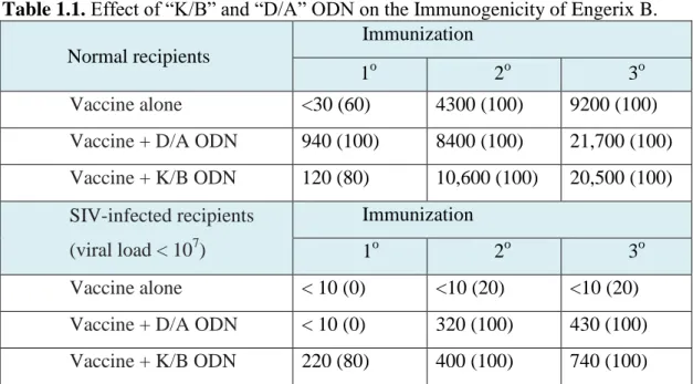
In this context, CpG ODNs have been shown to act as safe adjuvants and drive immune response to cell-mediated immunity (Table 1.1). These are oligodeoxynucleotides with cytosine-guanine motives, where cytosine is unmethylated.40 These motives are less frequent in vertebrate DNA than bacterial DNA, also higly methylated.40 Mammalian immune system recognizes bacterial/viral DNA or CpG ODNs in endosomes of certain immune cells like B-cells and plasmacytoid dendritic cells via TLR9 receptor.41 Binding of CpG ODN to TLR9 induce signaling pathways which end up with synthesis of pro-inflammatory cytokines, interferons and co-stimulatory molecules and maturation of dendritic cells.
Interactions of the immune system with pathogens are shaped not only by danger signals, but also by the physical nature of microbes, which are biological microparticles and nanoparticles.33 Soluble exogenous antigens are not displayed by cross-presentation (MHCI), while pathogenic or particlulate antigens can be displayed.37 Designing materials presenting these signatures to immune system on the same physical entity as pathogens, would allow us to exploit synergism between chemical and physical signals. Understanding effect of each signal on immune system will allow us to shape immune response by rationally changing concentration of each signal. While biochemical signals on pathogens are studied deeply, there is a lack of knowledge on how these perform in the context of physical signals such as size and shape. In Chapter 4, we studied how pathogenic CpG DNA signal acts in the context of shape of carrier. Our findings show that shape alters immune response to
19
CpG DNA, where nanofiber delivery is more relevant to induce cell-mediated immunity.
Table 1.1. Effect of “K/B” and “D/A” ODN on the Immunogenicity of Engerix B.
Normal recipients
Immunization
1o 2o 3o
Vaccine alone <30 (60) 4300 (100) 9200 (100)
Vaccine + D/A ODN 940 (100) 8400 (100) 21,700 (100)
Vaccine + K/B ODN 120 (80) 10,600 (100) 20,500 (100) SIV-infected recipients (viral load < 107) Immunization 1o 2o 3o Vaccine alone < 10 (0) <10 (20) <10 (20)
Vaccine + D/A ODN < 10 (0) 320 (100) 430 (100)
Vaccine + K/B ODN 220 (80) 400 (100) 740 (100)
Rhesus macaques (5–6/group) were immunized with 500 μl of Engerix B vaccine plus 300 μg of “K/B” or “D/A” ODN in alum. Serum anti-HepB Ab titers were monitored by ELISA. Average titers, and percent of animals with protective titers (in parenthesis) are shown. Note that the average response after both primary and secondary immunization was significantly higher in groups immunized with CpG ODN plus Engerix B vs. vaccine alone (Reproduced with permission from ref.42, copyright © 2009 Elsevier).42
20
CHAPTER 2
2. DESIGN OF HEPARIN-MIMETIC PEPTIDE NANOFIBERS
FOR INDUCTION OF ANGIOGENESIS.
This work is partially described in the following publication:
Mammadov R., Mammadov B., Toksoz S., Aydin B., Yagci R., Tekinay A.B. and Guler M.O. Biomacromolecules, 2011, (10), pp 3508–3519.43
21
2.1. Objective
Regeneration of tissues after damage requires formation of blood vessels for survival and performance of cells migrating into damaged area. Designing materials decorated with biological signals for induction of angiogenesis would be useful for tissue engineering purposes. However, recapitulating all the signals regulating blood vessel formation is not feasible. Simpler and still effective approaches in mimicking microenvironment of angiogenesis (new blood vessel formation) are required. Sulfated glycosaminoglycans (GAG), which are an essential part of basement membrane, a specialized extracellular matrix of endothelial cells, bind to growth factors critical for angiogenesis and regulate their activity. Chemical functional groups and their distribution on GAGs, especially on heparan sulfates, are known to be critical for growth factor binding and induction of angiogenesis. In this study, our objective was to design scaffold material with similar fiber size to natural ECM and GAG-mimicking chemical functional groups on fibers. We aimed to identify appropriate composition of simple functional groups on nanoscale fibers that would be sufficient to induce angiogenesis.
2.2. Introduction
Regenerative medicine studies offer promising therapeutic approaches for the repair of damaged tissues. Induction of angiogenesis is an important mechanism for tissue repair.10 The capillaries can only deliver oxygen and nutrients to the cells that are located at a distance of up to 200 μm, and thus angiogenesis is required for cells further away during new tissue formation.44 Angiogenesis is triggered by the integration of various neovascularization signals by endothelial cells, which in turn differentiate to form new capillaries. Structural proteins of the extracellular matrix (ECM) (laminin, collagen, etc.), growth factors (VEGF, FGF-2, etc.), and glycosaminoglycans (heparan sulfate, etc.) make up a framework of neovascularization signals for endothelial cells.45 Understanding the interactions between these biomolecules and endothelial cells and their roles in the regulation of
22
angiogenic processes paves the way to design effective synthetic biomaterials for induction of new blood vessel formation. Conventional tissue engineering strategies utilized some of the biological molecules mentioned above to provide bioactivity for promoting angiogenesis46 because synthetic biomolecules that have been produced so far lacked the ability to mimic the functions of all of these biological components. The main motivation for developing new synthetic ECM mimicking biomaterials is to minimize utilization of the above-mentioned natural biomacromolecules exogenously with the aim of reducing cost, preventing batch-to-batch variation, and avoiding biological contamination. Therefore, designing smart biomaterials that can harness endogenous factors for desired bioactivity is essential.
Among the basic components of the signaling framework for endothelial cells, heparan-sulfate proteoglycans (HSPGs) bind to angiogenesis promoting growth factors and their receptors through heparan sulfate chains and induce growth factor signaling (Figure 2.1.).47-50 Mice lacking heparan sulfate chain on HSPG molecule reveal defective angiogenesis and wound healing.51 Binding of growth factors to HSPGs, which strictly depends on the distribution of functional groups, such as sulfate, hydroxyl and carboxyl groups, on heparan sulfate chains, protects growth factors from degradation, increases local concentration of growth factors, and enhances growth factor-receptor interactions, which are important for long-term stimulation of signaling pathways in endothelial cells.8, 50, 52-53 Using glycosaminoglycans (e.g., heparin) within tissue engineering scaffolds has been shown to enhance angiogenesis significantly while reducing the need for exogenous growth factors at in vivo studies.5 A peptide amphiphile (PA) scaffold for angiogenesis was previously developed by mixing heparin-binding PA molecule and heparin.5 Heparin-binding PA molecule allowed growth factor binding and helped formation of various functional tissues.5, 54-55 However, being an animal-derived product, utilization of heparin in tissue engineering systems might have potential side effects (e.g., immune reactions).56 Designing heparin mimetic biomaterials will have high impacts in cellular therapy and regenerative medicine because they will enable us to avoid the use of heparin while minimizing the use of exogenous growth factors. Recent research efforts have focused on developing new scaffold materials with
23
proper functional groups that are sufficient to induce the desired physiological response in vitro without any need for growth factors or any other supplements.28
Figure 2.1. Co-receptor function of HSPG in VEGF signalling. a. VEGF receptor –
VEGFR2 is unable for signal transduction when cells lack HSPG. b. HSPG (GAG side chains are shown in blue and protein part in green) expressed on endothelial cells are engaged in the VEGF/VEGFR signaling complex and may affect signaling quantitatively (by stabilizing the complex) and qualitatively (by allowing transduction of signaling pathway not induced in the absence of coreceptors. c. Presentation of HSPGs in trans (i.e. by another cell) leads to further stabilization of the VEGF/VEGFR signaling complex, and prolonged signal transduction (red activity arrows) (Reproduced with permission from ref. 57, copyright © 2008 Springer Science+Business Media, LLC).57
The addition of functional groups inspired by heparin on peptide sequences and polymers has also been previously shown to enhance growth factor binding capacity.24, 26, 58-60 For example, sulfated alginate polymers gained growth factor binding capability and induced in vivo angiogenesis significantly better than
24
nonsulfated alginate in the presence of growth factors.24-25 Considering these findings, we hypothesized that chemical functional groups from heparan sulfates on peptide nanofibers, will recapitulate their function. As a result, heparin-mimetic PA nanofiber gel would bring together two distinct signal in ECM – biophysical signal of nanofiber network mimicking nanofibrous matrix of ECM and biochemical signal of chemical functional groups on heparan sulfates. Heparin-mimetic PA molecule functionalized with bioactive groups was designed and synthesized for mimicking functionality of heparan sulfates in ECM. The heparin-mimetic PA molecules self-assemble to form nanofibers with ability to bind to growth factors and to promote angiogenesis without the need for addition of exogenous heparin or growth factors. This chapter demonstrates that nanostructures with bioactive chemical groups inspired from biological macromolecules can be used to activate biological machinery for regenerative medicine applications.11, 28
2.3. Results and Discussion
To mimic natural extracellular environment that induces angiogenesis, we designed PA molecules which carry chemical features of heparan sulfate molecules in ECM to enable enhanced functioning of the growth factors that are crucial for angiogenesis. Heparan sulfates are sugar polymers with chemical functional groups on these sugar units. Key functional groups in heparan sulfate polymer chain are highlighted in Figure 2.2 for heparin – clinically used glycosaminoglycan molecule with similar structure to heparan sulfate. These functional groups are carboxylic acid (-COOH), hydroxyl (-OH), sulfate (-SO4), sulfonate and sulfamate or N-linked sulfonate
(-N-SO3). To assess the importance of these functional groups during angiogenesis
process, several PA molecules were designed that carry from three to zero of these functional groups (Figure 2.2). Heparin-mimetic PA (HM-PA after here) molecule is decorated with carboxylic acid, hydroxyl, and sulfonate groups. Carboxylic acid and hydroxyl groups are added through coupling aspartic acid/glutamic acid and serine amino acids (side chains of these amino acids), respectively. Sulfonate group is added through coupling sulfobenzoic acid to side chain of lysine amino acid.
25
Figure 2.2. Chemical structures of heparin and designed peptide amphiphiles.
Functional groups inspired from heparin are colored. Heparin-mimetic PA molecule SO3-PA, D-PA and H-PA carries 3, 2, 1 and 0 functional groups from heparin,
26
The other molecules have less functional groups than HM-PA: SO3-PA, has
sulfonate and carboxylic acid groups, D-PA only carboxylic acid groups and H-PA no functional groups from heparin. H-PA was designed to neutralize and self-assemble into nanofibers at physiological pH to control the effect of a nonbioactive PA gel during angiogenesis. Heparin was mixed with K-PA (K-PA/Heparin) to observe the effect of heparin on bioactivity of the PA gel.
All synthesized PA molecules were purified with High-Performance Liquid Chromatography (HPLC) and analyzed with liquid chromatography-mass spectrometry (LC-MS). In LC analysis, molecules were passed from hydrophobic stationary phase (c18 alkyl tails covalently bound to silica particles), which interacts with alkyl tails of PA molecules. By running gradient from an aqueous phase (water) to organic phase (acetonitrile), PA molecules were eluded according to hydrophilicity and detected with UV detector (at 220 nm wavelength). Indeed, elution time points given in LC chromatograms (Figures 2.3b – 2.7b) were consistent with hydrophilicity of molecules. More hydrophilic ones are eluded at earlier time point, such as HM-PA with the highest number of charged groups eluded at the earliest time point, while K-PA with the lowest number of charged group eluded at the latest time point. Mass spectra of peaks obtained in LC chromatograms indicated that all synthesized PA molecules have similar masses to the expected ones (Figures 2.3c – 2.7c). These purified molecules were used in further studies.