Digenic
DUOX1 and DUOX2 Mutations in Cases With
Congenital Hypothyroidism
Zehra Aycan,1* Hakan Cangul,2* Marina Muzza,3 Veysel N. Bas,1 Laura Fugazzola,4,5 V. Krishna Chatterjee,6 Luca Persani,5,7 and Nadia Schoenmakers6
1Division of Paediatric Endocrinology, Dr. Sami Ulus Woman Health and Children Research Hospital, 06080 Ankara, Turkey;2Department of Medical Genetics, Istanbul Medipol University, International School of Medicine, 34810 Istanbul, Turkey;3Endocrine Unit, Fondazione Istituto di Ricovero e Cura a Carattere Scientifico (IRCCS) Ca’ Granda Policlinico, 20122 Milan, Italy;4Department of Pathophysiology and Transplantation, University of Milan, 20122 Milan, Italy;5Division of Endocrinology and Metabolism, Istituto di Ricovero e Cura a Carattere Scientifico (IRCCS) Istituto Auxologico Italiano, 20149 Milan, Italy;6University of Cambridge Metabolic Research Laboratories, Wellcome Trust–Medical Research Council Institute of Metabolic Science, Addenbrooke’s Hospital, Cambridge CB2 0QQ, United Kingdom; and7Department of Clinical Sciences and Community Health, University of Milan, 20122 Milan, Italy
Context: The DUOX2 enzyme generates hydrogen peroxide (H2O2), a crucial electron acceptor for the thyroid peroxidase–catalyzed iodination and coupling reactions mediating thyroid hormone biosynthesis. DUOX2 mutations result in dyshormonogenetic congenital hypothyroidism (CH) that may be phenotypically heterogeneous, leading to the hypothesis that CH severity may be influenced by environmental factors (e.g., dietary iodine) and oligogenic modifiers (e.g., variants in the ho-mologous reduced form of NAD phosphate-oxidase DUOX1). However, loss-of-function mutations in DUOX1 have not hitherto been described, and its role in thyroid biology remains undefined. Case Description: We previously described a Proband and her brother (P1, P2) with unusually severe CH associated with a DUOX2 homozygous nonsense mutation (p.R434*); P1, P2: thyrotropin .100 mU/mL [reference range (RR) 0.5 to 6.3]; and P1: free T4 (FT4) ,0.09 ng/dL (RR 0.9 to 2.3). Subsequent studies have revealed a homozygous DUOX1 mutation (c.1823-1G.C) resulting in aberrant splicing and a protein truncation (p.Val607Aspfs*43), which segregates with CH in this kindred. Conclusion: This is a report of digenic mutations in DUOX1 and DUOX2 in association with CH, and we hypothesize that the inability of DUOX1 to compensate for DUOX2 deficiency in this kindred may underlie the severe CH phenotype. Our studies provide evidence for a digenic basis for CH and support the notion that oligogenicity as well as environmental modulators may underlie phenotypic variability in genetically ascertained CH. (J Clin Endocrinol Metab 102: 3085–3090, 2017)
C
ongenital hypothyroidism (CH), due to dyshormo-nogenesis, occurs due to defective thyroid hormone biosynthesis in a structurally normal gland, and causes include mutations in the reduced form of NAD phosphate (NADPH)-oxidase DUOX2, which generates the hy-drogen peroxide (H2O2) required for the organificationof iodide. DUOX2 is contiguous with DUOX1, which
encodes an additional thyroidal NADPH-oxidase on the long arm of chromosome 15, and their respective DUOXA maturation factor genes occupy the DUOX intergenic region [Supplemental Fig. 1(A)]. The DUOX1 and DUOX2 proteins exhibit 83% sequence homology; however, DUOX2 is thought to be the dominant iso-enzyme in the thyroid, as evidenced by its higher
ISSN Print 0021-972X ISSN Online 1945-7197 Printed in USA
This article has been published under the terms of the Creative Commons Attribution License (CC BY;https://creativecommons.org/licenses/by/4.0/), which permits unrestricted use, distribution, and reproduction in any medium, provided the original author and source are credited. Copyright for this article is retained by the author(s).
Received 28 February 2017. Accepted 13 June 2017. First Published Online 16 June 2017
*These authors contributed equally to this study.
Abbreviations: CH, congenital hypothyroidism; NADPH, reduced form of NAD phosphate; RR, reference range; TSH, thyrotropin.
doi: 10.1210/jc.2017-00529 J Clin Endocrinol Metab, September 2017, 102(9):3085–3090 https://academic.oup.com/jcem 3085
thyroidal expression levels and the observations that human mutations in both DUOX2 and DUOXA2, but not DUOX1, have been implicated in CH. Additionally, in murine models, only DUOX2 loss of function is as-sociated with hypothyroidism; thus, the role of DUOX1 in thyroid biology remains unclear (1).
DUOX2 mutations usually cause transient CH or permanent CH with partial iodide organification defect. Permanent and transient CH may result from both mono-and biallelic mutations, mono-and phenotypic heterogeneity may occur with similar mutations (2). The mechanisms modulating disease severity are unclear and may include genetic or epigenetic factors and environmental con-tributors, e.g., iodine intake. Because DUOX1 also generates H2O2in the thyroid, it has been suggested that
this isoenzyme may undergo variable upregulation to compensate for the DUOX2 deficiency, although no naturally occurring DUOX1 functional variants have hitherto been described.
We previously reported two Probands harboring a homozygous, known pathogenic nonsense mutation in DUOX2 (p.R434*), both of whom exhibited un-characteristically severe CH (3). Whole-exome sequencing in this kindred detected digenicity for a homozygous es-sential splice site DUOX1 mutation (c.1823-1G.C) in affected individuals, found to be pathogenic in vitro and likely contributing to the phenotypic severity.
Materials and Methods
All investigations were ethically approved and/or clinically indicated, being undertaken with patient or parental consent. Biochemical measurements
Hormone measurements were made using local automated assays.
Molecular genetic studies
Detailed methods for performing and analyzing data from whole-exome sequencing and Sanger sequencing of the DUOX1 variant are provided in Supplemental Material.
In vitro characterization of the DUOX1 splice site mutation
RNA extracted from peripheral leukocytes was reverse transcribed, and complementary DNA was polymerase chain reaction amplified using primers spanning translated exons 14–18, purified, and directly sequenced (Supplemental Material).
Results
Clinical and biochemical features
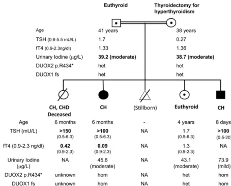
The patients’ clinical details have previously been published (3) (Fig. 1). Briefly, three patients with CH
were born to consanguineous Turkish parents; the first (female) was diagnosed aged 6 months with thyrotropin (TSH).150 mU/mL [reference range (RR) 0.5 to 6.3] and FT4 0.42 ng/dL (RR 0.9 to 2.3), but subsequently died due to congenital heart disease. The second (female, P1) presented aged 6 months with growth retardation (height 56 cm, ,3rd centile, weight 5.6 kg, ,3rd centile), somnolence, and constipation. She had coarse facial features, macroglossia, and severe biochemical hypo-thyroidism [TSH .100 mU/mL (RR 0.5 to 6.3) and FT4,0.09 ng/dL (RR 0.9 to 2.3)] and has severe learning difficulties aged 12 years. Thyroid ultrasound aged 3 years demonstrated a normally located thyroid gland (right lobe: 113 9 mm; left lobe: 13 3 8 mm), and more quantitative ultrasonography aged 11 years confirmed a normal thyroid volume (right lobe: 343 14 3 12 mm; left lobe: 323 13 3 14 mm; isthmus: 3.5 mm), although this may have been influenced by the fact that she was on levothyroxine treatment. Her younger brother (P2), who exhibited umbilical hernia, was diagnosed aged 8 days, with goitre and TSH .100 mU/mL (RR 0.5 to 20). Treatment was commenced with 25mg levothyroxine per day (10mg/kg/d); however, aged 1 month, TSH remained elevated despite good treatment compliance, suggesting severe CH [TSH 91mU/mL (RR 0.5 to 9) and FT4 1.2 ng/dL (RR 0.9 to 2.3)], and levothyroxine dose was rapidly in-creased to 50mg/d. Thyroid ultrasonography demonstrated a normally located, diffusely hyperplastic gland. Both chil-dren ultimately required significant doses ofL-thyroxine
(87.5mg, 1.9 micrograms/kg/d, P1 aged 12 years; 75 mg, 3.1 mg/kg/d, P2 aged 5.75 years). Their sister (S1) was unaffected aged 4 years [TSH 1.7mU/mL (RR 0.5 to 6.3); FT4 1.3 ng/dL (RR 0.9 to 2.3)].
Their father was euthyroid [TSH 1.7mU/mL (RR 0.6 to 5.5); FT4 1.33 ng/dL (RR 0.9 to 2.3)], and their mother, who had previously undergone thyroidectomy for autoimmune hyperthyroidism, was euthyroid on levothyroxine treatment [TSH 0.27 mU/mL (RR 0.6 to 5.5); FT4 1.36 ng/dL (RR 0.9 to 2.3); anti–thyroid peroxidase antibodies 51 IU/mL (RR 0 to 35)]. Her obstetric history also included two abortions and a hydatidiform mole. She had taken Propisyl for hyper-thyroidism during her pregnancies with P2, and S1 but not P1.
Molecular genetic studies
A previously described homozygous DUOX2 non-sense mutation (c.1300C.T, p. R434*) had initially been identified in P1 and P2, for which their parents and unaffected sibling were heterozygous (Fig. 1) (3). DNA was not available from the deceased sibling.
The severity of the CH prompted investigation for an additional genetic mutation using whole-exome sequencing
in P1 and P2. In addition to coding regions, significant intronic sequences were covered using this technique, enabling detection of a homozygous essential splice site change in DUOX1 (c.1823-1G.C), at the intron 14/ exon 15 boundary, validated by Sanger sequencing in both cases. This was absent from 400 ethnically matched control chromosomes and normal genome datasets [dbSNP, Exome Aggregation Consortium, Cambridge, MA (URL:
http://exac.broadinstitute.org), February 2017]; both par-ents and S1 were heterozygous, confirming the segregation of the mutation with congenital hypothyroidism in the family (Fig. 1). No additional mutations were detected in known causative CH genes.
In vitro confirmation of the pathogenicity of the DUOX1 splice site mutation
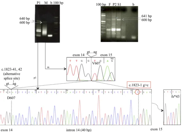
In the heterozygotes, a wild-type DUOX1 fragment of expected size (622 bp) was amplified from peripheral blood mononuclear cells. In contrast, in the homozygotes, a higher molecular weight band was detected and sequencing confirmed a 40-bp insertion in the 30portion of intron 14, indicating the activation of a cryptic acceptor site in intron 14 [r.(1823-40_1823-1ins; 1823-1G.C)] (Fig. 2). This alternative splicing generates a frameshift and a stop codon in exon 15 (p.Val607Aspfs*43) predicted to truncate DUOX1 within the transmembrane helices shortly after the peroxidase domain [Supplemental Fig. 1(B)]. In the heterozygotes, the high instability of this alternative splicing/nonsense transcript, derived from the mutant
DUOX1 allele, may have led to the preferential amplification of the correctly spliced wild-type transcript (Fig. 2). Further biochemical evaluation
Urine iodine measurements were not available at diagnosis, but subsequent spot measurements suggested mild (P2, 73.9mg/L) to moderate (P1, 45.6 mg/L) iodine deficiency (RR 100 to 700mg/L). Moderate iodine deficiency in associ-ation with double heterozygosity for DUOX1 and DUOX2 mutations (S1 and parents) did not result in hypothy-roidism (urinary iodine: mother 39.2mg/L; father 38.7mg/L; S1 43.1 mg/L; RR 100 to 700mg/L) (Fig. 1).
Discussion
We report CH cases harboring a ho-mozygous loss-of-function mutation in DUOX1 (c.1823-1G.C), inherited digenically with a homozygous DUOX2 nonsense mutation (c.1300 C.T, p. R434*) (3, 4). The tertiary structure of DUOX1 and -2 is summarized in Supplemental Fig. 1(B); aberrant splicing of DUOX1 (c.1823-1G.C) will generate a truncated protein (p.Val607Aspfs*43) lacking the C-terminal flavin adenine dinucleotide and NADPH binding domains and cytosolic Ca2+binding sites
(EF-hand motifs) [Supplemental Fig. 1(B)].
In vitro evaluation of a similarly truncated DUOX1 isoenzyme comprising amino acids 1 to 593 alone abolished H2O2-generating activity (5). Moreover, similar truncations
in the highly homologous DUOX2 [p.Q686*, p.R701*, p.(G418fsX482);(IVS19-2A.C), p.S965fsX994] are asso-ciated with CH or severely impaired H2O2-generating
activity in vitro (4, 6, 7). The c.1823-1G.C mutation would be predicted to generate a nonfunctional DUOX1 enzyme, and its digenic inheritance alongside the homo-zygous DUOX2 p.R434* will likely result in complete absence of functional DUOX isoenzyme in our patients.
It has been speculated that DUOX1 upregulation in the context of DUOX2 loss of function may at least partially compensate for defective H2O2 production. In
support of this notion, the majority of reported biallelic DUOX2 mutations, which are known to truncate the protein before the H2O2-generating domains, cause
transient or mild permanent CH, despite presumably abrogating DUOX2 activity completely (8, 9) (Table 1). Direct comparison of biochemistry from reported cases with measurements made in our kindred is precluded by lack of T4 measurement in P2, and the fact that CH was
Figure 1. Pedigree diagram summarizing clinical phenotype and genotype. Black:
homozygotes for the DUOX2 and DUOX1 mutations; central black dot: heterozygotes for the two mutations. The degree of iodine deficiency on the spot urinary measurement is classified according to World Health Organization criteria. DUOX1 fs, DUOX1 p.Val607Aspfs*43; het, heterozygous; hom, homozygous.
diagnosed in P1 and her deceased sibling aged 6 months, rather than neonatally. However, in cases with biallelic truncating mutations, there is a broad spectrum of FT4 measurements at diagnosis, ranging from undetectable to 1.5 ng/dL, and partial iodide organification defects in all except one evaluated case. As well as mandating that other enzymes besides DUOX2 are capable of thyroidal H2O2synthesis, this observation suggests heterogeneity
in the efficiency of this compensatory process, likely due to either genetic or environmental modulators. Digenic, homozygous DUOX2 and DUOX1 mutations in our patients are associated with uncharacteristically severe CH; therefore, we speculate that inability of DUOX1 to compensate for defective H2O2 production may be
contributing to disease severity. Unfortunately, the close chromosomal proximity of DUOX1 and DUOX2 mandates cosegregation of the two mutations, precluding evaluation of their individual contributions.
Urinary iodine was not measured contemporaneously with CH diagnosis in our kindred, and subsequent spot measurements did reveal mild–moderate iodine de-ficiency across the family, for which we cannot exclude a phenotypic contribution. Indeed, high dietary iodine intake in Japan is postulated to mitigate CH associated
with DUOX2 mutations, accounting for the high fre-quency of transient CH in Japanese cases harboring biallelic mutations (Table 1). However, individuals in our kindred who were digenic for heterozygous DUOX1 and DUOX2 mutations remained euthyroid, despite mod-erate iodine deficiency, supporting a digenic, rather than environmental cause for the severe phenotype in the homozygous offspring.
It is noteworthy that the only other reported case harboring the homozygous DUOX2 p.R434* mutation (also Turkish) is unique in manifesting both total iodide organification defect and uncharacteristically severe CH. Although the disease severity in both kindreds could reflect an intrinsic characteristic of the very proximal DUOX2 p.R434* mutation, this mutation is likely to be functionally identical to the Q202TfsX99, K530*, and T522PfsX64 truncating mutations, which will also truncate DUOX2 within the peroxidase-like domain before the first transmembrane region. Cases harboring such biallelic mutations have been associated with partial iodide organification defect and variable disease trajec-tory, including transient CH, suggesting that factors other than the p.R434* DUOX2 mutation itself are contributing to disease severity (6, 8, 9) (Table 1).
Figure 2. Complementary DNA amplification and sequencing from homozygous and heterozygous family members, demonstrating aberrant splicing of DUOX1. The electropherogram of the complementary DNAs and transcript sizes of the different DUOX1 variants detected in the family members are shown. Exons are numbered with the first translated exon as exon 1. P1, P2: children with CH and homozygous DUOX1 c.1823-1G.C mutation. M, mother; F, father; S1, sister; all unaffected and heterozygous for the DUOX1 c.1823-c.1823-1G.C mutation.
Mutations in coding regions and intron–exon boundaries of DUOX1 were excluded in the reported p.R434* mutation case, which argues against digenic inheritance of the same DUOX1 mutation as a founder effect in the Turkish population. However, oligogenic variants in other known hitherto undiscovered CH-associated genes (including the noncoding regions of DUOX1) may be contributing to disease severity. Alternative phenotypic modulators could include polygenic factors specific to the Turkish ethnic background, or environmental iodine de-ficiency, because iodine status was not evaluated (4). No DUOX1-sequencing results are reported for other cases with permanent CH associated with biallelic truncating DUOX2 mutations listed in Table 1, although variants in other CH-associated genes were occasionally sought (2, 6, 9, 10).
In the wider CH context, next-generation sequencing technologies are elucidating a role for oligogenicity in disease pathogenesis (10). We describe the first human cases with digenic DUOX mutations causing complete
DUOX isoenzyme deficiency in the context of likely io-dine deficiency. These individuals manifest severe CH, suggesting failure to compensate for defective thyroid H2O2 synthesis. Although limited subphenotype
infor-mation prevents definitive ascertainment of the relative roles of the two mutations in the thyroid dysfunction, we hypothesize that inability of DUOX1 to compensate for DUOX2 deficiency contributes to disease severity in this kindred. Further studies are required to interrogate the role of upregulation of DUOX1 and alternative H2O2-producing
enzymes in DUOX2-deficient cases and the contribution of variants in these genes to the phenotypic heterogeneity associated with DUOX2 mutations.
Acknowledgments
Address all correspondence and requests for reprints to: Nadia Schoenmakers, MB, BChir, PhD, University of Cambridge Metabolic Research Laboratories, Wellcome Trust–Medical Research Council Institute of Metabolic Science, Level 4, Box Table 1. Table Summarizing Clinical Phenotype and Genotype Information for Published Cases Harboring Biallelic, Confirmed Truncating Mutations in DUOX2
Reference Case DUOX2 Mutation (mU/L)bsTSH (mU/L)vTSH (ng/dL)FT4 US KClO(%)4 DurationCH
Current cases P1 p.[R434*];[R434*] — .100a ,0.09a — — P
P2 p.[R434*];[R434*] — .100b — G — P
Nicholas et al., 2016 (10) 1 p.[ L1028Afs*3];[ L1028Afs*3] — 55 — N —
Tan et al., 2016 (11) 2 p.[K530*];[K530*] 14.76 86.06 0.76 G — T 3 p.[K530*];[K530*] 111.45 .100 ,0.4 N — T 4 p.[K530*];[K530*] 20.25 .100 ,0.4 G — T 5 p.[K530*];[K530*] 122.66 23.9 0.92 N — MP 6 p.[K530*];[Q202Rfs*93] 9.3 .100 ,0.4 G — T 7 c.647-656del10ins15/p.K530* 54.64 92.38 0.60 G — MP/T 8 p.[K530*];[K530*] 14.05 9.58 0.92 G — T 9 p.[K530*];[K1174Sfs*12] 11.88 .100 0.49 N — — 10 p.[R701*];[K530*] 46.17 .100 0.43 G — MP/T 11 p.[Q202Tfs*99];[K530*] 14.47 12.1 1.03 G — — Fu et al., 2016 (9) 12 p.[K530*];[K530*] .8 .100 0.17 N — T 13 p.[L1114Sfs*56];[K530*]c .8 .100 0.32 N — T Fu et al., 2015 (12) 14 p.[L1114Sfs*56];[K530*]c .8 .100 0.32 N — T 15 p.[L1114Sfs*56;W301C];[K530*] .8 .100 0.63 H — P Muzza et al., 2014 (2) 16 p.[Q202Tfs*99];[T522Pfs*64] 18 180 — — 57 P 17 p.[Q202Tfs*99];[T522Pfs*64] 21 130 — — 66 P Maruo et al., 2008 (8) 18 p.[L479Sfs*2];[K628Rfs*10] 36.9 95.4 0.43 G — T 19 p.[L479Sfs*2];[K628Rfs*10] 21.4 233 0.19 G — T 20 p.[L479Sfs*2];[K628Rfs*10] 18.5 150 0.53 G — T 21 p.[L479Sfs*2];[K628Rfs*10] 10 25.7 1.5 G — T
Varela et al., 2006 (6) 22 p.[G418Efs*64];c.[2655-2A.C] — .100 ,1d G 60 P
23 p.[G418Efs*64];c.[2655-2A.C] — .100 0.8d G 68 P
Moreno et al., 2002 (4) 24 p.[R434*];[R434*] .50 1400 0.07 — 100 P
Abbreviations: bsTSH, blood spot screening TSH; G, goiter; H, hypoplastic; KClO4, perchlorate discharge; MP, mild permanent; N, normal; P, permanent; T, transient; US, ultrasound; vTSH, venous confirmatory TSH.
aBiochemistry aged 6 months (P1). bBiochemistry aged 8 days (P2). cCompound heterozygosity assumed.
dTotal T4,mg/dL, normal range 5.98 to 13.9, measured aged 8 months (case 22) and 1 month (case 23). Normal ranges: FT4 ng/dL: Moreno et al., 0.9 to 2.3; Fu et al., 0.9 to 1.7; Maruo et al., 0.97 to 1.7; Tan et al., 0.9 to 2.28.
289, Addenbrooke’s Hospital, Hills Road, Cambridge CB2 0QQ, United Kingdom. E-mail:[email protected].
This work was supported by Wellcome Trust Grants 100585/Z/12/Z (to N.S.) and 095564/Z/11/Z (to V.K.C.); National Institutes for Health Research Cambridge Biomedical Research Centre (to V.K.C. and N.S.); and Italian Ministry of Health Grant RF-2010-2309484 (to L.P.). H.C. is supported by TUBITAK: The Scientific and Technological Research Council of Turkey Grant 214S637.
Disclosure Summary: The authors have nothing to disclose.
References
1. O’Neill S, Brault J, Stasia MJ, Knaus UG. Genetic disorders coupled to ROS deficiency. Redox Biol. 2015;6:135–156.
2. Muzza M, Rabbiosi S, Vigone MC, Zamproni I, Cirello V, Maffini MA, Maruca K, Schoenmakers N, Beccaria L, Gallo F, Park SM, Beck-Peccoz P, Persani L, Weber G, Fugazzola L. The clinical and molecular characterization of patients with dyshormonogenic con-genital hypothyroidism reveals specific diagnostic clues for DUOX2 defects. J Clin Endocrinol Metab. 2014;99(3):E544–E553. 3. Cangul H, Aycan Z, Kendall M, Bas VN, Saglam Y, Barrett TG,
Maher ER. A truncating DUOX2 mutation (R434X) causes severe congenital hypothyroidism. J Pediatr Endocrinol Metab. 2014; 27(3-4):323–327.
4. Moreno JC, Bikker H, Kempers MJE, van Trotsenburg ASP, Baas F, de Vijlder JJM, Vulsma T, Ris-Stalpers C. Inactivating mutations in the gene for thyroid oxidase 2 (THOX2) and congenital hypo-thyroidism. N Engl J Med. 2002;347(2):95–102.
5. Meitzler JL, Ortiz de Montellano PR. Caenorhabditis elegans and human dual oxidase 1 (DUOX1)“peroxidase” domains: insights into heme binding and catalytic activity. J Biol Chem. 2009; 284(28):18634–18643.
6. Varela V, Rivolta CM, Esperante SA, Gru~neiro-Papendieck L, Chiesa A, Targovnik HM. Three mutations (p.Q36H, p.G418fsX482, and g.IVS19-2A.C) in the dual oxidase 2 gene responsible for
congenital goiter and iodide organification defect. Clin Chem. 2006; 52(2):182–191.
7. De Marco G, Agretti P, Montanelli L, Di Cosmo C, Bagattini B, De Servi M, Ferrarini E, Dimida A, Freitas Ferreira AC, Molinaro A, Ceccarelli C, Brozzi F, Pinchera A, Vitti P, Tonacchera M. Identification and functional analysis of novel dual oxidase 2 (DUOX2) mutations in children with congenital or subclini-cal hypothyroidism. J Clin Endocrinol Metab. 2011;96(8): E1335–E1339.
8. Maruo Y, Takahashi H, Soeda I, Nishikura N, Matsui K, Ota Y, Mimura Y, Mori A, Sato H, Takeuchi Y. Transient congenital hypothyroidism caused by biallelic mutations of the dual oxidase 2 gene in Japanese patients detected by a neonatal screening program. J Clin Endocrinol Metab. 2008;93(11):4261–4267.
9. Fu C, Luo S, Zhang S, Wang J, Zheng H, Yang Q, Xie B, Hu X, Fan X, Luo J, Chen R, Su J, Shen Y, Gu X, Chen S. Next-generation sequencing analysis of DUOX2 in 192 Chinese subclinical con-genital hypothyroidism (SCH) and CH patients. Clin Chim Acta. 2016;458:30–34.
10. Nicholas AK, Serra EG, Cangul H, Alyaarubi S, Ullah I, Schoen-makers E, Deeb A, Habeb AM, Almaghamsi M, Peters C, Nathwani N, Aycan Z, Saglam H, Bober E, Dattani M, Shenoy S, Murray PG, Babiker A, Willemsen R, Thankamony A, Lyons G, Irwin R, Padidela R, Tharian K, Davies JH, Puthi V, Park SM, Massoud AF, Gregory JW, Albanese A, Pease-Gevers E, Martin H, Brugger K, Maher ER, Chatterjee VK, Anderson CA, Schoenmakers N. Comprehensive screening of eight known causative genes in con-genital hypothyroidism with gland-in-situ. J Clin Endocrinol Metab. 2016;101(12):4521–4531.
11. Tan M, Huang Y, Jiang X, Li P, Tang C, Jia X, Chen Q, Chen W, Sheng H, Feng Y, Wu D, Liu L. The prevalence, clinical, and molecular characteristics of congenital hypothyroidism caused by DUOX2 mutations: a population-based cohort study in Guangz-hou. Horm Metab Res. 2016;48(9):581–588.
12. Fu C, Zhang S, Su J, Luo S, Zheng H, Wang J, Qin H, Chen Y, Shen Y, Hu X, Fan X, Luo J, Xie B, Chen R, Chen S. Mutation screening of DUOX2 in Chinese patients with congenital hypothyroidism. J Endocrinol Invest. 2015;38(11):1219–1224.