1 Experimental
& Clinical Article
Is Caesarean Myomectomy a Safe Procedure? A Comparative Study
Serdar BAŞARANOĞLU1, Elif AĞAÇAYAK2, Ayşegül DEREGÖZÜ3, İlknur İnegöl GÜMÜŞ1, Mustafa ACET4, Talip GÜL2Istanbul, Turkey ABSTRACT
OBJECTIVE: Uterine myomas are the most common benign pelvic tumours observed during the
repro-ductive period.Increased risks of haemorrhage and postoperative morbidity lead professionals to avoid myomectomy at the time of Cesarean (C-section). The present study retrospectively analysed the data of patients who had undergone C-section only and those that had undergone C-section and simultane-ous myomectomy.
STUDY DESIGN: The data of 42 patients (Group 1) who had underwent caesarean myomectomy and
of 50 patients underwent C-section only (Group 2) out of 92 patients that had been taken into C-section on the basis of obstetric indications were retrospectively analysed in this study. The relevant patient data were recorded with the inclusion of demographic data, gestational week, and preoperative and postop-erative laboratory findings. Types, locations and sizes (the largest diameter) of individual myomas were identified and noted.
RESULTS: The mean diameter of myomas was 66.3±30.2 mm. Ten patients that had underwent
cae-sarean myomectomy (23.8%) developed a need for intensive care. No statistically significant difference was found in laboratory parameters between Group 1 and Group 2.
CONCLUSION: Caesarean myomectomy, when performed by experienced obstetricians, does not lead
to a significant increase in maternal morbidity and mortality. Although the short-term effects of this pro-cedure are known, there is a need for the conduct of more comprehensive studies to establish its long-term effects on fertility or how it will affect the next pregnancy processes.
Keywords: Pregnancy, Cesarean, Myomectomy, Morbidity
1Fatih University Faculty of Medicine Department of Obstetrics and Gynecology, Istanbul.
2Dicle University Faculty of Medicine Department of Obstetrics and Gynecology, Diyarbakır.
3Bahçelievler State Hospital Obstetrics and Gynecology, Istanbul. 4Medipol University Faculty of Medicine Department of Obstetrics and
Gynecology, Istanbul.
Address of Correspondence: Serdar Başaranoğlu
Fatih University Faculty of Medicine Department of Obstetrics and Gynecology, Istanbul. [email protected] Submitted for Publication: 02.10.2015
Accepted for Publication: 06.11.2015
Obstetrics; Maternal-Fetal Medicine and Perinatology
Gynecol Obstet Reprod Med 2016;22:1-4
Introduction
Uterine myomas are the most common benign tumours of the uterus and female genital system. Their incidence in preg-nancy varies between 2.7 and 12.6% and increases with gesta-tional week.1,2As the age at conception and rate of C-section are on an increasing trend, obstetricians’ frequency of observ-ing myomas durobserv-ing C-section is also increasobserv-ing. Uterine
my-omas are usually asymptomatic during pregnancy, but may oc-casionally result in obstetric complications. This spectrum in-cludes; the location and size (>5cm) of the myoma that fre-quently presents with abortion, malpresentation, placental abruption, intrauterine growth restriction (IUGR), placenta previa, abnormal placental invasion, preterm labour, preterm rupture of membranes (PROM), post-partum haemorrhage or bleeding requiring blood transfusion.3,4 Caesarean myomec-tomy is avoided due to its fertility-reducing effects, including excessive post-partum bleeding and the resulting hysterec-tomy to control bleeding.5Specifically for myomas located in the lower segment or the posterior wall of the uterus, my-omectomy is recommended to be performed right after the C-section.6In recent years, studies undertaken with large series of patients to compare patients operated with myomectomy at the time of C-section to those that were operated with tion only demonstrated that myomectomy at the time of C-sec-tion did not increase the risk of intraoperative haemorrhage or uterine atony.7,8The present study aimed to identify and com-pare the maternal, demographic, clinical, laboratory and sur-gical findings belonging to patients that had and had not un-dergone myomectomy at the time of C-section.
2 Başaranoğlu S. Ağaçayak E. Deregözü A. Gümüş İİ. Acet M. Gül T.
Material and Method
A total of 92 patients admitted to our clinic between January 2010 and December 2014 were included in this study. The study group consisted of 42 patients that had been taken into C-section on the basis of different obstetric indications and underwent simultaneous myomectomy (Group 1), whereas the control group was composed of 50 patients with similar clinical and laboratory findings that had been taken into C-section for delivery at our clinic between the same time, but had not been subjected to any other surgical proce-dures (Group 2). In both groups, patients with multiple preg-nancies, placental abruption, abnormal placental invasion, bleeding-clotting disorders, HELLP syndrome and immune thrombocytopenic purpura (ITP) as well as patients with a his-tory of medicinal therapies causing bleeding disorders (as-pirin, heparin, LMWH, warfarin, etc.) and any additional sur-gical procedure other than intraoperative myomectomy were excluded from the study. At the time of presentation, certain data relating to the patients were recorded with the inclusion of age, gravida, parity, gestational week and myomas identi-fied during C-section (type, location and size - the largest di-ameter). The patients were then evaluated in terms of preop-erative and postoppreop-erative haemoglobin (Hb) and haematocrit (Htc) levels, need for blood transfusion, postoperative obstet-ric complications, need for intensive care and pathological di-agnoses. The data pertaining to the patients were obtained from files and electronic records of the hospital. Before initia-tion of the study, due approval was obtained from the Local Ethics Committee of Dicle University.
Myomectomy Technique: Once the baby had been deliv-ered, 60-90 drops/min of 30 IU oxytocin in 100 ml dextrose was administered to the patient, and this was followed by a linear incision on the myoma with electrocautery. Following the removal of the myoma from its pseudo-capsule, the my-ometrial bed was closed off with absorbable sutures. The serosa was closed off with absorbable sutures of 2-0 or 3-0 di-ameter. With bleeding taken under control, all patients were
put on prophylactic antibiotic regimens in the postoperative period.
Statistical analyses were undertaken with Statistical Package for Social Sciences for Windows 15.0 (SPSS Inc., Chicago, IL, USA) and Excel as software. The Kolmogorov-Smirnov test was used to establish whether or not numeric data exhibited a normal distribution, and the percentage was expressed as mean + standard deviation. Data demonstrating a normal distribution were analysed with the Student-t test. For the results thusly obtained, 95% confidence interval and p<0.05 were regarded as preconditions for statistical signifi-cance.
Results
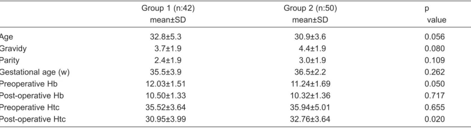
The present study included 42 patients (Group 1) and 50 control subjects (Group 2) amounting in total to 92 patients. Data on age, gravida, parity, gestational week and preopera-tive and postoperapreopera-tive hemogram levels of the patients and control subjects were demonstrated in Table 1. In this respect, no statistically significant difference was found between the two groups. The mean diameter of the removed myomas was 66.3±30.2 mm (40-150). Sizes, locations and types of myomas were given in Table 2. Myomas were generally subserous and most commonly had a diameter of ≤5 cm. They were most fre-quently localised in fundus, corpus or fundus and corpus. Indications of patients undergoing cesarean myomectomy were given in Table 3. A review of the groups led to the find-ing that none of the myomectomy patients had required addi-tional gynaecological operations, including hysterectomy. It was further identified that 10 of the patients in Group 1 (23.8%) had developed a need for intensive care in the preop-erative period, but none of the patients had required blood transfusion. Considering the histopathologic diagnoses of the patients that had undergone myomectomy, it was established that leiomyoma had been reported in 38 patients and degener-ated leiomyoma in 4 patients.
Table 1: Demographic data, preoperative and postoperative laboratory parameters of the patients
Group 1 (n:42) Group 2 (n:50) p
mean±SD mean±SD value
Age 32.8±5.3 30.9±3.6 0.056 Gravidy 3.7±1.9 4.4±1.9 0.080 Parity 2.4±1.9 3.0±1.9 0.109 Gestational age (w) 35.5±3.9 36.5±2.2 0.262 Preoperative Hb 12.03±1.51 11.24±1.69 0.050 Post-operative Hb 10.50±1.33 10.32±1.36 0.717 Preoperative Htc 35.52±3.64 35.94±5.01 0.655 Post-operative Htc 30.95±3.99 32.76±3.64 0.020
Gynecology Obstetrics & Reproductive Medicine 2016;22:1 3
Discussion
Increased oestrogen levels create a tendency for myomas to grow during pregnancy; however, myomas decrease in size in the post-partum period.9Myomectomy at the time of C-sec-tion is one of the currently debated matters in the field of ob-stetrics due to the risks of bleeding and the resulting hysterec-tomy. Recently, large-scale studies indicated that caesarean myomectomy could be safely performed in specifically se-lected cases (subserous, pedunculated or small-diameter my-omas <5cm). Patients who did not underwent myomectomy may later present with preterm labour, early delivery, in-trauterine growth restriction, morbidly adherent placenta and post-partum bleeding. Shavell et al. reported that pregnant women with myomas exceeding 5 cm in diameter were at a higher risk compared to patients with smaller or no myomas.4 Studies undertaken in recent years came to the conclusion that bleeding was not excessive and the procedure was safe if the caesarean myomectomy was undertaken by experienced
sur-geons.10,11In Turkey, Ortac et al.12reported no bleeding and no need for blood transfusion in patients with myomas exceeding 5 cm that had been operated with caesarean myomectomy. Kaymak et al.13 reported for a sample of 40 patients that 5 pa-tients had developed haemorrhage, but this finding was not statistically significant when compared to the control group.Another study comparing 111 patients that had under-gone caesarean myomectomy to 275 pregnant women that had undergone C-section only found no case that had been subject to hysterectomy and no significant difference between the groups in terms of preoperative and postoperative haematocrit (Hct) change and incidence of haemorrhage.14When the pre-operative and postpre-operative haemoglobin (Hb) and haemat-ocrit (Hct) levels of the patients in the present study were compared, no statistically significant difference was observed between the two groups. The recommendations for the reduc-tion of bleeding during myomectomy in the intraoperative process included the administration of high doses of oxytocin, uterine tourniquet, bilateral uterine arterial ligation and the use of electrocautery/argon-beam coagulator.15,16 All patients in our study underwent myomectomy with the use of electro-cautery and a standard dose of oxytocin during the procedure. Furthermore, it was found that no patient had been subject to the use of tourniquet or uterine arterial ligation. A look at the literature indicates that the length of operation is longer and blood loss is higher in caesarean myomectomy compared to C-section only. The present study could not obtain data on the length of operation, but experience shows that the length of operation will be longer in caesarean myomectomy. Kwon et al.17 conducted a study to compare caesarean myomectomy patients with myomas >5 cm to those with myomas ≤5 cm in terms of preoperative and postoperative haemoglobin changes, length of hospital stay, postoperative fever and length of operation and found no significant difference be-tween the groups. Park et al.5compared 97 pregnant women who underwent caesarean myomectomy and 60 pregnant women with myomas that had undergone C-section only and identified no complications that were independent from the lo-cation of the myoma. And they reported that myomectomy could be safely performed at the same time of C-section. The majority of the patients included in our study (78.6%) had subserous myomas, which were mainly localising in the fun-dus (21.4%), corpus (40.5%) or funfun-dus+corpus (7.1%). A re-view of the literature shows that size of the myoma matters in caesarean myomectomy patients, but does not exhibit any cut-off value for this assessment. Studies on the safety of removal of myomas ≥5cm during C-section reported no difference be-tween the groups in length of hospital stay, Hb changes and length of operation.18,19The mean diameter of myomas in our study was 66.3±30.2 mm. Among the operated patients, 24 (57.1%) had myomas with diameters ≤5 cm, 13 (30.9%) with diameters between >5cm and <10cm, and 5 (11.9%) with di-ameters ≥10cm. Furthermore, none of these patients with dif-ferent sizes of myomas exhibited haemorrhage or a need for
Table 2: Fibroids types, diameter and locations
Types n (%) Pedunculated 5 (11.9) Subserous 33 (78.6) Intramural 3 (7.1) Submucosal 1 (2.4) Diameter ≤5 cm 24 (57.1) >5cm. <10cm 13 (30.9) ≥10cm 5 (11.9) Locations Fundus 9 (21.4) Corpus 17 (40.5)
Lower uterine segment 7 (16.7) Posterior 6 (14.3) Fundus+corpus 3 (7.1)
Table 3: Indications of patients undergoing cesarean myomec-tomy (CM)
Indications n (42) % Previous cesarean 12 28.6 Malpresentation (breech, transverse, oblique) 7 17.5
CPD 5 11.9
Treatment of pregnancy (IVF, IUI) 3 7.1 Previous uterine surgery 4 9.5 Fetal distress 3 7.1 Unprogressive labor 3 7.1 Maternal request 2 4.8 Multiple pregnancy 2 4.8 Abruptio placenta 1 2.4
CPD: Cephalopelvic disproportion, IVF: In-vitro fertilization, IUI: Intra-uterine insemination
4 Başaranoğlu S. Ağaçayak E. Deregözü A. Gümüş İİ. Acet M. Gül T.
blood transfusion. The histopathological examination of the removed myoma materials revealed that all the materials were of benign character. Although short-term morbidities of cae-sarean myomectomy are known, there are not sufficient data regarding its effects on long-term fertility or the complications that may emerge in further pregnancies.20
In conclusion, considering the results of this study together with the knowledge in the literature, we think that perform-ance of myomectomy during C-section will not lead to a sig-nificant increase in maternal morbidity and mortality in the hands of experienced obstetricians, regardless of the size and location of the myoma. However, even though the short-term effects of this procedure are known, the literature does not provide sufficient data on its effects on endometrial and my-ometrial functions. Therefore, we believe that further studies are needed on how caesarean myomectomy affects the fol-lowing maternal processes to gain deeper understanding of the safety of this procedure.
References
1. Laughlin SK, Baird DD, Savitz DA, Herring AH, Hartmann KE. Prevalence of uterine leiomyomas in the first trimester of pregnancy: an ultrasound-screening study. Obstet Gynecol 2009;113(3):630-5.
2. Exacoustòs C, Rosati P. Ultrasound diagnosis of uterine myomas and complications in pregnancy. Obstet Gynecol 1993;82(1):97-101.
3. Klatsky PC, Tran ND, Caughey AB, Fujimoto VY. Fibroids and reproductive outcomes: a systematic litera-ture review from conception to delivery. Am J Obstet Gynecol 2008;198:357-66.
4. Shavell VI, Thakur M, Sawant A, Kruger ML, Jones TB, Singh M, et al. Adverse obstetric outcomes associated with sonographically identified large uterine fibroids. Fertil Steril 2012;97:107-10.
5. Park BJ, Kim YW. Safety of cesarean myomectomy. J Obstet Gynaecol Res 2009;35(5):906-11.
6. Kwawukume EY. Myomectomy during cesarean section. Int J Gynecol Obstet 2002;76:183-4.
7. Ma PC, Juan YC, Wang ID, Chen CH, Liu WM, Jeng CJ. A huge leiomyoma subjected to a myomectomy during a
cesarean section. Taiwan J Obstet Gynecol 2010;49:220-2. 8. Tinelli A, Malvasi A, Mynbaev OA, et al. The surgical outcome of intracapsular cesarean myomectomy: a match control study. J Matern Fetal Neonatal Med 2014;27:66-71.
9. Başaranoğlu S, Gül T. Miyoma Uterinin Kliniği ve Semp -tomları. T Klin J Gynecol Obst-Special Topics 2014;7(2): 18-23.
10. Sapmaz E, Celik H, Altungul A. Bilateral ascending uter-ine artery ligation vs. Tourniquet use for hemostasis in ce-sarean myomectomy: a comparison. J Reprod Med 2003; 48:950-4.
11. Li H, Du J, Jin L, Shi Z, Liu M. Myomectomy during ce-sarean section. Acta Obstet Gynecol Scand 2009;88:183-6. 12. Ortac F, Güngör M, Sönmezer M. Myomectomy during
cesarean section. Int J Gynecol Obstet 1999;67:189-90. 13. Kaymak O, Ustunyurt E, Okyay RE, Kalyoncu S,
Mollamahmutoglu L. Myomectomy during cesarean sec-tion. Int J Gynecol Obstet 2005;89:90-3.
14. Roman AS, Tabsh KM. Myomectomy at time of cesarean delivery: a retrospective cohort study. BMC Pregnancy Childbirth 2004;4(1):14.
15. Kwawukume EY. Cesarean myomectomy. Afr J Reprod Health 2002;6:38-43.
16. Topçu HO, İskender CT. Timur H. Kaymak O. Memur T. Danışman N. Outcome safter cesarean myomectomy ver-sus cesarean alone among pregnant women with uterine leiomyomas. Int J Gynecol Obstet 2015;130(3):244-46 17. Kwon DH, Song JE, Yoon KR, Lee KY. The safety of
ce-sarean myomectomy in women with large myomas. Obstet Gynecol Sci 2014;57(5):367-72.
18. Incebiyik A, Hilali NG, Camuzcuoglu A, Vural M, Camuzcuoglu H. Myomectomy during caesarean: a retro-spective evaluation of 16 cases. Arch Gynecol Obstet 2014;289(3):569-73.
19. Lee JH, Cho DH. Myomectomy using purse-string suture during cesarean section. Arch Gynecol Obstet 2011;283 (Supp l1):35-7.
20. Güney M, Oral B, Özsoy M, Mungan T. Sezaryen esnasında myomektomi: retrospektif matermal sonuçların değerlendirilmesi. SDÜ Tıp Fak Derg 2007;14(1):1-4.