CLINICAL DENTISTRY AND RESEARCH 2020; 44(2): 67-73 Original Research Article
CLINICAL DENTISTRY AND RESEARCH 2020; 44(2): 67-73 Orijinal Araştırma
Correspondence
Tan Fırat EYÜBOĞLU, DDS PhD
Department of Endodontics, School of Dentistry, Istanbul Medipol University, Atatürk Bulvarı No:27 Unkapanı, Fatih, 34083, Istanbul, Turkey ORCID: 0000-0002-0308-9579
Phone: +90 212 453 48 48 Fax:+90 212 521 04 26 E-mail: [email protected]
Tan Fırat Eyüboğlu, DDS PhD
Assistant Professor, Department of Endodontics, School of Dentistry, Istanbul Medipol University, Istanbul, Turkey ORCID: 0000-0002-0308-9579
Keziban Olcay, DDS, PhD
Assistant Professor, Department of Endodontics, School of Dentistry, Istanbul University-Cerrahpaşa, Istanbul, Turkey ORCID: 0000-0002-2168-710X
Didem Ekiz, DDS, PhD
Dr Ekiz Is A Freelance Endodontist. ORCID: 0000-0002-6407-9243
Arslan Terlemez, DDS, PhD
Assistant Professor, Department of Endodontics, School of Dentistry, Necmettin Erbakan University, Konya, Turkey ORCID: 0000-0002-6092-4817
DENTIN TUBULE PENETRATION DEPTH AND SEALER
PERCENTAGE OF AH 26, MTA FILLAPEX AND WELL-ROOT ST ROOT
CANAL SEALERS: A CONFOCAL LASER SCANNING MICROSCOPY
STUDY
ABSTRACTBackground and Aim: The aim of this study was to evaluate the dentin tubule penetration depth and sealer percentage along the root canal wall in root canals obturated with AH26, MTA Fillapex, and Well-Root ST using confocal laser scanning microscopy.
Materials and Methods: A total of 36 extracted human permanent canine teeth with root canal length fixed at 14 mm were shaped up to file F5 with a ProTaper rotary system. The teeth were divided into three groups (n=12) according to the sealer used: AH 26, MTA Fillapex, and Well-Root ST. All root canals were filled with their respective Rhodamine B-labeled sealers with F5 gutta-percha using the single-cone technique. Each root was sectioned horizontally 8-mm, 5-mm, and 2-mm above the apex. Confocal laser microscopy evaluation was performed at 10X magnification. Two-way ANOVA and Tukey’s post hoc test were used for the statistical analysis with a significance level of p < 0.05.
Results: The only significant difference between the sealers in terms of penetration depth was observed in the 8-mm cross section (p < 0.000). Well-Root ST group exhibited the lowest penetration depth values followed by MTA Fillapex and AH26 (p=0.000 and p=0.000, respectively) whereas the difference between the latter two was insignificant (p>0.237). No statistically significant difference was observed between the groups in terms of sealer percentage in the 2-mm, 5-mm, and 8-mm cross sections (p=0.482, p=0.888, and p=0.054, respectively).
Conclusion: Well-Root ST exhibited significantly lower penetration depths than MTA Fillapex and AH26.
Keywords: Calcium Silicate-Based Sealers, Confocal Laser Microscopy, Penetration Depth, Resin-Based Sealers, Sealer Percentage
Submitted for Publication: 11.01.2019 Accepted for Publication : 07.21.2020
CLINICAL DENTISTRY AND RESEARCH 2020; 44(2): 67-73 Orijinal Araştırma
Sorumlu Yazar
Tan Fırat Eyüboğlu
İstanbul Medipol Üniversitesi, Diş Hekimliği Fakültesi, Endodonti Anabilim Dalı, Atatürk Bulvarı No:27,Unkapanı, Fatih 34083 İstanbul, Türkiye ORCID: 0000-0002-0308-9579
Telefon: +90 212 453 48 48 Faks:+90 212 521 04 26 E-mail: [email protected]
Tan Fırat Eyüboğlu
Dr. Öğr. Üyesi, İstanbul Medipol Üniversitesi, Diş Hekimliği Fakültesi, Endodonti Anabilim Dalı, İstanbul, Türkiye ORCID: 0000-0002-0308-9579
Keziban Olcay
Dr. Öğr. Üyesi, İstanbul Üniversitesi-Cerrahpaşa, Diş Hekimliği Fakültesi, Endodonti Anabilim Dalı, İstanbul, Türkiye ORCID: 0000-0002-2168-710X
Didem Ekiz
Dr. Ekiz Serbest Çalışan Bir Endodontistir ORCID: 0000-0002-6407-9243
Arslan Terlemez
Dr. Öğr. Üyesi, Necmettin Erbakan Üniversitesi, Diş Hekimliği Fakültesi, Endodonti Anabilim Dalı, Konya, Türkiye ORCID: 0000-0002-6092-4817
AH 26, MTA FİLLAPEX AND WELL-ROOT ST KÖK KANAL
PATLARININ DENTİN TÜBÜLÜ PENETRASYON DERİNLİĞİ VE PAT
ORANI: BİR KONFOKAL LAZER TARAMA MİKROSKOBİ ÇALIŞMASI
ÖZ
Amaç: Bu çalışmanın amacı, konfokal lazer tarama mikroskobu kullanarak AH26, MTA Fillapex ve Well-Root ST ile doldurulmuş kök kanallarında kök kanal duvarındaki dentin tübül penetrasyon derinliğini ve pat oranını değerlendirmektir.
Gereç ve Yöntem: 14 mm’ye sabitlenmiş kök kanal uzunluğuna sahip toplam 36 adet çekilmiş insan kalıcı köpek dişi, bir ProTaper döner sistemiyle F5’e kadar şekillendirildi. Kullanılan kök kanal patına göre dişler üç gruba (n=12). Grup 1: AH 26, Grup 2: MTA Fillapex, Grup 3: Well-Root ST. Tüm kök kanalları, F5 gutta-perka ile tek kon tekniği kullanılarak Rhodamine B ile etiketlenmiş kök kanal patı ile dolduruldu. Her kökün apeksinin 2 mm, 3 mm ve 5 mm üstünden horizontal kesitler alındı. Konfokal lazer mikroskopi değerlendirmesi, 10X büyütmede yapıldı. İstatistiksel analiz için iki yönlü ANOVA analizi ve post hoc Tukey testi anlamlılık düzeyi 0,05 olarak kullanılmıştır.
Bulgular: Kanal patları arasındaki tek önemli fark 8 mm kesitte gözlendi (p<0,000). Well-Root ST grubu, MTA Fillapex ve AH26’nın (p1=0,000, p2=0,000) ardından anlamlı derecede düşük penetrasyon derinliği sonuçları gösterirken, son iki grup arasındaki fark önemsizdi (p>0.237). Gruplar arasında, 2 mm, 5 mm ve 8 mm kesitlerde, ortalama kanal patı yüzdesi değerleri anlamında istatistiksel olarak anlamlı bir fark gözlemlenmemiştir (p1=0,482, p2=0,888, p3=0,054).
Sonuç: Well-Root ST, hem MTA Fillapex hem de AH26 ile karşılaştırıldığında daha düşük penetrasyon derinliği sonuçları sergilemiştir.
Anahtar Kelimeler:Kalsiyum Silikat Esaslı Dolgu Maddeleri, Konfokal Lazer Mikroskopisi, Penetrasyon Derinliği, Rezin Esaslı Dolgu Maddeleri, Pat Oranı
Yayın Başvuru Tarihi : 01.11.2019 Yayına Kabul Tarihi :21.07.2020
Penetration DePth of Different root Canal SealerS
CLINICAL DENTISTRY AND RESEARCH 2020; 44(2): 67-73 Orijinal Araştırma
INTRODUCTION
Achieving a bacteria free root canal space is not possible even after thorough chemo-mechanical preparation of the root canal system.1 Therefore, the purpose of the root canal filling is to both establish a three-dimensional hermetic seal and entomb the bacterial remnants inside the root canal system.2,3 To achieve this, the endodontic sealer should penetrate the root canal walls, creating a physical barrier.4 Various factors, such as the presence of smear layer,5 dentin permeability, the physicochemical properties of the obturation materials,6 and filling technique,7 affect the root canal sealer’s penetration into the dentinal tubules.
Although calcium silicate-based sealers, were introduced into the market almost a decade ago, new calcium silicate-based sealers are still emerging. Well-Root ST (Vericom, Gangwon-Do, South Korea) is a recently introduced tricalcium silicate-based sealer. It is a premixed, injectable bioceramic sealer for permanent obturation. It contains zirconium oxide, calcium silicate, a filler, and hydrophilic thickening agents that use moisture inside the root canal system to complete the setting reaction. Its setting time is 2.5 hr.8
MTA Fillapex (Angelus, Londrina, Brazil) is a well-studied paste-catalyst root canal sealer which has been available for eight years. Paste A contains salicylate resin (methyl salicylate, butylene glycol, and colophony), bismuth oxide, and silica, whereas Paste B contains base resin (pentaerythritol, rosinate, and toluene sulfonamide), silicone dioxide and titanium dioxide. The filler part includes 13.2% set mineral trioxide aggregate particles. Its complete setting time is 2 hr with a working time of 23 min.8,9
Epoxy resin-based sealer, AH26 (Dentsply DeTrey Konstanz, Germany) is another well-known, commonly used root canal sealer. It has excellent physicochemical properties as well as adaptability to the root canal walls.10
The purpose of this study was to evaluate and compare the maximum sealer penetration depth and sealer percentage along the root canal wall in the apical, middle, and coronal thirds of teeth obturated with AH26, MTA Fillapex, and Well-Root ST using confocal laser scanning microscopy.
MATERIALS AND METHODS
A total of 36 extracted human permanent canine teeth were used in this in vitro study following the approval of the University’s Non-interventional Clinical Research Ethics Board (approval number:
10840098-604.01.01-E.17832/402). The external surfaces of the teeth were cleaned using a periodontal curette. All teeth were checked for cracks under a stereomicroscope (Zeiss Axio Zoom V16, Carl Zeiss, Jena, Germany) at 25X magnification. After removing the crowns from the cemento-enamel junction using diamond discs (Komet, Gebr Brasseler, Lemgo, Germany) under water cooling, the length of the roots was fixed at 14 mm. The apical patency of the root canals was confirmed using a #15 K-file (Mani, Utsunomiya, Tochigi, Japan). Instrumentation of the root canals was performed up to file F5 with a nickel-titanium rotary system (ProTaper Universal, Dentsply Maillefer, Ballaigues, Switzerland) at a 13-mm working length. During instrumentation 2 ml of sodium hypochlorite (NaOCl; Promida, Eskişehir, Turkey) at 5% was applied between each file using a 27-gauge endodontic irrigation needle (Ultradent Products, South Jordan, UT, USA). In total 16 ml of NaOCl was used for each root canal. Following the completion of the preparation, 2.5 ml of 17% ethylene diamine-tetra acetic acid (EDTA; Endo-Solution, Cerkamed, Stalowa Wola, Poland) for 1 min, 2.5 ml of 5% NaOCl for 30 s and 5 ml of distilled water for 30 s were applied as the final irrigation protocol. The root canals were then dried with paper points (DiaDent Group International, Chungcheongbuk-do, South Korea) and the specimens were randomly divided into three groups of 12 according to the sealer used during the root canal filling: AH 26, MTA Fillapex, and Well-Root ST.
Before filling, to achieve fluorescence for confocal laser microscopy examinations, all root canal sealers were tagged with a final concentration of 0.1% fluorescent Rhodamine B isothiocyanate (Bereket Chemical Industry, Istanbul, Turkey) prior to mixing. The root canals were obturated with F5 size gutta percha (ProTaper Universal, Dentsply Maillefer, Ballaigues, Switzerland) and gutta-percha cones size 20 with a .02 taper (DiaDent, South Korea). The filled specimens were stored at 37°C and 100% humidity for seven days to ensure complete setting of the sealers. The roots were then horizontally sectioned using a diamond saw (IsoMet 1000, Buehler, Lake Bluff, IL, USA) under water cooling. Dentine slices 1 mm thick were cut from the coronal, middle and apical thirds of each root 8, 5, and 2 mm above the apex, respectively.
The dentine samples were examined under a confocal laser microscope (Zeiss LSM 800; Carl Zeiss, Jena, Germany) using 561-nm wavelength laser excitation at 10X magnification. Images were taken in fluorescent mode. The dimensions of the images were 2994.84×3555.13 µm
CLINICAL DENTISTRY AND RESEARCH
for the apical third, 4089.21×5239.73 µmfor the middle third and 6457.63×7030.40 µmfor the coronal third. Their resolution was 5175×5634 pixels. The maximum sealer penetration depth into the dentinal tubules was measured using ImageJ V1.52p software (Wayne Rasband National Institutes of Health, USA). Each image was imported into ImageJ program and the system was spatially calibrated using the “Set Scale” tool with the aid of a line drawn on an established scale in the image equivalent to 500 µm. The “Straight Line” tool was used to measure the maximum sealer penetration point between the root canal wall and the external root surface at four dimensions (buccal, lingual, mesial, and distal), and the mean of the obtained data was used for the analysis. The “Oval Selection” tool was used to measure the circumference of the root canal, and the “Free Hand Line” selection tool was used to outline the endodontic sealer perimeter around the root canal. The percentage of the sealer perimeter integrity was calculated as follows: endodontic sealer perimeter integrity×100/root canal circumference.
For the statistical analysis, IBM SPSS Statistics 22 software (IBM, Armonk, NY, USA) was used. The Shapiro-Wilk test was used to confirm the normal distribution of the data. Two-way analysis of variance was used to determine the significance of difference between groups and the percentage of the sealer perimeter integrity. Tukey’s post hoc test was used to compare root levels within the same group. Mean differences were considered statistically significant at a p-value less than 0.05l.
RESULTS
The differences between the three root canal sealers in terms of average sealer penetration depth was statistically significant (p=0.000). The difference between cross
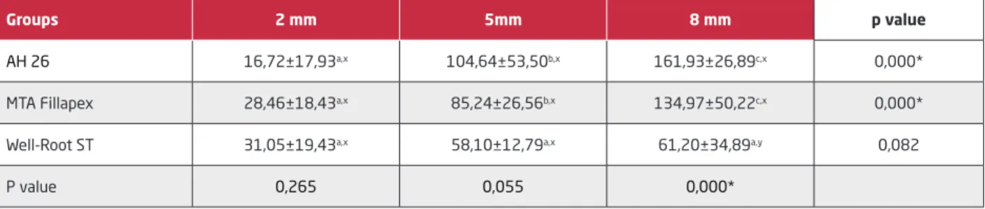
sections was also significant (p=0.000). No significant difference in average penetration depth was observed between the sealers in 2-mm (p=0.265) and 5-mm cross sections (p=0.055). In contrast, in 8-mm cross sections, the Well-Root ST group had significantly lower average penetration depth values than the AH26 and MTA Fillapex groups (p=0.000 and, p=0.000, respectively). Although AH 26 exhibited the greatest penetration depths in 8-mm cross sections, there was no significant difference between the AH 26 and MTA Fillapex groups (p=0.237; Table 1, Figure 1). The average penetration depth within each sealer group was greatest in 8-mm followed by 5-mm and 2-mm cross sections.
In the AH 26 group, the penetration depth in 8-mm cross sections was significantly greater than in 2-mm and 5-mm cross sections (p=0.000 and, p=0.002, respectively), and that in 5-mm cross sections was significantly greater than in 2-mm cross sections (p=0.000).
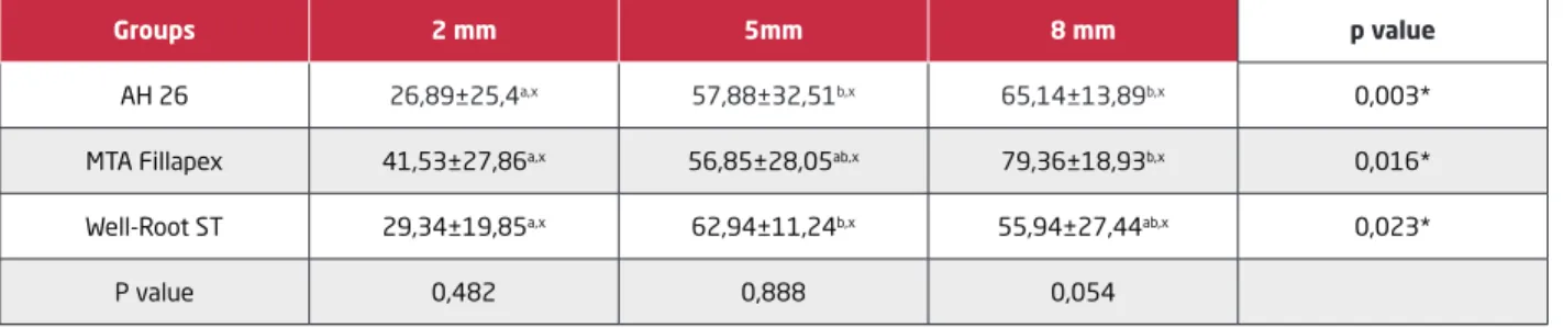
The penetration depth in the MTA Fillapex group was also significantly greater in 8-mm cross sections than in 2-mm and 5-mm cross sections (p=0.000 and, p=0.033, respectively), and that in5-mm cross sections was also significantly greater than in 2-mm cross section (p=0.024). In contrast, the average penetration depth differences between all cross sections in the Well-Root ST group were statistically insignificant (p=0.082; Table 1, Figure 1). No statistically significant differences were observed between the three sealer groups in 2-mm (p=0.482), 5-mm (p=0.888), and 8-mm (p=0.054) cross sections in terms of sealer percentage (Table 2).
In the AH26 group, there was a significant difference in sealer percentage between the three cross sections (p=0.003). In 2-mm cross sections sealer percentages were lower than in 5-mm and 8-mm cross sections (p=0.019 and,
Table 1. The data related the avarage sealer penetration (µm) into dentinal tubules (mean±Std. Dev.) 2-mm, 5-mm and 8-mm above the apex and p values.
Groups 2 mm 5mm 8 mm p value
AH 26 16,72±17,93a,x 104,64±53,50b,x 161,93±26,89c,x 0,000*
MTA Fillapex 28,46±18,43a,x 85,24±26,56b,x 134,97±50,22c,x 0,000*
Well-Root ST 31,05±19,43a,x 58,10±12,79a,x 61,20±34,89a,y 0,082
P value 0,265 0,055 0,000*
a,b,c Different superscript letters show statistically significant groups in the same line. x,y Different superscript letters show statistically significant groups in the same column.
Penetration DePth of Different root Canal SealerS
p=0.003, respectively), whereas there was no significant difference between 5-mm and 8-mm cross sections (p=0.761).
In the MTA Fillapex group, the sealer percentages were also significantly different between the three cross sections (p=0.016). The sealer percentage was significantly lower in 2-mm cross sections than in 8-mm cross sections (p=0.013). However, there were no significant differences either between 2-mm and 5-mm cross sections (p=0.483), or between 5-mm and 8-mm cross sections (p=0.174). The sealer percentages were also significantly different between the three cross sections in the Well-Root ST group (p=0.023). The values in 2-mm cross sections were significantly lower than in 5-mm cross sections (p=0.022). Conversely, no significant differences were observed either between 2-mm and 8-mm (p=0.063) or between 5-mm and 8-mm cross sections (p=0.773; Table 2).
DISCUSSION
In this study, confocal laser scanning microscopy was used due to its nondestructive processing ability, lack of special requirements for the specimens, and low possibility of artifact production.11
The aim was to compare the sealer percentage and maximum dentinal tubule penetration depth between three root canal sealers: AH 26, MTA Fillapex and Well-Root ST. The results showed that the penetration depth differed significantly between the three sealers and between the three cross sections. The sealer percentage also differed significantly between the three sealers.
Several factors, such as the presence of smear layer, dentin permeability, and the sealer’s chemical and physical properties, affect the sealer penetration depth.12,13 The presence of smear layer has been reported to either Table 2. The data related the percentage (%) of the sealer integrity in the root canal perimeter (mean±Std. Dev.) 2-mm, 5-mm and 8-mm above the apex and p values.
Groups 2 mm 5mm 8 mm p value
AH 26 26,89±25,4a,x 57,88±32,51b,x 65,14±13,89b,x 0,003*
MTA Fillapex 41,53±27,86a,x 56,85±28,05ab,x 79,36±18,93b,x 0,016*
Well-Root ST 29,34±19,85a,x 62,94±11,24b,x 55,94±27,44ab,x 0,023*
P value 0,482 0,888 0,054
a,b,c Different superscript letters show statistically significant groups in the same line. x,y Different superscript letters show statistically significant groups in the same column.
Figure 1. Representative CLSM images from all groups: a) AH 26 group-apical third; b) AH 26 group-middle third; c) AH 26 group-coronal third; d) MTA Fillapex group-apical third; e) MTA Fillapex group-middle third; f) MTA Fillapex group-coronal third; g) Well-Root ST group-apical third; h) Well-Root ST group-middle third; ı) Well-Root ST group-coronal third.
CLINICAL DENTISTRY AND RESEARCH
completely block6,14 or at least limit the sealer’s penetration into the dentinal tubules.15 It has also been reported to prevent the penetration of irrigation solutions into the dentinal tubules to achieve adequate disinfection and to possibly be a source of microleakage after the completion of root canal filling due to its physical instability.16,17 For this reason, in this study, the smear layer was removed completed using 17% EDTA. Flow, viscosity, surface tension, working and setting time, solubility, and chemical composition of root canal sealers may also influence the penetration depth.18,19 Especially flow, has been suggested to be a major factor.19
In this study maximum penetration depth was greater in the coronal and middle cross sections and lowest in the apical cross sections. These findings are consistent with the results of previous studies.17,20 This might be due to the higher number of open dentinal tubules with a larger diameter in the coronal and middle thirds of the root canal compared to the apical third.17
Several studies have compared the sealer penetration depth of epoxy resin and calcium silicate-based root canal sealers, reporting results significantly favoring the latter.17,20,21 These results, however, are not in line with our study’s findings. Although both AH 26 and MTA Fillapex are characterized as pseudoplastic,22 which means that they exhibit low viscosity and high flow under compaction, Kuçi et al. reported that MTA Fillapex exhibits greater flow than AH26.17 However, in our study AH 26 exhibited higher penetration depth than MTA Fillapex and Well-Root ST at 5 and 8 mm, there was no significant difference in any cross sections between AH 26 and MTA Fillapex. This might be because of the single cone obturation technique used in our study, which reduced the sealers’ pseudoplastic ability due to the absence of compaction. Moreover, the film thickness of MTA Fillapex was greater than that of epoxy resins,9 which may have resulted in similar penetration depths between the three sealers within each cross section. In a confocal microscopic analysis, MTA Fillapex and AH Plus showed similar flow rates and penetration depths, which is in agreement with the results of our study.23
Well-Root ST showed lower penetration depth values than AH 26 and MTA Fillapex in 5-mm and 8-mm cross sections, but higher values were observed in 2-mm cross sections. The difference in 8-mm cross sections was significant. This might be due to this materials’ flow rate and film thickness. Well-Root ST is a new material and no studies to date have compared it to other root canal sealers in terms
of physical properties such as flow rate, film thickness, and viscosity although it has been found to meet ISO standards Cetinkaya and Bodrumlu found that Well-Root ST and epoxy resin–based sealer AD Seal had similar flow rates in room temperature.24
In this study, the difference in terms of sealer percentage were not significant between the three sealers in any of the cross sections. This is consistent with the results of a previous study that found similar sealer percentages in AH 26, MTA Fillapex, and BioRoot RCS, regardless of the presence or absence of smear layer.20 In contrast an in vitro study investigating the effects of agitation methods on sealer penetration percentage reported that MTA Fillapex was superior to AH Plus irrespective of the agitation method.2 Using agitation methods to enhance the sealers’ penetration ability would affect their flow rate, hence the difference in our sealer percentage results compared to the findings of that present study.2
CONCLUSIONS
MTA Fillapex and AH 26 exhibited similar performance in the root canal and similar penetration depths whereas Well-Root ST showed lower penetration depth values. All three root canal sealers showed similar sealer percentages in all three cross sections evaluated. Further studies on Well-Root ST are required to better understand its physical properties and performance within the root canal.
REFERENCES
1. Wu M-K, Wesselink PR. Endodontic leakage studies reconsidered. Part 1. Methodology, application and relevance. Int Endod J 1993; 26: 37–43.
2. Gharib SR, Tordik PA, Imamura GM, Baginski TA, Goodell GG. A confocal laser scanning microscope investigation of the epiphany obturation system. J Endod 2007; 33: 957-961.
3. Ordinola-Zapata R, Bramante CM, Graeff MS, del Carpio Perochena A, Vivan RR, Camargo EJ et al. Depth and percentage of penetration of endodontic sealers into dentinal tubules after root canal obturation using a lateral compaction technique: a confocal laser scanning microscopy study. Oral Surg Oral Med Oral Pathol Oral Radiol Endod 2009; 108: 450-457.
4. Pommel L, Jacquot B, Camps J. Lack of correlation among three methods for evaluation of apical leakage. J Endod 2001; 27: 347– 350.
5. White RR, Goldman M, Lin PS. The influence of the smeared layer upon dentinal tubule penetration by endodontic filling materials. Part II. J Endod 1987; 13, 369–374.
Penetration DePth of Different root Canal SealerS
6. Oksan T, Aktener BO, Sen BH, Tezel H. The penetration of root canal sealers into dentinal tubules. A scanning electron microscopic study. Int Endod J 1993; 26, 301–305.
7. De Deus GA, Gurgel-Filho ED, Maniglia-Ferreira C, Coutinho-Filho T. The influence of filling technique on depth of tubule penetration by root canal sealer: A study using light microscopy and digital image processing. Aust Endod J 2004; 30, 23–28.
8. Reszka P, Nowicka A, Lipski M, Dura W, Droździk A, Woźniak K. A Comparative Chemical Study of Calcium Silicate-Containing and Epoxy Resin-Based Root Canal Sealers. Biomed Res Int 2016; 2016: 9808432.
9. Zhou HM, Shen Y, Zheng W, Li L, Zheng YF, Haapasalo M. Physical properties of 5 root canal sealers. J Endod 2013; 39: 1281-1286. 10. Marciano MA, Guimaraes BM, Ordinola-Zapata R, Bra-mante CM, Cavenago BC, Garcia RB et al. Physical properties and interfacial adaptation of three epoxy resin-based sealers. J Endod 2011; 37: 1417-1421.
11. Van Meerbeek B, Vargas M, Inoue S, Yoshida Y, Perdigão J, Lambrechts P et al. Microscopy investigations. Techniques, results, limitations. Am J Dent 2000; 13: 3–18.
12. De Deus GA, Gurgel-Filho ED, Maniglia-Ferreira C, Coutinho-Filho T. The influence of filling technique on depth of tubule penetration by root canal sealer: A study using light microscopy and digital image processing. Aust Endod J 2004; 30: 23–28. 13. Ørstavik D. Materials used for root canal obturation: Technical, biological and clinical testing. Endod Topics 2005; 12: 25–38. 14. Kokkas AB, Boutsioukis ACh, Vassiliadis LP, Stavrianos CK. The influence of the smear layer on dentinal tubule penetration depth by three different root canal sealers: an in vitro study. J Endod 2004; 30: 100–102.
15. Kara Tuncer A, Tuncer S. Effect of different final irrigation solutions on dentinal tubule penetration depth and percentage of root canal sealer. J Endod 2012; 38: 860–863.
16. Violich DR, Chandler NP. The smear layer in endodontics—a review. Int Endod J 2010; 43: 2–15.
17. Kuçi A, Alaçam T, Yavaş O, Ergul-Ulger Z, Kayaoglu G. Sealer penetration into dentinal tubules in the presence or absence of smear layer: a confocal laser scanning microscopic study. J Endod 2014; 40: 1627-1631.
18. Ferrari M, Mannocci F, Vichi A, Cadigiaco MC, Mjor IA. Bonding to root canal: Structuralcharacterstics of the substrate. Am J Dent 2000; 13: 255–260.
19. Nikhil V, Bansal P, Sawani S. Effect of technique of sealer agitation on percentage and depth of MTA Fillapex sealer
penetration: A comparative in-vitro study. J Conserv Dent 2015; 18: 119-123.
20. Aktemur Türker S, Uzunoğlu E, Purali N. Evaluation of dentinal tubule penetration depth and push-out bond strength of AH 26, BioRoot RCS, and MTA Plus root canal sealers in presence or absence of smear layer. J Dent Res Dent Clin Dent Prospects 2018; 12: 294-298.
21. Piai GG, Duarte MAH, Nascimento ALD, Rosa RAD, Só MVR, Vivan RR. Penetrability of a new endodontic sealer: A confocal laser scanning microscopy evaluation. Microsc Res Tech 2018; 81: 1246-1249.
22. Silva EJ, Rosa TP, Herrera DR, Jacinto RC, Gomes Brenda PFA, Zaia AA. Evaluation of cytotoxicity and physicochemical properties of calcium silicate-based endodontic sealer MTA Fillapex. J Endod 2013; 39: 274–277.
23. Amoroso-Silva PA, Guimarães BM, Marciano MA, Duarte MA, Cavenago BC, Ordinola-Zapata R et al. Microscopic analysis of the quality of obturation and physical properties of MTA Fillapex. Microsc Res Tech 2014; 77: 1031-1036.
24. Cetinkaya I, Bodrumlu E. Evaluation of the flow properties of two different root canal sealers with three different temperatures. Atatürk Üniv Diş Hek Fak Derg 2019; 29: 7-11