Evaluation Of Maxillary Sinus Findings In Children Using CBCT
KADER CESUR AYDIN 1*, BEYZA BALLI AKGOL 2, BARIS CAGRI DELILBASI 3, GOKHAN GURLER 3.
1*
, Istanbul Medipol University, School of Dentistry, Department of Dento Maxillofacial Radiology; 2 , Istanbul Medipol University, School of Dentistry, Department of Pediatric Dentistry; 3 , Istanbul Medipol University, School of Dentistry, Department of Maxillofacial Surgery.
Corresponding Author: KADER CESUR AYDIN
Istanbul Medipol University, School of Dentistry, Department of Dento Maxillofacial Radiology. Ataturk Bulvarı no: 27 Unkapanı- Istanbul – TURKEY 34083
[email protected] Tel: 090212 4534951
PEER REVIEW COPY
Minerva Pediatrica
7 8 9 10 11 12 13 14 15 16 17 18 19 20 21 22 23 24 25 26 27 28 29 30 31 32 33 34 35 36 37 38 39 40 41 42 43 44 45 46 47 48 49 50 51 52 53 54 55 56 57 58 59 60 61 62 63 64 65 66 67 68 69 70 81 82 83 84 85 86 87 88 89 90 91 92 93 94 95
ABSTRACT
BACKGROUND: Evaluation of the maxillary sinus is usually overlooked in children. The aim of this study was to evaluate incidental findings in the maxillary sinus in a group of children using cone beam computerized tomography (CBCT).
MATERIALS AND METHODS: CBCTs obtained from 74 children (148 sinuses) were retrospectively evaluated by the same dento-maxillofacial radiologist. Presence of antral septum, pseudocyst, mucosal thickening, and sinusitis were recorded. Involvement of the right and left sinuses, as well as age and gender correlation with the findings was analyzed.
RESULTS: Within the age group of 5 to15, 43 out of 74 subjects revealed different sorts of maxillary sinus findings. Anthral septum was the most common (26.9 %) finding among all subjects.
CONCLUSION: Maxillary sinus evaluation is essential for both dentists and pediatricians. CBCT is a novel and advantageous technique to view pathologic lesions and anatomic variations in the maxillary sinus.
Key Words: CBCT, Maxillary sinus, diagnosis.
PEER REVIEW COPY
Minerva Pediatrica
7 8 9 10 11 12 13 14 15 16 17 18 19 20 21 22 23 24 25 26 27 28 29 30 31 32 33 34 35 36 37 38 39 40 41 42 43 44 45 46 47 48 49 50 51 52 53 54 55 56 57 58 59 60 61 62 63 64 65 66 67 68 69 70 81 82 83 84 85 86 87 88 89 90 91 92 93 94 95
INTRODUCTION
Maxillary sinus is one of the four sets of air- filled cavities of the craniofacial complex1. The shape and size of the maxillary sinuses may vary among individuals, but they are usually pyramidal in shape and the walls correspond to orbital, maxillary alveolar, facial and infratemporal aspects of the maxillary bone 2,3. Maxillary sinus is the largest among all paranasal sinuses. It is important in dental practice owing to its close proximity to orodental structures. Furthermore, symptoms of sinus pathologies may be confused with dental symptoms. Detailed radiological evaluation of the maxillary sinus is essential in both dentistry and pediatrics. Conventional radiographs such as periapical and panoramic radiographs as well as Water’s graph are commonly used in dental practice; however, they may sometimes come short to visualize anatomical structures due to 2-D nature of these techniques. Cone beam computed tomography (CBCT) is the latest improvement in craniofacial imaging, also presenting the best imaging for the paranasal sinuses via acquired multiplanar images 4,5. CBCT provides accurate images with a short exposure time. Recently most attention is focused on CBCT findings in the craniofacial region.
Maxillary sinus pathologies may be intrinsic or extrinsic (mostly dental) in origin. Intrinsic ones may include; inflammatory diseases (mucositis, sinusitis, retention pseudocyst, polyp, antrolith, and mucocele) and neoplasms (papilloma, osteoma, tumors and pseudotumors). Extrinsic ones may also involve inflammatory diseases (periostitis) and odontogenic cysts, tumors, trauma and bone dysplasia 1.
Incidental findings of the maxillary sinuses in adult population using CBCT have been addressed earlier 6,7; however, data involving children is scarce. The aim of this retrospective study was to determine maxillary sinus findings in a group of children, who have undergone maxillary sinus CBCT evaluation. for several reasons ( dental trauma, presence of a impacted supernumerary tooth or dental trauma).
MATERIALS AND METHOD Study Sample
This study included 74 maxillary sinus CBCT images obtained from pediatric patients who have referred to Department of Dento-Maxillofacial Radiology, Istanbul Medipol University, School of Dentistry. Study group consisted of 28 girls and 46 boys with an age range of 5 to
PEER REVIEW COPY
Minerva Pediatrica
7 8 9 10 11 12 13 14 15 16 17 18 19 20 21 22 23 24 25 26 27 28 29 30 31 32 33 34 35 36 37 38 39 40 41 42 43 44 45 46 47 48 49 50 51 52 53 54 55 56 57 58 59 60 61 62 63 64 65 66 67 68 69 70 81 82 83 84 85 86 87 88 89 90 91 92 93 94 95
15 years. Age and gender distributions were shown in Table 1. This study was approved by the local ethical committee of Istanbul Medipol University.
CBCT Images
CBCT images were obtained using an I-CAT Next Generation (Imaging Sciences International, Hatfield, USA) 3D volume scanner with 6 x 16 cm FOV, 120 kV and 20.27 mA energy, 14.7 seconds exposure and 0.25 mm slice thickness and isotropic voxels. All CBCTs which displayed the entire maxillary sinuses bilaterally were included in the study.
Maxillary sinuses were evaluated by both sagittal and coronal planes using Vision software. 1920 x 1020 pixel resolution was selected for evaluation on 24 inch Dell displaying screen. CBCT scans were reviewed by a single dento-maxillofacial radiologist and the diagnostic criteria were mainly developed on the basis of published literature 7. Ten CBCT samples were evaluated twice by the same radiologist at 20 day-intervals for intra-observer agreement. The following findings in the maxillary sinus were evaluated:
Maxillary sinus septa (Anthral Septum):
Maxillary sinus septum was described as an inverted gothic arch shaped walls of cortical bone within the maxillary sinus; arising from the inferior or lateral walls of the sinus, and may even divide the sinus into two or more cavities 8.
Mucosal thickening
Sinus mucosa thickness may increase due to infectious or allergic response. Image is detected as a band paralleling to the sinus wall which is more radiopaque than the air in the sinus. For statistical analysis sinus mucosal thickness was evaluated being between 1-3 mm thickness (mild thickening) or higher than 3 mm thickness (moderate thickening) in both sinuses.
Retention Pseudocyst
Radiographic interpretation displays full or partial involvement of the maxillary sinus with a well-defined, non-corticated opacity, being dome shaped and homogenous mass.
Sinusitis
Radiographic interpretation displays reduced airy content and increased radioopacity. Ostium may be blocked. The shape of the mucosa may be uniform or polipoid due to infection or allergy. Sinus periosteum may become sclerotic in chronic sinusitis.
PEER REVIEW COPY
Minerva Pediatrica
7 8 9 10 11 12 13 14 15 16 17 18 19 20 21 22 23 24 25 26 27 28 29 30 31 32 33 34 35 36 37 38 39 40 41 42 43 44 45 46 47 48 49 50 51 52 53 54 55 56 57 58 59 60 61 62 63 64 65 66 67 68 69 70 81 82 83 84 85 86 87 88 89 90 91 92 93 94 95
Statistical analysis
For evaluation of the statistical findings IBM SPSS Statistics 22 (IBM SPSS, Turkey) was used. Normal distribution of the parameters was evaluated with the use of Shapiro Wilks test. One-way Anova test was used for descriptive statistical methods (mean, standart deviation,frequency) and inter group relationships of normally distributed quality measurements. Inter group relationships of abnormally distributed quality measurements of parameters were evaluated by Mann Whitney U test. Quantitative data comparison was evaluated by Chi- Square test. The level of significance was set as p<0.05.
RESULTS
This study was conducted on 74 CBCTs (148 maxillary sinuses) obtained from 74 patients. There were 28 (37.8%) girls and 46 (62.2%) boys. Age ranged 5 to 15 years with a mean of 9.24±2.12 years. Age and gender distribution of the patients were given in Table 1. Sinus findings were detected in 43 (58.1%) patients whereas 31 (41.9 %) patients did not reveal any sinus findings. There were findings in the right maxillary sinus of 10 (13.5%) patients, in the left maxillary sinus of 8 (10.8%) patients, whereas there were findings in the both sinuses of 25 (33.8%) patients. (Figure 1.)
Antral septum was detected in 21 patients (26.9 %) and in 28 sinuses (17.9 %). Right maxillary sinus affected in 15 (20.3%) patients, and left maxillary sinus affected in 8 (10.8%) patients. (Figure 2.)
Mucosal thickening was observed in 9 patients (11.5 %) and 16 sinuses (10.2 %). In the right maxillary sinus, mild thickening was observed in 5 (6.8%) patients, moderate thickening was observed in 4 (5.4%) patients. In the left maxillary sinus, mild thickening was observed in 4 patients (5.4%), and moderate thickening was observed in only one (1.4%) patient. Figure 3.) Retention pseudocysts were detected in 9 (11.5 %) patients and 10 sinuses (6.4%). Right maxillary sinus was involved 4 (5.4% patients), and left maxillary sinus was involved in 3 (4.1%) patients. Sinusitis was detected in 12 (21.4%) sinuses of 10 (12.8%) patients. Right maxillary sinus was affected in 4 (5.4%) patients and left maxillary sinus was affected in 7 (9.5%) patients.
In 5 (6.75%) patients, multiple findings were detected. Antral septum and mild mucosal thickening association was observed in two (2.7%) patients. Antral septum and pseudocyst
PEER REVIEW COPY
Minerva Pediatrica
7 8 9 10 11 12 13 14 15 16 17 18 19 20 21 22 23 24 25 26 27 28 29 30 31 32 33 34 35 36 37 38 39 40 41 42 43 44 45 46 47 48 49 50 51 52 53 54 55 56 57 58 59 60 61 62 63 64 65 66 67 68 69 70 81 82 83 84 85 86 87 88 89 90 91 92 93 94 95
association was observed in two (2.7%) patients. Antral septum and sinusitis association was observed in one (1.3%) patient. There is not a significant difference between girls and boys regarding presence of unilateral or bilateral sinus findings (p=0.915). There is not a significant difference regarding gender and right maxillary sinus (p=0.578) and left maxillary sinus (p=0.186) findings. The incidental findings in the maxillary sinuses in children were given in Table 2.
DISCUSSION
Maxillary sinus is an important anatomical region since it has close proximity to dental structures and it is prone to infections. The degree of the maxillary sinus extension into the alveolar process is variable. The floor of the maxillary sinus is detectable on radiographs around the pubertal age. Premolar and molar root apices may project into the floor of the sinus, thus dental originated pathologies may influence the maxillary sinus 5.
Inflammatory sinus disease is the most encountered disease of the paranasal sinuses 4. When the maxillary sinus is involved, symptoms may be similar to that of dental origin; therefore, dentist and pediatrician should seek to differential diagnosis. Nearly 10-12% of the maxillary sinus infection is of dental origin. Pediatricians should be familiar with interpreting radiographs to find out whether the symptoms arise from the maxillary sinus or dental origin. The continuity of maxillary sinus floor, periodontal ligament space, lamina dura, bone trabeculae are landmarks for differential diagnosis 4. Paranasal sinuses are lined by respiratory epithelium and have a 1 mm of thickness in general. In case of inflammation the thickness may increase up to 10-15 folds . According to Ruprecht and Larn, mucosal thickening more than 3 mm; and according to MacDonalds thickening more than 4 mm is regarded as pathologic 1,3. Inflammatory sinus disease manifest itself as a bone thickening in the maxillary sinus, sinus opacification and air-fluid level 6. Odontogenic sinusitis may occur after disruption of the sinus membrane by periapical or periodontal infection, tooth extraction, or pathological lesions in children 6. Therefore, pediatricians need to rule out dental factors in case of maxillary rhinosinusitis.
Conventional radiographs such as Waters and panoramic radiographies are commonly preferred to examine the maxillary sinus. Advanced techniques are required in order to examine pathological entities or anatomical variations in detail. Computed tomography (CT) is often required for this purpose, but recently CBCT has been introduced being more advantageous. CBCT uses cone-shaped beam to obtain multiple projections in only one
PEER REVIEW COPY
Minerva Pediatrica
7 8 9 10 11 12 13 14 15 16 17 18 19 20 21 22 23 24 25 26 27 28 29 30 31 32 33 34 35 36 37 38 39 40 41 42 43 44 45 46 47 48 49 50 51 52 53 54 55 56 57 58 59 60 61 62 63 64 65 66 67 68 69 70 81 82 83 84 85 86 87 88 89 90 91 92 93 94 95
rotation whereas CT uses fan-shaped beams rotating around the patient. Moreover, CBCT has lower radiation dose than that of CT 9. The image quality of CBCT is nearly the same with that of conventional CT for the maxillofacial region 10. In adult patients maxillary sinus evaluation using CBCT is generally preferred for implants and augmentation procedures, whereas in children it is preferred for the detection of impacted supernumerary teeth, odontoma or facial trauma. The noteworthy advantage of CBCT about having adequate image quality with highly diminished radiation doses in comparison with CT, should be used for adaptation of the technique to be safely used in childhood pathologies of the head and neck region.
There are discrepancies among the articles considering rate of the maxillary sinus anomalies and variations. This may be due to sampling criteria, imaging techniques, and diagnostic criteria. In this study 74 maxillary CBCT images obtained from pediatric patients were retrospectively evaluated. We detected sinus findings in 53.6 % of girls and in 60.1% of boys. Maxillary sinus pathologies are almost neglected in children thus, limited literature is available on this subject10. Pseudocysts commonly occur on the floor of the maxillary sinus, are generally noticed at routine radiographic examination, and need no treatment 4. Some of them show spontaneous remission, or no change in volume over time, thus only follow-up is recommended.
Gracco et al. reported CBCT evaluation of orthodontic patients with an age group of 12-60 years and reported sinus findings in 50.3% of the patients 7. They also reported that 10.1 % of the patients presented pseudocysts and 40.1 % presented mucositis. These findings are almost in accordance with this study. Gender and age were significantly associated with pseudocysts and mucosal thickening. In our study age group was limited to between 5-15 years, and mean age was 9.2 years. In this age group both mucositis and pseudocyst rate was 11.5 %, which can be interpreted as nearly the same result found for pseudocysts but 4 times lower than that of mucositis findings of Gracco et al. This result may indicate that relative risk of mucositis increases with age when compared with the results other researches.
Rege et al. observed maxillary sinus abnormalities diagnosed in 68.2% cases using CBCT. Their distribution of the pathologic entities were; 66% mucositis, 10% retention pseudocyst, 7.8% sinusitis and 5.6% polyps, respectively 9. They excluded patients aged under 12 years, thus direct comparison with this study would be irrelevant.
PEER REVIEW COPY
Minerva Pediatrica
7 8 9 10 11 12 13 14 15 16 17 18 19 20 21 22 23 24 25 26 27 28 29 30 31 32 33 34 35 36 37 38 39 40 41 42 43 44 45 46 47 48 49 50 51 52 53 54 55 56 57 58 59 60 61 62 63 64 65 66 67 68 69 70 81 82 83 84 85 86 87 88 89 90 91 92 93 94 95
A study conducted by Orhan et al. displayed that prevalence of septa has no relation with age and gender but increases with edentolousim 10. The study conducted by Neugebauer et al. 11 searched for sinus septa and found no correlation with different age groups. The incidence of septum in at least one sinus was found to be 47%, whereas this study displayed a rate of 26.9 %. Regarding the smaller age group of this sample, the outcome of Neugebauer et al. 11 may be due to edentolousim, rather than age relation.
Antral septum has a significant importance in adult patients for whom lateral sinus augmentation is planned. Presence of septum increases the risk of membrane perforation and thus complication rate. Presence and morphology of antral septa are also important for planning endoscopic sinus surgery in children. Precise planning will help decrease the surgical complications with the aid of CBCT.
Conclusions
CBCT imaging of the maxillary sinuses in childhood will reveal qualified images for diagnostic approach.
PEER REVIEW COPY
Minerva Pediatrica
7 8 9 10 11 12 13 14 15 16 17 18 19 20 21 22 23 24 25 26 27 28 29 30 31 32 33 34 35 36 37 38 39 40 41 42 43 44 45 46 47 48 49 50 51 52 53 54 55 56 57 58 59 60 61 62 63 64 65 66 67 68 69 70 81 82 83 84 85 86 87 88 89 90 91 92 93 94 95
REFERENCES
1. Horner K, Islam M, Flygare L, Tsiklakis K, Whaites E. Basic principles for use of dental cone beam computed tomography: consensus guidelines of the European Academy of Dental and Maxillofacial Radiology. Dentomaxillofac Radiol. 2009 May;38(4):187-95. 2. Hidalgo Rivas JA, Horner K, Thiruvenkatachari B, Davies J, Theodorakou C.
Development of a low-dose protocol for cone beam CT examinations of the anterior maxilla in children. Br J Radiol. 2015 Oct;88(1054):20150559.
3. Ruprecht A, Larn EWN. Paranasal Sinuses. In: White SC, Pharoah MJ. Oral Radiology. Principles and Interpretation. Missoury: Mosby & Elsevier; 6th ed. 2009. p. 506-525. 4. Poleti ML, Paes da Silva Ramos Fernandes LM, Oliveira-Santos C, Capelozza ALA,
Chinellato LEM, Rubira-Bullen IRF. Anatomical Variation of the Maxillary Sinus in Cone Beam Computed Tomography. Case Reports in Dentistry. 2014;2014:707261. doi:10.1155/2014/707261.
5. MacDonald D. Maxillary antrum: in MacDonald D. Oral & Maxillofacial Radiology: Wiley & Blackwell; 1st ed. 2011.p.195-224.
6. Ritter L, Lutz J, Neugebauer J, Scheer M, Dreiseidler T, Zinser MJ, Rothamel D, Mischkowski RA. Prevalence of pathologic findings in the maxillary sinus in cone-beam computerized tomography. Oral Surg Oral Med Oral Pathol Oral Radiol Endod. 2011 May;111(5):634-40.
7. Gracco A, Incerti Parenti S, Ioele C, Alessandri Bonetti G, Stellini E. Prevalence of incidental maxillary sinus findings in Italian orthodontic patients: a retrospective cone-beam computed tomography study. Korean J Orthod. 2012 Dec;42(6):329-34.
8. Underwood AS: An inquiry into the anatomy and pathology of the maxillary sinus. J Anat Physiol 1910;44:354–369.
9. Rege IC, Sousa TO, Leles CR, Mendonça EF. Occurrence of maxillary sinus abnormalities detected by cone beam CT in asymptomatic patients. BMC Oral Health.2012 Aug 10;12:30.
10. Orhan K, Kusakcı Seker B, Aksoy S, Bayındır H, Berberoglu A, Seker E. Cone Beam CT Evaluation of Maxillary Sinus Septa Prevalence, Height, Location and Morphology in Children and an Adult Population. Med Princ Pract 2013;22:47–53.
11. Neugebauer J, Ritter L, Mischkowski RA, Dreiseidler T, Scherer P, Ketterle M, Rothamel D, Zöller JE: Evaluation of maxillary sinus anatomy by cone-beam CT prior to sinus floor elevation. Int J Oral Maxillofac Implants 2010;25 :258- 265.
PEER REVIEW COPY
Minerva Pediatrica
7 8 9 10 11 12 13 14 15 16 17 18 19 20 21 22 23 24 25 26 27 28 29 30 31 32 33 34 35 36 37 38 39 40 41 42 43 44 45 46 47 48 49 50 51 52 53 54 55 56 57 58 59 60 61 62 63 64 65 66 67 68 69 70 81 82 83 84 85 86 87 88 89 90 91 92 93 94 95
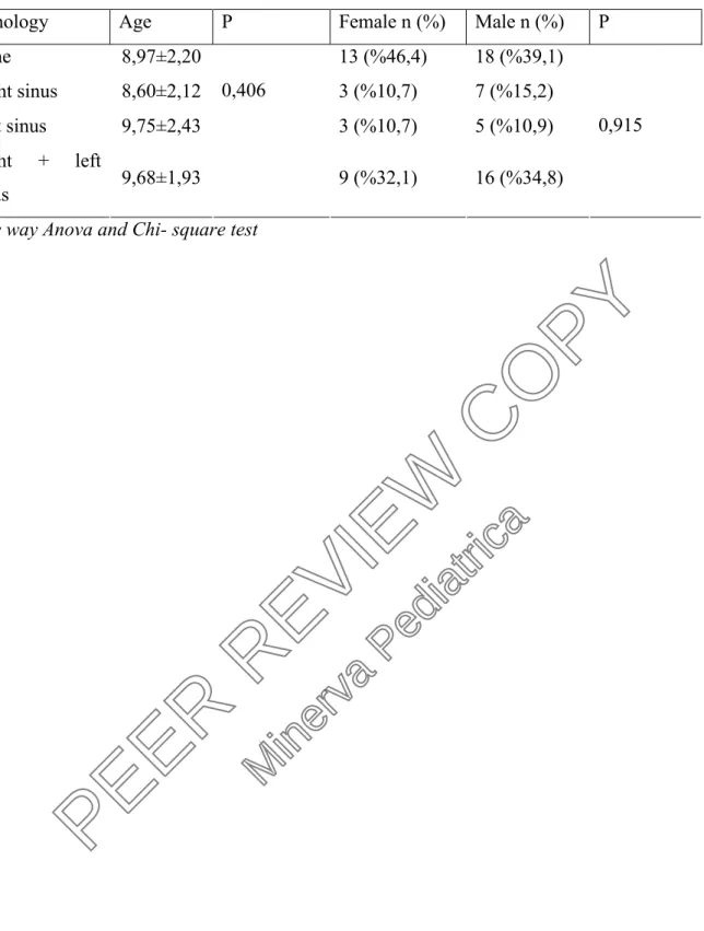
Table 1: Evaluation of the pathologies via age and gender.
Table 2: Distribution of the pathologies via gender.
PEER REVIEW COPY
Minerva Pediatrica
7 8 9 10 11 12 13 14 15 16 17 18 19 20 21 22 23 24 25 26 27 28 29 30 31 32 33 34 35 36 37 38 39 40 41 42 43 44 45 46 47 48 49 50 51 52 53 54 55 56 57 58 59 60 61 62 63 64 65 66 67 68 69 70 81 82 83 84 85 86 87 88 89 90 91 92 93 94 95
Table 1: Evaluation of the pathologies via age and gender
Pathology Age P Female n (%) Male n (%) P
None 8,97±2,20 13 (%46,4) 18 (%39,1) Right sinus 8,60±2,12 3 (%10,7) 7 (%15,2) Left sinus 9,75±2,43 3 (%10,7) 5 (%10,9) Right + left sinus 9,68±1,93 0,406 9 (%32,1) 16 (%34,8) 0,915
One way Anova and Chi- square test
PEER REVIEW COPY
Minerva Pediatrica
7 8 9 10 11 12 13 14 15 16 17 18 19 20 21 22 23 24 25 26 27 28 29 30 31 32 33 34 35 36 37 38 39 40 41 42 43 44 45 46 47 48 49 50 51 52 53 54 55 56 57 58 59 60 61 62 63 64 65 66 67 68 69 70 81 82 83 84 85 86 87 88 89 90 91 92 93 94 95
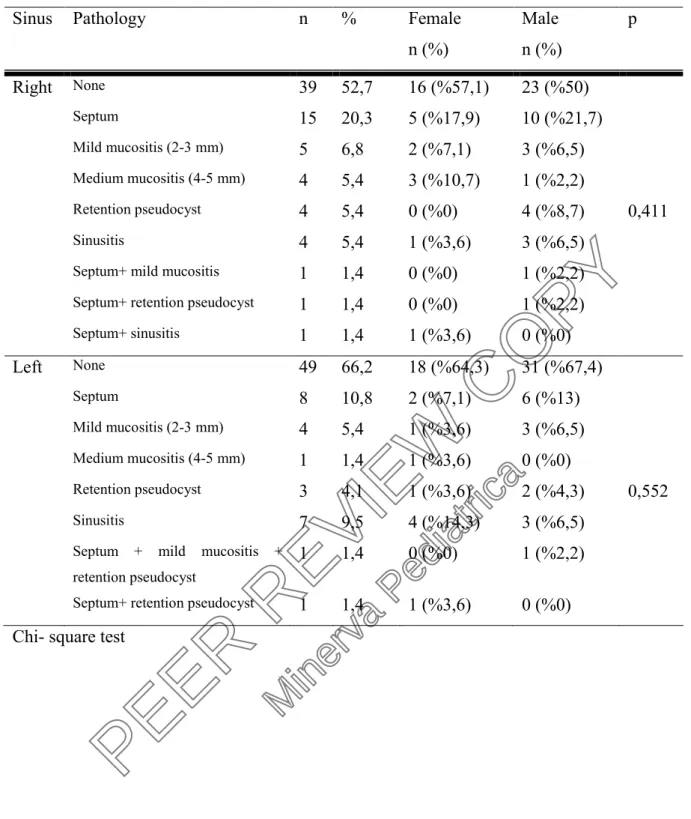
Table 2: Distribution of the pathologies via gender
Sinus Pathology n % Female
n (%) Male n (%) p Right None 39 52,7 16 (%57,1) 23 (%50) Septum 15 20,3 5 (%17,9) 10 (%21,7) Mild mucositis (2-3 mm) 5 6,8 2 (%7,1) 3 (%6,5) Medium mucositis (4-5 mm) 4 5,4 3 (%10,7) 1 (%2,2) Retention pseudocyst 4 5,4 0 (%0) 4 (%8,7) 0,411 Sinusitis 4 5,4 1 (%3,6) 3 (%6,5)
Septum+ mild mucositis 1 1,4 0 (%0) 1 (%2,2)
Septum+ retention pseudocyst 1 1,4 0 (%0) 1 (%2,2)
Septum+ sinusitis 1 1,4 1 (%3,6) 0 (%0) Left None 49 66,2 18 (%64,3) 31 (%67,4) Septum 8 10,8 2 (%7,1) 6 (%13) Mild mucositis (2-3 mm) 4 5,4 1 (%3,6) 3 (%6,5) Medium mucositis (4-5 mm) 1 1,4 1 (%3,6) 0 (%0) Retention pseudocyst 3 4,1 1 (%3,6) 2 (%4,3) 0,552 Sinusitis 7 9,5 4 (%14,3) 3 (%6,5)
Septum + mild mucositis + retention pseudocyst
1 1,4 0 (%0) 1 (%2,2)
Septum+ retention pseudocyst 1 1,4 1 (%3,6) 0 (%0)
Chi- square test
PEER REVIEW COPY
Minerva Pediatrica
7 8 9 10 11 12 13 14 15 16 17 18 19 20 21 22 23 24 25 26 27 28 29 30 31 32 33 34 35 36 37 38 39 40 41 42 43 44 45 46 47 48 49 50 51 52 53 54 55 56 57 58 59 60 61 62 63 64 65 66 67 68 69 70 81 82 83 84 85 86 87 88 89 90 91 92 93 94 95
PEER REVIEW COPY
Minerva Pediatrica
7 8 9 10 11 12 13 14 15 16 17 18 19 20 21 22 23 24 25 26 27 28 29 30 31 32 33 34 35 36 37 38 39 40 41 42 43 44 45 46 47 48 49 50 51 52 53 54 55 56 57 58 59 60 61 62 63 64 65 66 67 68 69 70 81 82 83 84 85 86 87 88 89 90 91 92 93 94 95PEER REVIEW COPY
Minerva Pediatrica
7 8 9 10 11 12 13 14 15 16 17 18 19 20 21 22 23 24 25 26 27 28 29 30 31 32 33 34 35 36 37 38 39 40 41 42 43 44 45 46 47 48 49 50 51 52 53 54 55 56 57 58 59 60 61 62 63 64 65 66 67 68 69 70 81 82 83 84 85 86 87 88 89 90 91 92 93 94 95Page 16 of 16
PEER REVIEW COPY
Minerva Pediatrica
7 8 9 10 11 12 13 14 15 16 17 18 19 20 21 22 23 24 25 26 27 28 29 30 31 32 33 34 35 36 37 38 39 40 41 42 43 44 45 46 47 48 49 50 51 52 53 54 55 56 57 58 59 60 61 62 63 64 65 66 67 68 69 70 81 82 83 84 85 86 87 88 89 90 91 92 93 94 95