http://journals.tubitak.gov.tr/medical/ © TÜBİTAK
doi:10.3906/sag-1307-30
Surgery for intractable temporal lobe epilepsy: experience of a single institution
Gökhan KURT1, Mehmet TÖNGE2, Emrah ÇELTİKÇİ1,*, Irem ÇAPRAZ3, Ayşe SERDAROĞLU4, Erhan BİLİR3
1Department of Neurosurgery, Faculty of Medicine, Gazi University, Ankara, Turkey 2Department of Neurosurgery, Faculty of Medicine, Medipol University, İstanbul, Turkey
3Department of Neurology, Faculty of Medicine, Gazi University, Ankara, Turkey 4Department of Pediatric Neurology, Faculty of Medicine, Gazi University, Ankara, Turkey
1. Introduction
As one of the world’s oldest recognized conditions, epileptic disorder affects approximately 50 million people all around the world. Whereas 90% of patients are diagnosed in developing countries, 40 to 70 per 100 million people are annually diagnosed in developed countries (1). Currently, surgical treatment has an important place in the management of epilepsy in accordance with the technical improvements in modern medicine, which leads to patients’ quality of life improving (2–4). Encouraging results have been achieved, especially for surgical treatment of temporal lobe epilepsy (TLE).
The first attempts for surgical treatment of TLE, including hippocampectomy, were reported by Penfield in 1950. Penfield and Baldwin also reported the first anterolateral temporal lobectomy including the hippocampus and amygdalae in 3 patients in 1952, and they initially mentioned the pathological term “incisural
sclerosis” as an atrophic lesion in 2 of them (5). Falconer et al. in 1964 and Margerison and Corsellis in 1966 showed that hippocampal sclerosis is a common finding in TLE specimen series (6). Research for less invasive approaches led surgeons to develop more conservative approaches. Niemeyer described transcortical amygdalohippocampectomy in 1958, followed by Yasargil’s microsurgical transsylvian amygdalohippocampectomy in 1973, which are commonly known as “selective amygdalohippocampectomy” (SAH) (7,8). Mesial TLE is the most common form of partial epilepsies in adults. Even though benign cases were reported by Labate et al., the seizures are often resistant to antiepileptic drugs (AEDs) (9,10). Drug resistance must be diagnosed as early as possible, because the ensuing seizures can be eliminated surgically via temporal lobe surgery in a high percentage (70%–90%) of patients (11). However, mesial temporal sclerosis (MTS) is the most common pathology identified
Background/aim: In the treatment of epilepsy, encouraging results have been achieved with surgical treatment, especially for temporal
lobe epilepsy (TLE). Drug resistance must be diagnosed as early as possible, because the ensuing seizures can be eliminated surgically via temporal lobe surgery in a high percentage (70%–90%) of patients. In this study we share our experience, in a single institution, of surgical treatment of intractable TLE.
Materials and methods: Between March 2006 and November 2010 we performed 127 corticoamygdalohippocampectomy (CAH)
procedures. All CAH surgical procedures were done as described by Niemeyer’s technique. Resection lengths were 4–4.5 cm from the temporal pole.
Results: At the end of 24 months, 79.7% (n = 94) patients were still on antiepileptic medications, with 55 of them on a decreased number
or dose of drugs, and 20.3 (n = 24) patients were antiepileptic drug-free. Postoperative Engel’s classes were 1, 2, and 3 in 87.2%, 5.08%, and 7.6%, respectively. There was no mortality in follow-up, and dysphasia in 1 patient (0.84%) was the only morbidity.
Conclusion: In our series we found that the outcome of surgery is associated with careful patient selection, which requires a detailed
investigation of the patients. Our final conclusion is that outcome scores are independent of age, pathology, or sex but are dependent on correct patient selection.
Key words: Corticoamygdalohippocampectomy, epilepsy surgery, hippocampal sclerosis, mesial temporal lobe, outcome, temporal
lobectomy
Received: 08.07.2013 Accepted: 09.09.2013 Published Online: 15.08.2014 Printed: 12.09.2014
in these patients and the exact role of this lesion in the pathogenesis of refractory epilepsy remains unclear (10).
The aim of this study is to share our experience in a single institution, the Gazi University School of Medicine’s Department of Neurosurgery, of the surgical treatment of intractable TLE.
2. Materials and methods
We performed surgical procedures for epileptic disorders in the Gazi University Hospital between March 2006 and November 2010 including vagal nerve stimulation (n = 68), callosotomy (n = 2), lesionectomy via stereotactic procedures (n = 22), extratemporal resection (n = 6), and corticoamygdalohippocampectomy (CAH) (n = 127). All corticoamygdalohippocampectomy surgical procedures were performed as described by Niemeyer’s technique. Resection lengths were 4–4.5 cm from the temporal pole. A descriptive retrospective cohort study was carried out using data acquired from the epilepsy surgery database of our center, conducted by analyzing patients who had undergone CAH between March 2006 and November 2010 for intractable TLE. Only patients who reliably attended outpatient follow-up visits for at least 24 months were included (n = 118). Engel’s scale was used to assess the patients, systematically applied by a neurologist and neurosurgeon during outpatient follow-up appointments.
Patients who did not attend outpatient appointments reliably during the follow-up period were excluded (n = 9). Patient records were collected from our institution’s database (AviCenna, DataSel Computing Systems Corp.) and statistical analysis was performed using SPSS 11.0.0 for Windows (SPSS Inc.) via one-sample t-test, one-way ANOVA, and paired-samples test. Surgical specimens were studied by neuropathologists at our institution. Cases were classified as mesial sclerosis, cortical dysplasia, without any pathological finding, and other pathologies (e.g., vascular malformation) according to histopathological examinations. All patients were evaluated pre- and postoperatively via standard electroencephalography (EEG) and long-term video EEG monitoring, and, if required, invasive EEG monitoring was performed for some patients by epileptologists. Even though the results
were not analyzed in this study, all patients were evaluated by neuropsychiatric testing by neuropsychiatrists pre- and postoperatively. Additionally, 3-T cerebral and high-resolution temporal magnetic resonance imaging (MRI), which included coronal sections of the hippocampus in FLAIR and SPACIR sequences (3 mm) for anatomic view, and fluorodeoxyglucose-positron emission tomography (FDG-PET) scan and magnetic resonance spectroscopy for hippocampal metabolism, were performed on all patients. A Wada test was not performed for those with language-dominant-side epilepsy; instead, we performed functional MRI. All patients had a control brain computed tomography scan within the first 24 h following surgery. Outpatient follow-ups were set at the 2nd, 6th, 12th, and 24th months consecutively and Engel’s scores were used in outcome classifications routinely as follows: class 1, no seizures or simple partial seizures only; class 2, 90% or more seizure frequency reduction; class 3, seizure frequency reduction from 50% to 90%; class 4: seizure frequency reductions lower than 50% or no worthwhile reduction. Continuation status of medications was also recorded in detail.
3. Results
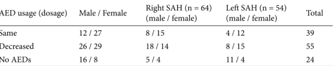
Of the 118 patients, 54 were male and 64 were female. Mean follow-up period was 30.08 ± 12.44 (range: 12–55) months after surgery. Mean age at the time of the surgery was 26.99 ± 9.1 (range: 6–59) years. At the end of 24 months, 79.7% (n = 94) of patients were still on antiepileptic medications, with 55 of them using a decreased number or dose of drugs, and 20.3% (n = 24) were AED-free (Table 1). Pathological examination findings were mesial sclerosis in 59.3% (n = 70), cortical dysplasia in 4.2% (n = 5), no pathological finding in 25.4% (n = 30), and other pathologies (cavernous malformation, glioma, etc.) in 11% (n = 13). Postoperative Engel’s class was 1, 2, and 3 in 87.2%, 5.08%, and 7.6%, respectively. No patient had an Engel’s class of 4 (Table 2). There was no mortality in follow-up, and dysphasia in 1 patient (0.84%) was the only morbidity. Surgery-related complications were not seen. One-sample t-test showed no significant difference between sexes for having intractable TLE (P = 0.054).
Table 1. Clinical data of the SAH patient groups with regards to AED usage at the end of 24 months.
AED usage (dosage) Male / Female Right SAH (n = 64)(male / female) Left SAH (n = 54)(male / female) Total
Same 12 / 27 8 / 15 4 / 12 39
Decreased 26 / 29 18 / 14 8 / 15 55
No AEDs 16 / 8 5 / 4 11 / 4 24
One-way ANOVA showed no significant relation between age and postoperative Engel’s score (P = 0.943) and no significant relation between sex and postoperative Engel’s score (P = 0.516). Paired-samples test showed no relation between pathology and postoperative Engel’s score (P = 0.235). There was also no significant relation between pathology and postoperative drug usage (P = 0.046). Engel class 1 patients were considered as having effective surgical outcome and the surgical treatment was found to be significantly effective within the cohort (P < 0.01).
4. Discussion
Surgical treatment for intractable TLE is a very effective modality for carefully selected candidates at experienced centers. Patient selection and management require collaboration among the neurosurgeon, epileptologist, neuroradiologist, neuropsychiatrist, and nuclear medicine physician. When deciding on surgery, the criteria for the selection of the appropriate surgical technique remain unclear. Some tend to perform SAH, especially for left-sided TLE, and CAH is reserved for right-left-sided cases (12). In our series, all CAH procedures were performed by the same neurosurgeon (the first author), and a more conservative corticectomy was performed for dominant-sided patients. A review by Schramm showed similar seizure outcomes for both SAH and CAH, with probable better cognitive outcomes in SAH (8). Another study comparing SAH, anterior temporal lobectomy, lesionectomy, and neocorticectomy found that SAH is related to better seizure outcome (13). The terms “anterior temporal lobectomy” and “corticoamygdalohippocampectomy” seem to be synonyms in the literature. We prefer to use “corticoamygdalohippocampectomy”, which describes the technique more accurately. However, studies comparing SAH and CAH by Ozkara et al. and Bate et al. found CAH more effective on seizure control, while other studies by Sagher et al. and Tanriverdi et al. found no significant difference (12,14–16). There is also still no consensus on the extent of resection. The main goal of CAH is to excise the amygdalae, hippocampus, and entorhinal cortex (12). However, Yasargil et al. proposed not excising the posterior one-third of the hippocampus and the parahippocampus because of the possible harm to short-term memory and
probable visual loss due to lack of vascular supply to the lateral geniculate body via damage on anterior choroidal artery branches. They also suggested that this causes the least damage to neopallial cortical-subcortical regions (T1–4) owing to a transsylvian approach (7). Spencer et al. showed that 20% of patients have seizures arising from the posterior hippocampus itself (17). Chabardés et al. divided TLE patients into 2 groups according to ictal EEG records. The first group of patients had simultaneously arising seizures both in the mesial temporal lobe (hippocampus) and temporal pole, while the second group had seizures arising within the hippocampus and spreading over the temporal pole seconds after. This led to the conclusion that SAH might be less effective for the first group of patients (18). An MRI-based volumetric analytic study by Sagher et al. showed rates of the extent of resection of the mesial structures as 98% and 91% for CAH and SAH, respectively, which was statistically slightly higher for the CAH group. On the other hand, their study did not show any seizure outcome differences between the 2 techniques (12). Many authors accept the sum of Engel class 1 and 2 patients as “favorable outcome”, which ranges between 35% and 90% in different studies and was found to be 93.2% in our study. Our study showed no relation between age and Engel’s score, similar to the study of McIntosh et al., while Jeong et al. found age as one of the predictors of seizure outcome (19,20). The most common pathological finding in our series was MTS (59.3%), similar to various other studies. However, we found no relation between seizure outcome and histopathology, and some studies suggest better seizure control results with MTS (13). Schijins et al. found no relation between hippocampal sclerosis and normal pathology patients in terms of seizure control, similar to our study. They found that the seizure onset in hippocampal sclerosis patients was significantly lower than in patients with normal pathology results (21). Yasargil et al. divided MTS into 4 categories in terms of underlying pathology: macrolesions (tumors, mass-like vascular lesions), microlesions (sclerosis, gliosis, volume reduction, hypometabolism), no visible lesions (not shown on MRI or FDG-PET scan, but proven on EEG), and infectious (e.g., viral encephalitis). Seizure control rates were reported as 96%, 90%, and 42.8% for macrolesions, microlesions, and no visible lesions, respectively (7).
Only 1 out of 118 patients had a postoperative complication: dysphasia following a left-sided CAH. Pre- and postoperative neuropsychiatric assessment results were not included in this study. We did not observe any previously described complications related to SAH including infection, hemorrhagic infarct, cranial nerve palsies, subdural effusion, hemiparesis, or clinical psychiatric syndromes in our entire series (22). Mostly, verbal memory deficits following left-sided CAH and
Table 2. Relation between pathology and outcome.
Engel’s class 1 2 3 Mesial sclerosis (n = 70) 66 4 0 Cortical dysplasia (n = 5) 2 1 2 No pathological findings (n = 30) 25 0 5 Other pathologies (n = 13) 10 1 2
spatial memory deficits following right-sided CAH have been reported (23). On the other hand, recurrent seizures might also result in deterioration of both verbal and spatial memory functions over time. Some reports show improvement in both verbal and spatial memory functions following SAH (1,24). Despite the risk of morbidity and mortality of surgery, epileptic seizures themselves have a higher risk, and surgical treatment has favorable and cost-effective results in comparison with AEDs (25).
In conclusion, temporal lobe epilepsy is a common type of epileptic disorder, and some patients are strictly intractable to AEDs. In carefully selected patients, discontinuation rates of AEDs reach up to 90%, which is
related to a better quality of life with fewer costs and side effects. The outcome of surgical treatment of TLE has been established for more than half of a century and it has been proven as very effective and safe. Optimal management of epilepsy patients should be done in specialized centers with a collaborative epilepsy team. In our series, we found that surgery outcome was associated only with careful patient selection, which requires a detailed investigation of patients by the team work of the neuroradiologist, neurologist, neuropsychiatrist, and neurosurgeon. As a result, our final conclusion is that outcome scores are independent of age, pathology, or sex, while correct patient selection is the key point.
References
1. Morino M, Ichinose T, Uda T, Kondo K, Ohfuji S, Ohata K. Memory outcome following transsylvian selective amygdalohippocampectomy in 62 patients with hippocampal sclerosis. J Neurosurg 2009; 110: 1164–1169.
2. Engel J Jr, Driver MV, Falconer MA. Electrophysiological correlates of pathology and surgical results in temporal lobe epilepsy. Brain 1975; 98: 129–156.
3. Spencer SS, Berg AT, Vickrey BG, Sperling MR, Bazil CW, Shinnar S, Langfitt JT, Walczak TS, Pacia SV, Ebrahimi N et al. Initial outcomes in the Multicenter Study of Epilepsy Surgery. Neurology 2003; 61: 1680–1685.
4. Engel J Jr, Wiebe S, French J, Sperling M, Williamson P, Spencer D, Gumnit R, Zahn C, Westbrook E, Enos B et al. Practice parameter: temporal lobe and localized neocortical resections for epilepsy: report of the Quality Standards Subcommittee of the American Academy of Neurology, in association with the American Epilepsy Society and the American Association of Neurological Surgeons. Neurology 2003; 60: 538–547.
5. Bingaman W, Najm I. Standard temporal lobectomy. In: Winn HR. editor. Youmans Neurological Surgery. 6th ed. Philadelphia, PA, USA: Elsevier; 2011. pp. 767–773.
6. Falconer MA, Serafetinides EA, Corsellis JA. Etiology and pathogenesis of temporal lobe epilepsy. Arch Neurol 1964; 10: 233–248.
7. Yasargil MG, Krayenbühl N, Roth P, Hsu SPC, Yasargil DCH. The selective amygdalohippocampectomy for intractable temporal limbic seizures. Historical vignette. J Neurosurg 2010; 112: 168–185.
8. Schramm J. Temporal lobe epilepsy surgery and the quest for optimal extent of resection: a review. Epilepsia 2008; 49: 1296–1307.
9. Engel J Jr, Williamson PD, Wieser HG. Mesial temporal lobe epilepsy. In: Engel J, Pedley TA, editors. Epilepsy: A Comprehensive Textbook. Philadelphia, PA, USA: Lippincott-Raven Publishers; 1997. pp. 2417–2426.
10. Labate A, Ventura P, Gambardella A, Le Piane E, Colosimo E, Leggio U, Ambrosio R, Condino F, Messina D, Lanza P et al. MRI evidence of mesial temporal sclerosis in sporadic “benign” temporal lobe epilepsy. Neurology 2006; 66: 562–565. 11. Thadani VM, Williamson PD, Berger R, Spencer SS, Spencer
DD, Novelly RA, Sass KJ, Kim JH, Mattson RH. Successful epilepsy surgery without intracranial EEG recording: criteria for patient selection. Epilepsia 1995; 36: 7–15.
12. Sagher O, Thawani JP, Etame AB, Gomez-Hassan DM. Seizure outcomes and mesial resection volumes following selective amygdalohippocampectomy and temporal lobectomy. Neurosurg Focus 2012; 32: E8.
13. Dunlea O, Doherty CP, Farrell M, Fitzsimons M, O’Brien D, Murphy K. The Irish epilepsy surgery experience: long-term follow-up. Seizure 2010; 19: 247–252.
14. Ozkara C, Uzan M, Benbir G, Yeni N, Oz B, Hanoglu L. Surgical outcome of patients with mesial temporal lobe epilepsy related to hippocampal sclerosis. Epilepsia 2008; 49: 696–699. 15. Tanriverdi T, Olivier A, Poulin N, Andermann F, Dubeau F.
Long-term seizure outcome after mesial temporal lobe epilepsy surgery: corticalamygdalohippocampectomy versus selective amygdalohippocampectomy. J Neurosurg 2008; 108: 517–524. 16. Bate H, Eldridge P, Varma T, Wieshmann UC. The seizure
outcome after amygdalohippocampectomy and temporal lobectomy. Eur J Neurol 2007; 14: 90–94.
17. Spencer DD, Spencer SS, Mattson RH, Williamson PD, Novelly RA. Access to the posterior medial temporal lobe structures in the surgical treatment of temporal lobe epilepsy. Neurosurgery 1984; 15: 667–671.
18. Chabardés S, Kahane P, Minotti L, Tassi L, Grand S, Hoffmann D. The temporopolar cortex plays a pivotal role in temporal lobe seizures. Brain 2005; 128: 1818–1831.
19. Jeong SW, Lee SK, Kim KK, Kim JY, Chung CK. Prognostic factors in anterior temporal lobe resection for mesial temporal lobe epilepsy: multivariate analysis. Epilepsia 1999; 40: 1735– 1739.
20. McIntosh AM, Wilson SJ, Berkovic SF. Seizure outcome after temporal lobectomy: current research practice and findings. Epilepsia 2001; 42: 1288–1307.
21. Schijins O, Bien CG, Majores M, Lehe M, Urbach H, Becker A. Presence of temporal gray-white matter abnormalities does not influence epilepsy surgery outcome in temporal lobe epilepsy with hippocampal sclerosis. Neurosurgery 2011; 68: 98–107. 22. Ipekdal HI, Karadas O, Erdogan E, Gokcil Z. Spectrum of
surgical complications of temporal lobe epilepsy surgery: a single-center study. Turk Neurosurg 2011; 21: 147–151.
23. Dulay MF, Levin HS, York MK, Li X, Mizrahi EM, Goldsmith I. Changes in individual and group spatial and verbal learning characteristics after anterior temporal lobectomy. Epilepsia 2009; 50: 1385–1395.
24. Paglioli E, Palmini A, Portuguez M, Paglioli E, Azambuja N, Costa JC. Seizure and memory outcome following temporal lobe surgery: selective compared with nonselective approaches for hippocampal sclerosis. J Neurosurg 2006; 104: 70–78. 25. Schmidt D, Stavem K. Long-term seizure outcome of surgery
versus no surgery for drug-resistant partial epilepsy: a review of controlled studies. Epilepsia 2009; 50: 1301–1309.