TRAUMA
Pediatric open-globe injuries: clinical characteristics and factors
associated with poor visual and anatomical success
Sabahattin Sul1,4&Gökhan Gurelik2&Safak Korkmaz3&
Sengül Ozdek2&Berati Hasanreisoglu2
Received: 24 November 2014 / Revised: 2 June 2015 / Accepted: 9 June 2015 / Published online: 5 July 2015 # Springer-Verlag Berlin Heidelberg 2015
Abstract
Purpose We determine the clinical characteristics, visual and anatomical outcomes, and factors associated with poor visual outcome and anatomical success in pediatric open-globe inju-ries (OGI).
Methods The records of patients aged 17 years and younger who had undergone surgical intervention after OGI between January 2000 and June 2010 were reviewed retrospectively. Results In total, 182 patients were enrolled in the study (70.9 % males, 29.1 % females; mean age: 8.6±4.5 years [range 1– 17 years]). Injuries occurred mostly at home (45.1 %). Objects most often causing the ocular trauma were sharp ob-jects (74.1 %). Over one-third of the eyes (39.7 %) had final visual acuities of 20/200 or better. Anatomical success was achieved in 76.9 % of eyes. Time between primary globe repair and secondary surgical intervention was 61.4±144 days. Visual outcomes were poor in eyes with initial visual acuity less than for counting fingers, retinal detachment, proliferative vitreoretinopathy, and endophthalmitis. Anatomical success rates were low with retinal detachment.
Conclusion Visual outcomes seem to be poor in more than half of the patients despite advanced microsurgical techniques.
Keywords Open globe injury . Retinal detachment . Endophthalmitis
Introduction
Ocular trauma is the leading cause of monocular vision loss in children [1,2]. Although eye injuries occur suddenly and un-expectedly, previous studies have indicated that up to 90 % of eye injuries are preventable with education, adult supervision, and protective eyewear [3]. Despite advanced medical and surgical techniques and increased understanding of the mech-anisms of trauma, amblyopia seems to be a challenging prob-lem during rehabilitation after ocular trauma [4].
Ocular trauma may cause either open-globe injuries (OGI) or closed-globe injuries (CGI). CGI constitute a large propor-tion of mechanical globe injuries in childhood, but visual and anatomical outcomes seem to be poorer in OGI than CGI [5–9]. There have been limited and conflicting studies on the prognosis of OGI and factors affecting the prognosis [4–6,
10–16]. Therefore, there is still a need for detailed reports to increase our understanding of this potentially sight-threatening injury.
The aim of this study is to determine the clinical character-istics, visual and anatomical outcomes, and factors associated with poor visual and anatomical outcomes in OGI.
Materials and methods
We reviewed the records of the Gazi University Faculty of Medicine (Ankara, Turkey), Department of Ophthalmology, from the period January 2000 until June 2010 for consecutive patients aged 17 years and younger who needed surgical in-tervention after OGI.
* Sabahattin Sul [email protected]
1
Department of Ophthalmology, Muğla Sıtkı Koçman University
School of Medicine, Muğla, Turkey
2
Department of Ophthalmology, Gazi University Medical School, Ankara, Turkey
3
Department of Ophthalmology, Düzce State Hospital, Düzce, Turkey
4 Göz Hastalıkları Anabilim Dalı, Muğla Sıtkı Koçman University,
Written informed consent was obtained from all of the pa-tients or their relatives. Our study was reviewed and approved by the ethics committee of the Gazi University Faculty of Medicine.
Epidemiological characteristics such as age, gender, caus-ative agent, and place were reviewed. We determined clinical characteristics (anterior and posterior segment findings), ini-tial and final visual acuities (VAs), mean number of surgeries, follow-up time, and mean age at last follow-up from each patient’s records. Initial VA was defined as VA at presentation before the primary globe repair. Initial VAs of the referrals were accessed from the records of other clinics. VAs were measured using a Snellen chart. OGI was classified according to Birmingham Eye Trauma Terminology [10]. The time be-tween the trauma and the primary repair of the globe could be determined only in eyes that were operated on at Gazi University. The time between the primary surgical repair and the secondary surgical intervention was identified in each pa-tient. All patients who were under the age of 10, had strict amblyopia treatment with best-spectacle correction and occlu-sion therapy after surgical interventions.
Poor visual outcome was defined as a final VA less than 20/ 200, according to a previous study [4]. Anatomical success was defined as persisting retinal attachment and/or no phthisis bulbi at the last follow-up visit. We analyzed the factors asso-ciated with poor visual outcome and anatomical success.
We summarized the data using descriptive statistics (e.g., mean, standard deviation, frequency, percentage). We used Student’s t-test to determine and compare the means, and we used the chi-squared (χ2)
test for the categorical data. The clinical variables that were associated with visual outcome and anatomical success in theχ2test were included in a re-gression model to better delineate the effects of the clinical variables on visual outcome and anatomical success. The Statistical Package for the Social Sciences (SPSS) software (version 16.0 for Windows, SPSS Inc., Chicago, Illinois, USA) was used for all of the analysis. A p value of less than 0.05 was considered to indicate statistical significance.
Results
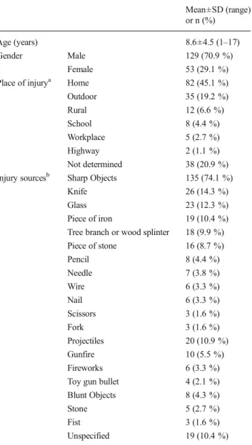
We identified 182 eyes in 182 patients during the 10-year period. There were 129 males (70.9 %) and 53 females (29.1 %) (male:female ratio: 2.4:1). The mean age of the co-hort was 8.6±4.5 years (range; 1–17 years). The mean age at the last follow-up was 10.1±4.3 years (range; 1–19 years).
The place of injury was determined in 144 patients (79.1 %). Most of the injuries occurred at home (45.1 %) (Table1). In 163 patients, the injury sources were identified: 135 sharp objects (74.1 %) [the most frequent sharp objects were knives in 26 injuries (14.3 %)], 20 projectiles (10.9 %), and 8 blunt objects (4.3 %) (Table1).
Forty-three eyes with OGI (23.6 %) had undergone prima-ry surgical repair at our clinic. Twenty-one out of 43 eyes underwent only primary surgical repair (20 eyes underwent primary surgical repair and one eye underwent primary surgi-cal repair combined with lensectomy and intraocular lens im-plantation). Twenty-two out of 43 eyes needed further surgical management. The remaining 139 eyes underwent primary sur-gical repair at other clinics and were referred to our hospital for further surgical management.
All patients underwent primary surgical repair within 48 hours of initial trauma. One hundred and sixty-three pa-tients (89.5 %) presented within 24 hours, 19 (10.5 %) papa-tients between 24–48 hours of trauma. The mean time between the trauma and the primary surgical repair was 12.3±5.5 hours
Table 1 Demographical characteristics of patients (n=182)
Mean±SD (range) or n (%)
Age (years) 8.6±4.5 (1–17)
Gender Male 129 (70.9 %)
Female 53 (29.1 %)
Place of injurya Home 82 (45.1 %)
Outdoor 35 (19.2 %) Rural 12 (6.6 %) School 8 (4.4 %) Workplace 5 (2.7 %) Highway 2 (1.1 %) Not determined 38 (20.9 %)
Injury sourcesb Sharp Objects 135 (74.1 %)
Knife 26 (14.3 %)
Glass 23 (12.3 %)
Piece of iron 19 (10.4 %)
Tree branch or wood splinter 18 (9.9 %)
Piece of stone 16 (8.7 %) Pencil 8 (4.4 %) Needle 7 (3.8 %) Wire 6 (3.3 %) Nail 6 (3.3 %) Scissors 3 (1.6 %) Fork 3 (1.6 %) Projectiles 20 (10.9 %) Gunfire 10 (5.5 %) Fireworks 6 (3.3 %)
Toy gun bullet 4 (2.1 %)
Blunt Objects 8 (4.3 %)
Stone 5 (2.7 %)
Fist 3 (1.6 %)
Unspecified 19 (10.4 %)
a
The place of injury was determined in 144 patients
b
(range; 5–35 hours) in 43 patients who were operated on at Gazi University. We could not determine the mean time be-tween the trauma and the primary surgical repair in referrals due to lack of information for the exact primary surgical repair time in some patients.
One hundred and sixty-one eyes underwent secondary sur-gical interventions (surgeries for cataract, secondary lens im-plantation, vitreous hemorrhage, retinal detachment [RD], in-traocular foreign body [IOFB] removal and endophthalmitis). The mean time between the primary repair of the globe and the secondary surgical interventions was 61.4±144 days (range 2–1460 days). The mean follow-up time was 18.2 ± 17.8 months (range; 6–120 months).
Ocular findings and visual outcomes
One hundred and forty-three eyes (78.5 %) had penetrating injuries, 12 eyes (6.7 %) had ruptures, and 27 eyes (14.8 %) had IOFB. There were no perforating injuries.
The most common ocular findings were corneal lacerations or scars (58.7 %) and RD (51.6 %) (Table2).
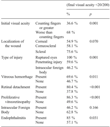
Initial VA was determined in 166 patients and final VA was determined in 174 patients at the last follow-up (Table3). The initial VA was 20/200 or better in ten eyes (5.4 %). At the last follow-up visit, 69 eyes (37.9 %) had a final VA of 20/200 or better. Patients who had initial VAs counting fingers (CF) or greater had significantly better final VAs than patients who had initial VAs less than CF (63.4 % vs. 32 %) (Table4).
The mean patient age was 9.9±4.6 years in eyes that had final VAs of 20/200 or better and 8.3±4.1 years in eyes that had final VAs less than 20/200; the difference was statistically significant (t-test, p=0.013).
One hundred and thirty-four eyes (73.6 %) had posterior seg-ment involveseg-ment, and 48 eyes (26.4 %) had isolated anterior
segment injury. Final VA was less than 20/200 in 96 out of 130 eyes (73.8 %) with posterior segment involvement and in nine out of 44 eyes (20.5 %) with isolated anterior segment injury (p<0.001). All patients who needed secondary vitreoretinal in-terventions were operated on within two weeks of the primary repair of the globe or admission to our hospital. The mean num-ber of vitreoretinal surgeries in eyes with posterior segment in-jury was 1.52±0.68 (range: 1–3). Five eyes underwent primary globe repair in our clinic but they were concluded to be inoper-able for the secondary reconstructive surgery.
Vitreous hemorrhage was detected in 56 eyes. There was accompanying RD in 36 eyes and proliferative vitreoretinopathy
Table 2 Ocular findings of open globe injuries
Type of injury Ocular findings n (%)
Open globe injury Corneal injury 107 (58.7 %)
Retinal detachment 94 (51.6 %) Traumatic cataract 74 (40.7 %) Vitreous hemorrhage 56 (30.8 %) Proliferative vitreoretinopathy 52 (28.6 %) Scleral injury 44 (24.2 %) Iris injury 36 (19.7 %) Corneoscleral injury 31 (17 %)
Intraocular foreign body 27 (14.8 %)
Endophthalmitis 22 (12.1 %)
Lens subluxation 2 (1.1 %)
Hyphema 20 (10.9 %)
Choroidal injurya 11 (6 %)
a
Choroidal hemorrhage and choroidal detachment
Table 3 Initial and final visual acuity in open globe injury
Visual acuity Initial Last Follow-up
n % n % NLP 11 6.0 % 23 12.6 % LP 45 24.7 % 19 10.4 % HM 69 37.9 % 15 8.2 % CF 31 17.0 % 48 26.4 % 20/200 and above 10 5.4 % 69 37.9 % Not determined 16 18.8 % 8 4.4 % Total 182 100 % 182 100 %
NLP No light perception, LP Light perception, HM Hand move, CF Counting fingers
Table 4 Effects of clinical variables on visual outcome (Univariate
analysis-x2)
(final visual acuity <20/200)
% p
Initial visual acuity Counting fingers
or greater 36.6 % 0.001 Worse than counting fingers 68 % Localization of the wound Corneal 54.9 % 0.070 Corneoscleral 58.1 % Scleral 75.6 %
Type of injury Ruptured eyes 100 % 0.001
Penetrating injury 59.6 %
Intraocular foreign body
46.2 %
Vitreous hemorrhage Present 69.6 % 0.011
None 46.7 %
Retinal detachment Present 80.4 % <0.001
None 37.8 %
Proliferative vitreoretinopathy
Present 86.3 % <0.001
None 49.6 %
Intraocular Foreign Present 46.2 % 0.166
Body None 62.8 %
Endophthalmitis Present 85 % 0.031
(PVR) in 15 eyes. Final VA was 20/200 or better in 17 eyes (30.4 %).
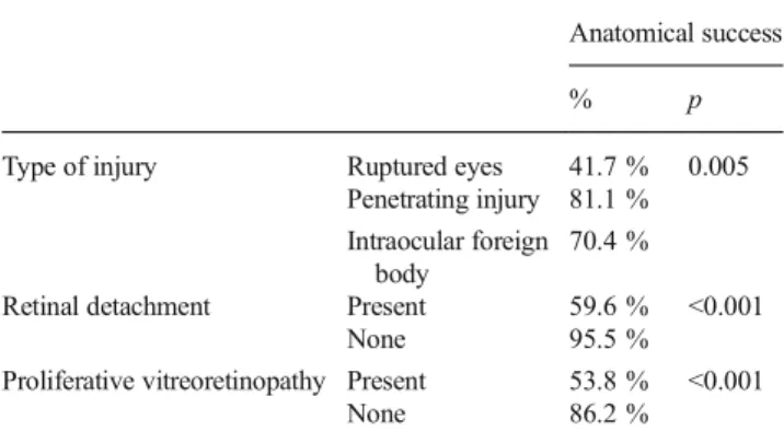
RD was seen in 94 eyes (51.6 %). Eighty eyes (85 %) had rhegmatogen RD and 14 eyes (15 %) had combined rhegmatogen and tractional RD. Macula was involved in all patients. RD was detected immediately after primary surgical repair in 22 eyes (23.4 %) and during the control examinations in the remaining 72 eyes (76.4 %). In 31 eyes (32.9 %), there was total RD. Retinal incarceration was detected in 13 eyes (13.8 %) and retinal dialysis in 11 eyes (11.7 %). PVR was detected in 52 eyes (55.3 %). The mean number of surgeries in eyes with RD was 1.69±0.72 (range; 1–3). The mean number of surgeries in eyes with PVR was 1.78±0.75 (range; 1–3). In 18 eyes (19.6 %), the final VA was 20/200 or better. In 56 eyes (59.6 %), ical success was achieved. In 28 out of 56 eyes (50 %), anatom-ical success was achieved with one surgery.
The following types of IOFB were identified; nine metallic foreign bodies, four gunshots , four pieces of stone, three nails, two pieces of fireworks, two pieces of wire, one air-gun shot, and one piece of glass. Two eyes underwent primary repair at our clinic, and 25 eyes underwent primary repair at other clinics and were sent for IOFB removal. The mean time for IOFB removal was 21.3±14.7 days (range; 1–60 days). Of the eyes with IOFB, endophthalmitis was seen in four eyes (14.8 %). All of these eyes had been injured with a metallic foreign body. In 14 eyes (53.8 %), the final VA was 20/200 or better.
Endophthalmitis was seen in 22 patients (12.1 %). Four eyes had undergone primary surgical repair at Gazi University. Three out of four eyes had undergone primary surgical repair more than 24 hours after the trauma. In these four eyes, mean time between the trauma and the primary surgical repair was 27.7±5.9 hours (range; 22–35 hours). Of the referrals, 13 out of 18 eyes underwent primary surgical repair more than 24 hours, and five out of 18 eyes within 24 hours after the trauma. The mean time between the trauma and the primary surgical repair could not be determined due to lack of information about the exact primary repair time in some patients. In three eyes, intra-vitreal tap and vancomycin (1 mg/ml)-ceftazidime (2.25 mg/ml) injections were performed. In 19 eyes vitrectomy combined with intravitreal vancomycin-ceftazidime was performed for the reasons of IOFB removal, RD, and vitritis. The cultures were positive in four eyes; two were staphylococcus aureus, and two were staphylococcus epidermidis. RD was detected in 11 eyes (50 %); in three eyes, endophthalmitis was present, in eight eyes RD developed after endophthalmitis treatment. In three eyes (15 %), the final VA was 20/200 or greater at the final follow-up.
Factors associated with functional and anatomical success
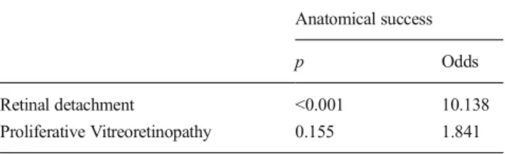
Posterior segment involvement, glob rupture, an initial VA less than CF, RD, PVR and endophthalmitis were associated with poor visual outcome (Tables4 and5). RD and glob
rupture were also associated with anatomical failure (Tables 6and 7). Furthermore, phthisis bulbi was more fre-quent in ruptures (3/12 eyes; 25 %) than penetrating injuries (8/145 eyes; 5.3 %), and the difference was statistically sig-nificant (p=0.04).
Discussion
In this retrospective study, we summarized the clinical char-acteristics of pediatric OGI in our clinic. We found that OGIs were mostly seen in males (male:female ratio: 2.4:1), as re-ported by other studies [11, 12,17]. In the literature, OGI occurs in up to 60 % of cases at home; 32–79 % of injuries caused by sharp objects [4,5,18–20]. Our findings are con-sistent with literature results that most of the injuries occurred at home (45.1 %) and were caused by sharp objects (71 %). A lack of safety, preventative measures, and adult supervision are thought to be the main reasons for the high incidence of OGI at home and injuries due to sharp objects.
A young age and low initial VA were shown to be poor prognostic factors in OGI [4,12–14]. Younger patients are more susceptible to amblyopia and are expected to have a worse visual outcome. We compared the mean age between the patients with final VA better or worse than 20/200 with the t-test (univariate analysis). The patients with worse final VA were significantly
Table 5 Effects of clinical variables on visual outcome (Multivariate
analysis-Logistic regression)
(final visual acuity <20/200)
p Odds
Initial visual acuity (<Counting fingers) 0.034 2.584
Vitreous hemorrhage 0.052 0.428
Retinal detachment 0.008 0.281
Proliferative vitreoretinopathy 0.031 0.271
Endophthalmitis 0.009 0.154
Table 6 Effects of clinical variables on anatomical success (Univariate
analysis- X2)
Anatomical success
% p
Type of injury Ruptured eyes 41.7 % 0.005
Penetrating injury 81.1 %
Intraocular foreign body
70.4 %
Retinal detachment Present 59.6 % <0.001
None 95.5 %
Proliferative vitreoretinopathy Present 53.8 % <0.001
younger than patients with better final VA. Furthermore, children with an initial VA less than CF had significantly worse final VA than children with better initial VA.
Wound localization did not significantly affect functional success, although scleral injuries (i.e., more posterior lacera-tions) were associated with poorer visual outcomes, as sug-gested by previous studies [6,15,16,21]. Injuries involving the posterior segment had a significantly worse final VA, as in the reports of Bunting et al. and Tok et al. [22,23].
Other factors associated with poor visual outcome were globe rupture, RD, PVR, and endophthalmitis. Factors asso-ciated with anatomical failure were globe rupture and RD.
Globe ruptures had poor anatomical and visual outcomes in our study, consistent with the results of Farr et al. [11] that 41 % of OGI caused by blunt trauma had a final VA less than 5/200. Furthermore, the number of phthisis bulbi was signif-icantly higher for ruptured globes.
Trauma is the most frequent cause of RD in childhood [24]. In the present study, despite the high anatomical success due to improved surgical techniques, the visual outcome was low, similar to previous studies [21,24,25]. Delayed diagnosis is a significant feature of RD. Only 23.4 % eyes had immediate diagnosis, in the remaining patients RD was detected in the control examinations. In other words, we do not definitely know how many patients had diagnoses of RD and were promptly referred for surgical management . An RD diagnosis after OGI in a pediatric population is difficult due to their lack of awareness of visual deterioration owing to young age, cor-neal scar, lens opacification, and vitreous hemorrhage. Delays in surgical intervention, which can be connected with high preoperative PVR rates in our study, may be associated with delayed diagnoses. Furthermore, half of the eyes had two or more surgeries in anatomically successful cases. Additionally, macular attachment before surgery has been associated with a good visual outcome by some previous reports [24, 26]. However in our study, all patients had macula off RD. Therefore, delays in surgical intervention, anatomical failures after surgical intervention and macula off status before surgery can cause irreversible damage to the macula and amblyopia that are thought to be the main reasons for the low functional successes in the treatment of traumatic RD [4,24,26].
PVR is seen in up to 64 % of traumatic pediatric RD cases and is the most frequent cause of anatomical failure after
surgical intervention [26]. In our study, however, anatomical success was not associated with PVR, visual outcomes were significantly lower in eyes with PVR.
In this study, the majority of the IOFBs were metallic, similar to the literature [27,28]. Some authors have advised that IOFB be removed within 24 hours due to the high risk of endophthalmitis and the risk of PVR [29,30]. However, in a recent study, there was no correlation with the time of IOFB removal and functional success [31]. In this same study, en-dophthalmitis did not develop with oral and topical antibiotic use. In our investigation, the mean time for IOFB removal seems to be long (21 days). Most of the IOFB cases were referrals and the prolonged removal time may be due to a failure to diagnose IOFB or the delay in referring patients. Despite the mean long removal time, the rate of endophthal-mitis (14.8 %) is considered to be acceptable. We consider that IOFB removal can be delayed for a while unless acute intox-ication or endophthalmitis is present.
The reported frequency of endophthalmitis after OGI varies from 6.8 % to 54.2 % [6,31,32]. In our study, endoph-thalmitis was detected in 22 patients (12.1 %). As reported in previous studies, the final VA was worse in eyes with endoph-thalmitis in this study. The cultures were positive in four vitreus specimens and all of them were staphylococcus spe-cies. Delayed repair was associated with the development of endophthalmitis [33]. We also found that, 16 out of 22 eyes had undergone primary surgical repair more than 24 hours after trauma, which reveals the importance of immediate re-pair of OGI (as shown by the literature). RD is seen in up to 57 % of eyes with endophthalmitis [34]. In our study, 11 eyes (50 %) with endophthalmitis had RD, and in eight of them RD developed after endophthalmitis treatment (late RD). This re-sult highlights the importance of following these eyes for RD development even after endophthalmitis treatment.
In conclusion, more than half of the eyes seem to be legally blind despite advanced microsurgical techniques combined with strict amblyopia treatment after OGI that were shown to be 90 % preventable. The high PVR rates in eyes with RD and the prolonged time for IOFB removal may be the result of delays at first surgical intervention after the primary repair of the globe. These high rates can be attributed to the inadequate compliance of patients in terms of follow-up examinations, delays in referring or admission to our hospital. The relatives or parents should be strictly warned and clinicians should take care to have timely examinations and surgical interventions, which is important for removing amblyogenic factors such as cata-racts, vitreous hemorrhage, and RD.
All authors certify that they have no affliations with or involvement in any organization or entity with any financial interest (such as honoraria; educational grants; participation in speaker’s bureaus; membership employment, consultancies, stock ownership, or other equity interest; and expert testimony
Table 7 Effects of clinical variables on anatomical success
(Multivariate analysis)
Anatomical success
p Odds
Retinal detachment <0.001 10.138
or patient-licensing arrangements), or non-financial interest (such as personal or professional relationships, affliations, knowledge or beliefs) in the subject matter or materials discussed in this manuscript.
References
1. May DR, Kuhn FP, Morris RE et al (2000) The epidemiology of
serious eye injuries from the United States Eye Injury Registry.
Graefes Arch Clin Exp Ophthalmol 238:153–157
2. Sternberg P, deJuan E, Michels R (1984) Penetrating ocular injuries
in young patients. Initial injuries and visual results. Retina 4:5–8
3. Coody D, Banks JM, Yetman RJ, Musgrove K (1997) Eye trauma
in children: epidemiology, management, and prevention. J Pediatr
Health Care 11:182–188
4. Jandeck C, Kellner U, Bornfeld N et al (2000) Open globe injuries
in children. Graefes Arch Clin Exp Ophthalmol 238:420–426
5. Serrano JC, Chalela P, Arias JD (2003) Epidemiology of childhood
ocular trauma in a northeastern Colombian region. Arch
Ophthalmol 121:1439–1445
6. Lee CH, Su WY, Lee L, Yang ML (2008) Pediatric ocular trauma in
Taiwan. Chang Gung Med J 31:59–65
7. Strahlman E, Elman M, Daub E et al (1990) Causes of pediatric eye
injuries. A population based study. Arch Ophthalmol 108:603–606
8. Karaman K, Znaor L, Lakos V, Olujic I (2009) Epidemiology of
pediatrice eye injury in Split-Dalmatia County. Ophthalmic Res 42: 199–204
9. Liu ML, Chang YS, Tseng SH et al (2010) Major pediatric ocular
trauma in Taiwan. J Pediatr Ophthalmol Strabismus 47:88–95
10. Kuhn F, Morris R, Witherspoon D et al (1996) A standard
classifi-cation of ocular trauma. Graefes Arch Clin Exp Ophthalmol 234: 399–403
11. Farr AK, Hairston RJ, Humayun MU et al (2001) Open globe
injuries in children: a retrospective analysis. J Pediatr Ophthalmol
Strabismus 38:72–77
12. Baxter RJ, Hodgkins PR, Calder I, Morrell AJ, Vardy S, Elkington
AR (1994) Visual outcome of childhood anterior perforating eye injuries: prognostic indicators. Eye 8:349–352
13. Gupta A, Rahman I, Leatherbarrow B (2009) Open globe injuries in
children: factors predictive of a poor final visual acuity. Eye 23:
621–625
14. Rostomianm K, Thach AB, Isfahani A, Pakkar A, Pakkar A,
Borchert M (1998) Open globe injuries in children. J AAPOS 2:
234–238
15. De Juan E, Sternberg P, Michels RG (1983) Penetrating ocular
injuries: types of injuries and visual results. Ophthalmology 90:
1318–1322
16. Lesniak SP, Bauza A, Son JH et al (2012) Twelve-year review of
pediatric traumatic open globe injuries in an urban U.S. population.
J Pediatr Ophthalmol Strabismus 49:73–79
17. Rudd JC, Jaeger EA, Freitag SK, Jeffers JB (1994) Traumatically
ruptured globes in children. J Pediatr Ophthalmol Strabismus 31:
307–311
18. Hill JR, Crawford BD, Lee H, Tawansy KA (2006) Evaluation of
open globe injuries of children in the last 12 years. Retina 26:65–68
19. MacEwen CJ, Baines PS, Desai P (1999) Eye injuries in children:
the current picture. Br J Ophthalmol 83:933–936
20. Soylu M, Demircan N, Yalaz M, Isiguzel (1998) Etiology of
pedi-atric perforating eye injuries in southern Turkey. Ophthalmic Epidemiol 5:7–12
21. Moisseiev J, Vidne O, Treister G (1998) Vitrectomy and silicone oil
injection in pediatric patients. Retina 18:221–227
22. Bunting H, Stephens D, Mireskandar K (2013) Prediction of visual
outcomes after open globe injury in children: a 17-year Canadian experience. J AAPOS 17:43–48
23. Tok O, Tok L, Ozkaya D, Eraslan E, Ornek F, Bardak Y (2011)
Epidemiological characteristics and visual outcome after open globe injuries in children. J AAPOS 15(6):556–561
24. Sarrazin L, Averbukh E, Halpert M, Hemo I, Rumelt S (2004)
Traumatic pediatric retinal detachment: a comparison between open and closed globe injuries. Am J Ophthalmol 137:1042–1049
25. Sheard RM, Mireskandari K, Ezra E, Sullivan PM (2007)
Vitreoretinal surgery after childhood ocular trauma. Eye 21:793– 798
26. Wang NK, Chen YP, Yeung L et al (2007) Traumatic pediatric
retinal detachment following open globe injury. Ophthalmologica 221:255–263
27. Greven CM, Engelbrecht NE, Slusher MM, Nagy SS (2000)
Intraocular foreign bodies: management, prognostic factors, and visual outcomes. Ophthalmology 107:608–617
28. Williams DF, Mieler WF, Abrams GW et al (1988) Results and
prognostic factors in penetrating ocular injuries with retained intra-ocular foreign bodies. Ophthalmology 95:911–916
29. Jonas JB, Budde WM (1999) Early versus late removal of
intraoc-ular foreign bodies. Retina 19:193–197
30. Jonas JB, Knorr HL, Budde WM (2000) Prognostic factors in
oc-ular injuries caused by intraococ-ular or retrobulbar foreign bodies.
Ophthalmology 107:823–828
31. Colyer MH, Weber ED, Weichel ED et al (2007) Delayed
intraoc-ular foreign body removal without endophthalmitis during Operations Iraqi Freedom and Enduring Freedom. Ophthalmology
114:1439–1447
32. Essex RW, Yi Q, Charles PG et al (2004) Post-traumatic
endoph-thalmitis. Ophthalmology 111:2015–2022
33. Narang S, Gupta V, Simalandhi P et al (2004) Paediatric open globe
injuries: visual outcome and risk factors for endophthalmitis. Indian
J Ophthalmol 52:29–34
34. Thordsen JE, Harris L, Hubbard GB (2008) Pediatric